Method and device for dividing area of image of particle in urine

a technology of image region and segmentation method, which is applied in the field of image region segmentation, can solve the problems of time-consuming examination, and achieve the effects of preventing erroneous classification of object particles, accurate binary images, and accurate calculation of feature parameters

Active Publication Date: 2011-01-06
HITACHI HIGH-TECH CORP
View PDF2 Cites 18 Cited by
  • Summary
  • Abstract
  • Description
  • Claims
  • Application Information

AI Technical Summary

Benefits of technology

[0023]With the method for region segmentation of urine particle images according to the present invention, stable region segmentation can be performed for each particle image even for a urine specimen in which urine particles having different sizes and tones coexist and thus a more accurate binary image can be obtained. This effect consequently allows accurate calculation of feature parameters of an object region, prevention of erroneous classification of an object particle, and therefore improvement of accuracy in identifying urine particles of various types.

Problems solved by technology

Such microscopic examination has such problems that (1) the result depends on the proficiency of the technician and (2) examination takes time.

Method used


Image

  • Method and device for dividing area of image of particle in urine
  • Method and device for dividing area of image of particle in urine
  • Method and device for dividing area of image of particle in urine

Examples


embodiment 1

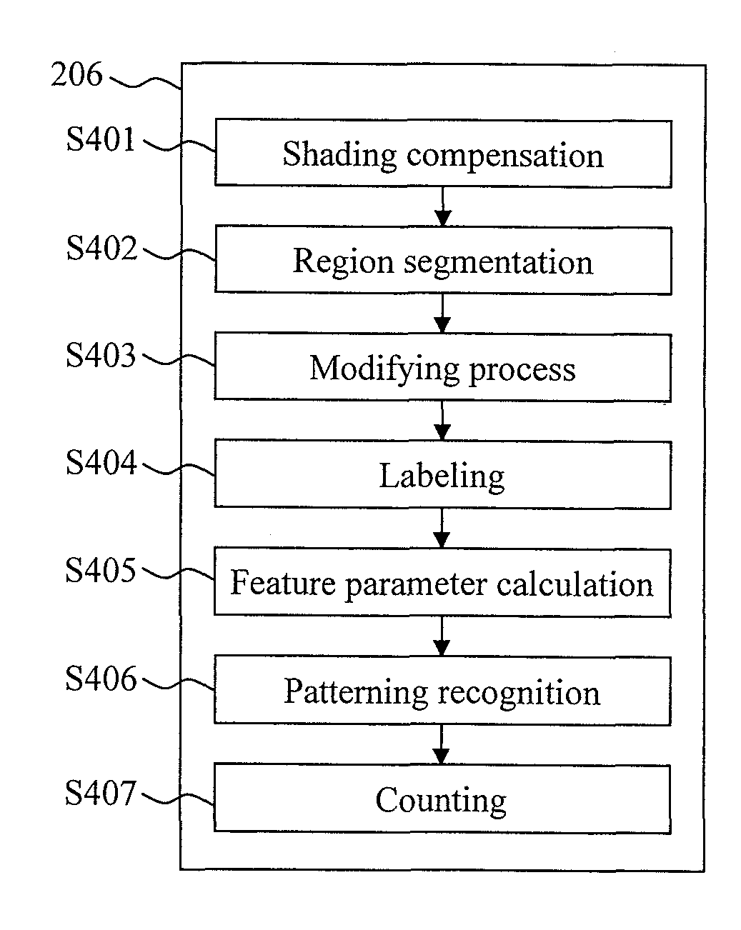
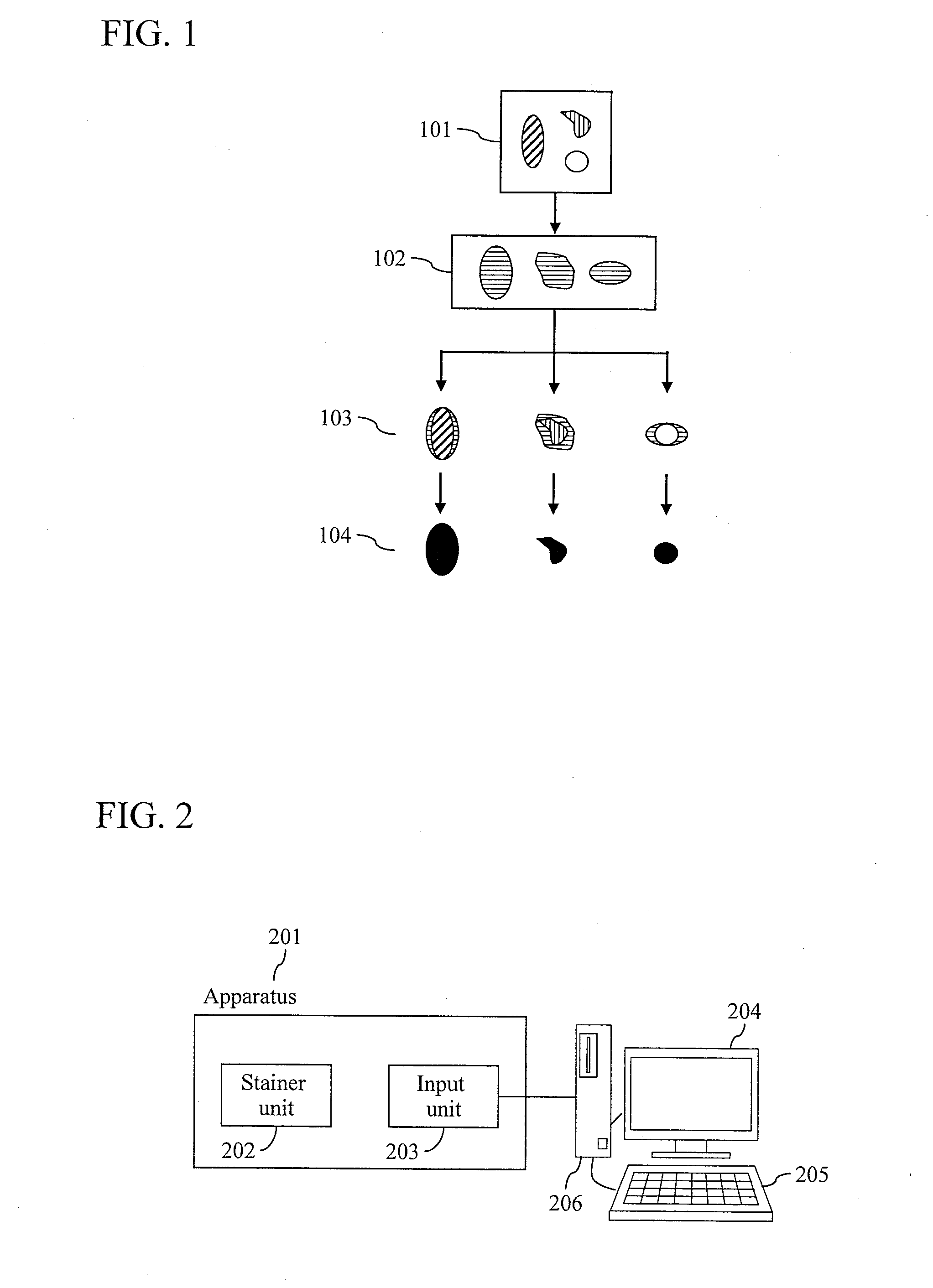
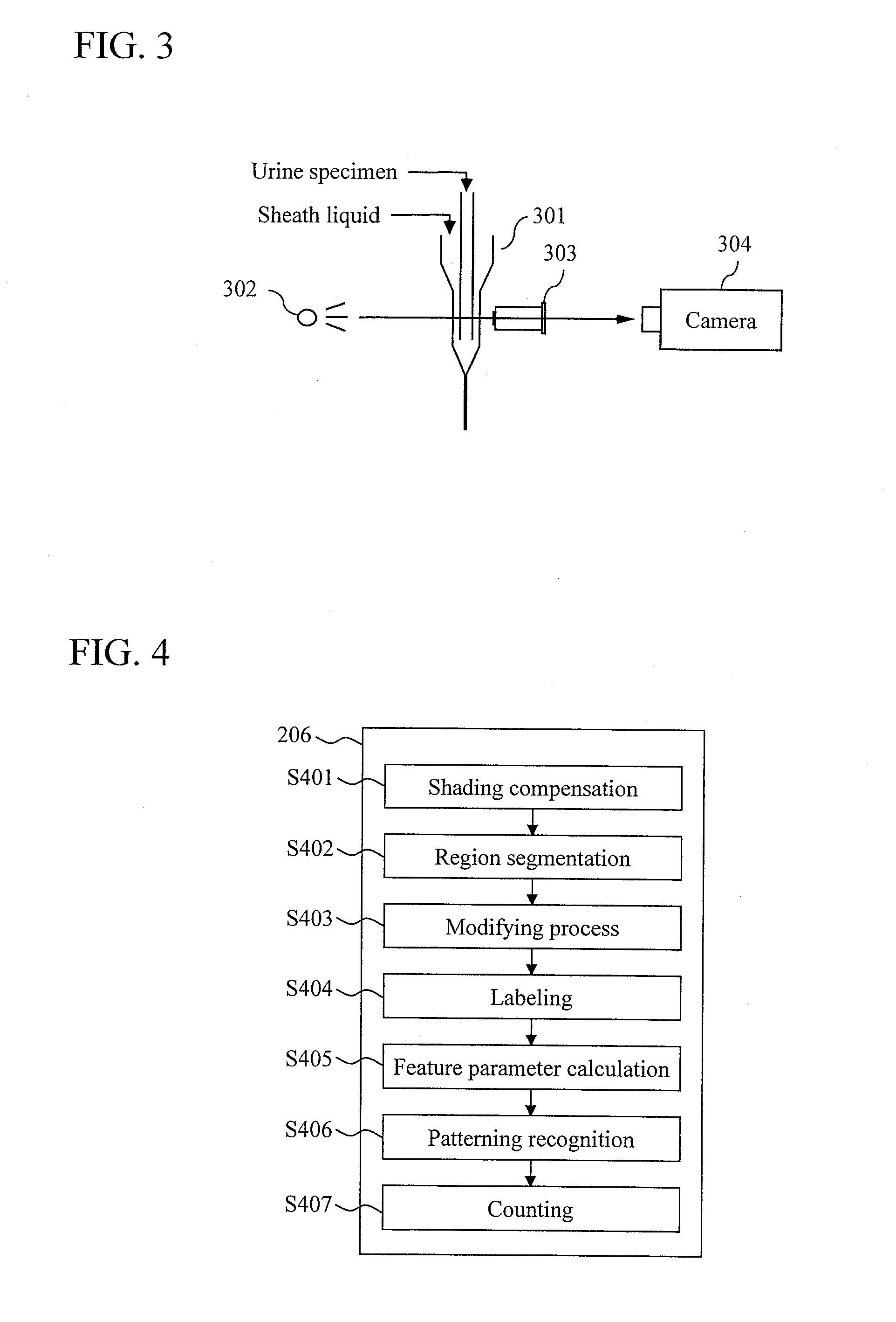
[0067]FIG. 2 is a diagram illustrating an apparatus for automatic analysis of urinary sediments, to which the present invention is applied. In an apparatus 201, a stainer unit 202 stains a urine specimen with a stain, and after a certain period of time, an input unit 203 takes an enlarged static image of particles in the urine. The image thus taken is transferred to a processing unit 206, where the urine particles are classified through image pattern recognition. The processing unit 206 then counts the types of the urine particles included in a single specimen as well as the appearance frequency of each type. For example, a general-purpose personal computer having a display 204 and a keyboard 205 is used as the processing unit 206. The operator is informed of the counting results through the display 204. The image taken by the input unit 203, data obtained by the processing unit 206, such as a measurement result, a classification result for each object region, and image feature para...

embodiment 2

[0125]FIG. 18 is a diagram illustrating the configuration of an apparatus for region segmentation of urine particle images according to a second embodiment of the present invention.

[0126]An original image taken by an input device, such as a camera, is transferred to a memory 1801. The original data is then transferred to a first region segmentation device 1802, where first region segmentation is carried out. For the first region segmentation, the method described in Embodiment 1 may be used. Images of first object regions obtained through the region segmentation are transferred to the memory 1801.

[0127]Next, the first object regions are transferred to a grouping device 1803, where feature parameters are calculated for each first object region, and the first object region is classified into a predetermined number of groups. For the grouping, the method described in Embodiment 1 may be used. Results of the grouping are transferred to the memory 1801.

[0128]A group-specific second regio...


PUM


Abstract

An objective is to provide a method and an apparatus for accurately extracting a region of an object particle from a urine particle image obtained by taking an image of urine particles in a urine specimen having varying properties. First, a first object region is extracted using one or more of an R image, a G image, and a B image of a urine particle image taken by an image input optical system configured to input particle images. Then, a density distribution and a size of the first object region of one or more of the R image, the G image, and the B image are calculated. Based on these feature parameters, the first object region is classified into a predetermined number of groups. A second object region is extracted from a local region including the first object region, by using one or more of the R image, the G image, and the B image, depending on each of the groups. This configuration allows stable region segmentation for each particle image even for a urine specimen in which urine particles having different sizes and tones coexist.

Description

TECHNICAL FIELD[0001]The present invention relates to a method for image region segmentation that uses density information, and particularly to a method and an apparatus for region segmentation favorable for segmentation of a urine particle image.BACKGROUND ART[0002]In a conventional method for morphological examination of particles in urine, an examination is performed by centrifugal separation of urine particles from collected urine, and then by direct observation of the urine particles by a laboratory technician through a microscope. Such microscopic examination has such problems that (1) the result depends on the proficiency of the technician and (2) examination takes time. Accordingly, a more efficient way is demanded.[0003]In recent years, automation of an examination of urine sediments (urine particles) has been in progress. For example, “Particle Analysis Apparatus and Particle Analysis Method (Patent documents 1 and 2)” describes a method in which a urine specimen is passed...

Claims


Application Information

Patent Timeline
Patent Type & Authority Applications(United States)
IPC IPC(8): G06T7/00G06K9/34
CPCG01N15/147G01N2015/1472G01N15/1475
Inventor MANRI, CHIHIROBAN, HIDEYUKIMITSUYAMA, SATOSHIOWADA, NORIO
Owner HITACHI HIGH-TECH CORP
Why Eureka
Try Eureka