Medical apparatus and method for attaching a suture to a bone

a technology of medical equipment and bone, applied in the field of medical equipment, can solve the problems of prone to rupture, less stability, muscle and tendons becoming thinner, etc., and achieve the effect of avoiding rupture, avoiding bruising, and avoiding bruising

Active Publication Date: 2011-01-13
T MEDICAL DEVICES AGRI COOP
View PDF18 Cites 69 Cited by
  • Summary
  • Abstract
  • Description
  • Claims
  • Application Information

AI Technical Summary

Benefits of technology

[0060]In both described preferred embodiments, the hook includes a loop movable to an extended position with respect to the hook for facilitating entry of the first end of the suture through the loop into the first bore of the bone, and to a retracted position towards the hook for clamping the first end of the suture to enable pulling-out the first end of the suture through the first bore of the bone. In addition, the handle includes a second manually manipulatable member carried by the proximal end of the handle and coupled to the handle for moving the loop to its extended and retracted positions.
[0062]According to another aspect of the present invention, there is provided a medical kit useful for attaching a suture to a bone, the kit including a medical implement as described above, and a drill receivable within the passageway of the handle for drilling the second bore in the bone; the drill including a stop engageable with the proximal end of the handle to fix the length of the second bore to extend for a slight distance past the first bore, to thereby permit the first end of the suture to traverse and extend past the first bore in order to facilitate its access and extraction via the first bore.
[0064]As will be described more particularly below, the invention enables the surgeon to perform bone tunnels in the bone in exactly the required locations to receive sutures, and thereby to achieve reattachment of the torn tendon without the use of bone anchors, in a manner similar to that of the open surgical procedure. The invention is therefore particularly useful in an arthroscopic surgical procedure, but may also be used in an open surgical procedure.

Problems solved by technology

While the shoulder joint has a great range of motion, it is not very stable.
As one ages, these muscles and tendons become thinner and prone to rupture.
In a younger patient, rupture is usually associated with significant trauma.
It is difficult to achieve exactly the desired exit points for the drill.
Drilling at a more acute angle for safety may result in being too close to the surface of the tubercle.
However, the conventional arthroscopic procedure uses anchors resulting in points or lines of attachment, rather than in attachments over a significant surface.

Method used


Image

  • Medical apparatus and method for attaching a suture to a bone
  • Medical apparatus and method for attaching a suture to a bone
  • Medical apparatus and method for attaching a suture to a bone

Examples


Embodiment Construction

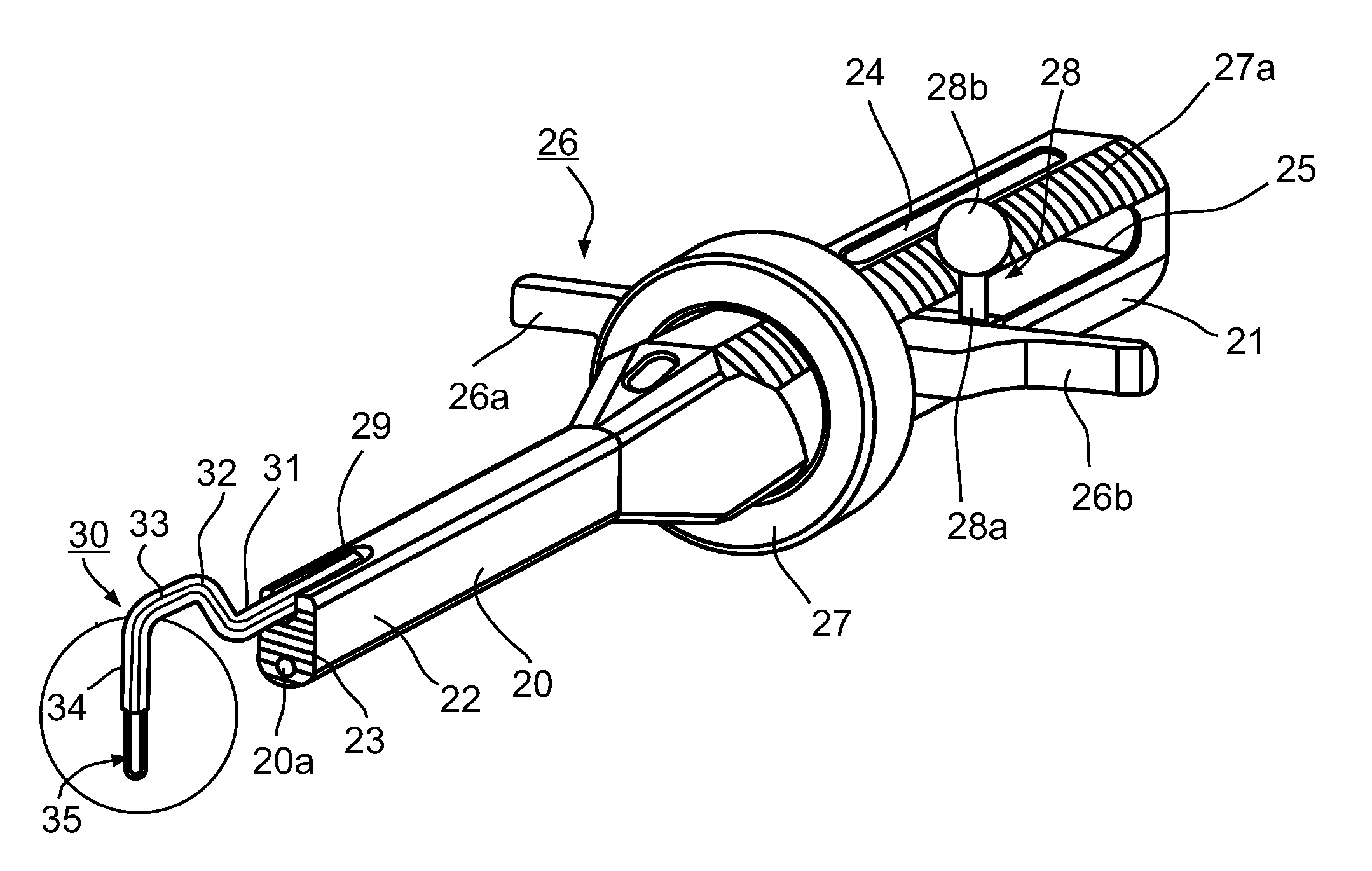
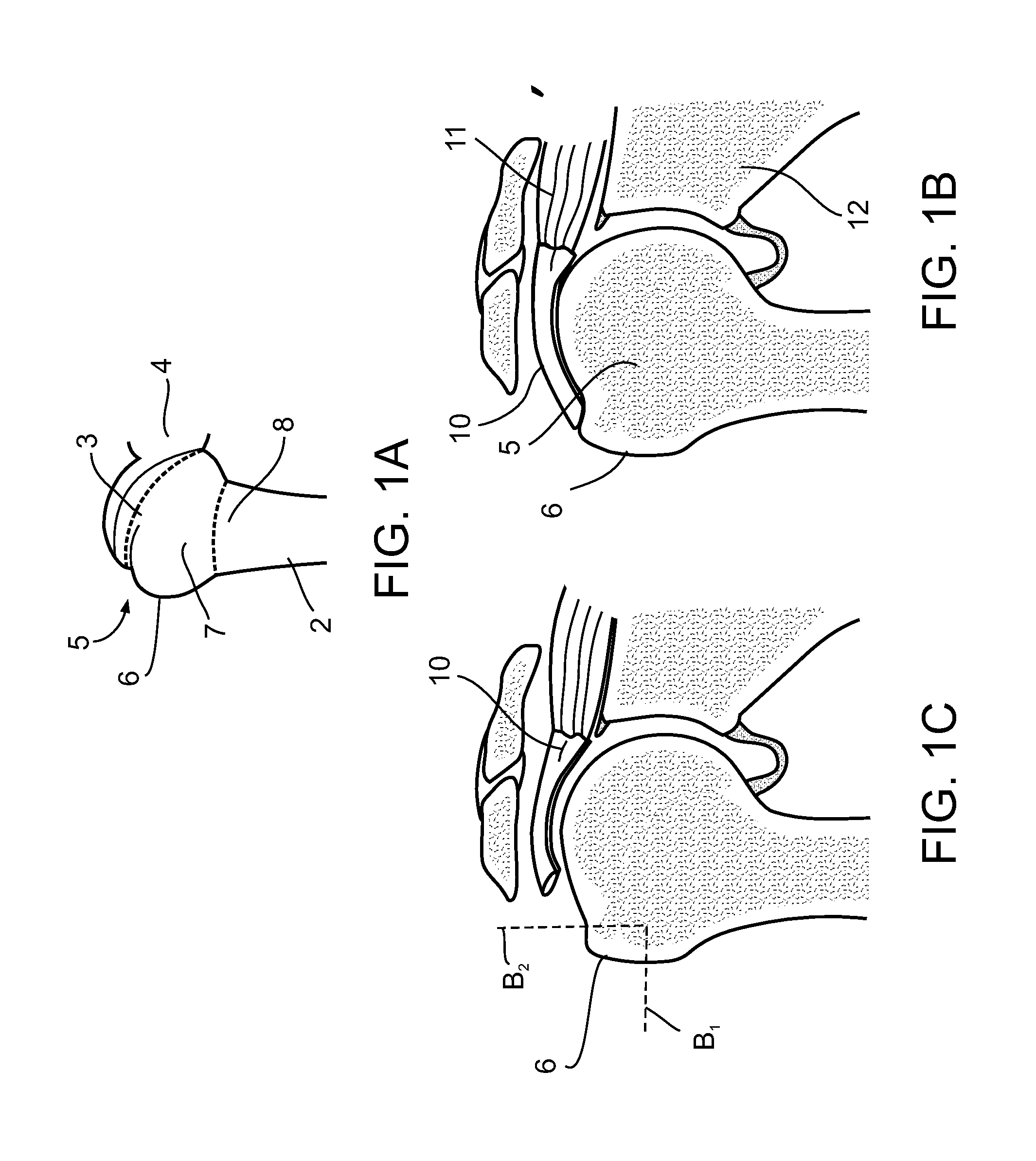
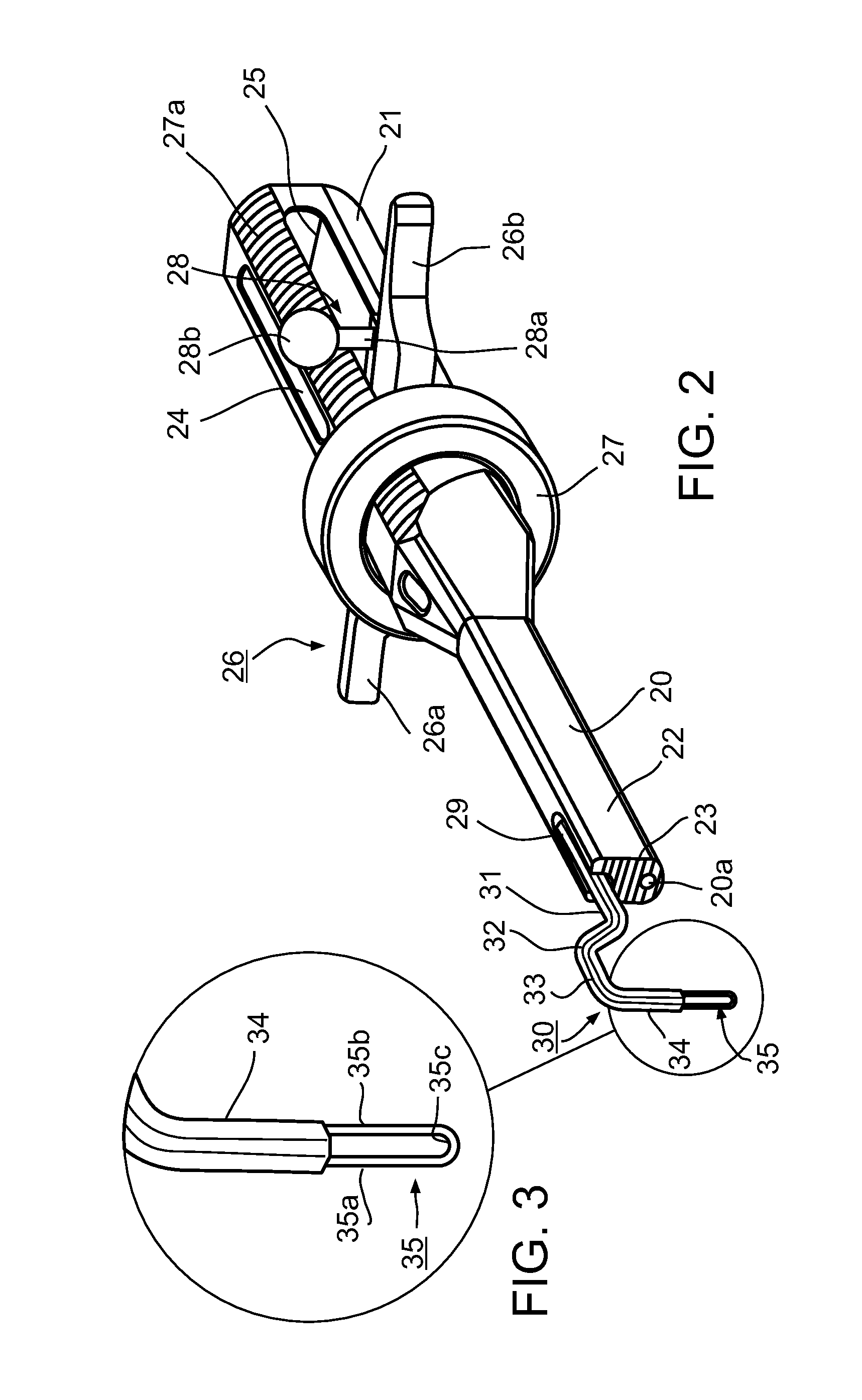
[0091]An aspect of some embodiments of the invention relates to attaching a tendon to a bone by threading a suture through a channel in the bone and through the tendon. In an exemplary embodiment of the invention, the channel comprises of a first and a second bore intersecting in the bone. In an exemplary embodiment of the invention, the intersection of the bores define a predetermined angle between them, suitable for attaching a tendon to the bone by threading a suture through the bores and the tendon. Preferably, the first and second bores are formed in an arthroscopic procedure.

[0092]In an exemplary embodiment, the first bore is formed first and is then used to assist forming the second bore. Optionally, the first bore is used as a reference point for determining the location and / or alignment of the second bore in the bone. Alternatively or additionally, the reference point is used for determining the depth of the second bore such that the first and second bores intersect in the ...


PUM


Abstract

A method of forming a channel in a bone, the method comprising: providing a first bore in the bone; and forming, a second bore in the bone at a predefined angle from said first bore, using said first bore as a reference point fordefining the location of the second bore in the bone,wherein the first and second bores intersect in the bone.

Description

FIELD AND BACKGROUND OF THE INVENTION[0001]The present invention relates to medical apparatus, particularly to a medical implement and a kit including such an implement, and also to a method, for attaching a suture to a bone. The invention is especially useful in an arthroscopic surgical procedure for attaching a tendon of a rotator cuff muscle to the humerus bone for repairing a damaged shoulder joint, and is therefore described below with respect to such a procedure.[0002]While the shoulder joint has a great range of motion, it is not very stable. Four rotator cuff muscles (supraspinatus, infraspinatus, subscapularis and teres minor) surround the shoulder joint and provide the power to lift and rotate the arm while keeping the head of the upper arm bone (humerus) in approximation to the socket in the shoulder blade (glenoid) for stability. Each of these muscles is attached by a tendon to the humerus bone. The supraspinatus muscle is attached by the supraspinatus tendon to the supe...

Claims


Application Information

Patent Timeline
Patent Type & Authority Applications(United States)
IPC IPC(8): A61B17/16A61B17/04
CPCA61B17/1796A61B17/1739A61B17/0485A61B17/0469A61B2017/1778A61B17/0482A61B17/1684A61B17/1778A61B17/0401A61B2017/00358A61B2017/00424A61B2017/0409A61B2017/0445
Inventor OREN, RANKRISHNAN, SUMANT G.RANON, LEENAHMIAS, SHAIMIROCHINIK, ARYEH
Owner T MEDICAL DEVICES AGRI COOP
Why Eureka
Try Eureka