Methods for direct biomolecule identification by matrix-assisted laser desorption ionization (MALDI) mass spectrometry

Inactive Publication Date: 2011-03-17
NEW YORK UNIV
View PDF11 Cites 7 Cited by
  • Summary
  • Abstract
  • Description
  • Claims
  • Application Information

AI Technical Summary

Benefits of technology

[0017]In one embodiment, the biological molecule is selected from the group consisting of a protein, a peptide, or fragments thereof, a nucleic acid, including both DNA and RNA, an oligonucleotide or polynucleotide, an antisense molecule, a small interfering nucleic acid molecule, such as a siRNA or shRNA, a carbohydrate, a lipid, a lipoprotein and the like. In another particular embodiment, the chemical used for dissociation of the tissue or cells is formic acid or cyanogen bromide. In another embodiment, the method may be used to study the disposition of a new chemical entity (NCE), such as a small organic molecule (synthetic or naturally derived), or new biological entity (NBE) in vitro or in vivo, whereby pre-clinical or clinical studies are necessary prior to FDA approval of a new chemical or biological entity for therapeutic use. In this manner, the methods of the present invention may be used to track the location of the NCE or NBE in vitro eg. within the cell or tissue. Alternatively, the methods of the present invention may be used to track the disposition of the NCE or NBE within the body, thus providing a new means to perform ADME studies (Absorption, Distribution, Metabolism and Excretion). In another embodiment, the methods of the present invention may prove useful when it is of interest in determining whether two or more therapeutics agents, when delivered to a cell, tissue or organ, result in a toxic effect on the cell. Accordingly, the methods of the present invention allow for potentially determining whether the use of two or more therapeutic agents may be contraindicated, based on the presence or absence of particular biomarkers after administration of the two or more therapeutic agents to a subject. Once such an effect is realized, an in vitro screen may be developed that could assess such an effect on particular cells, or tissues in culture. Thus, in vitro screening for potential toxic profiling of drugs may be envisioned using the methods of the present invention.

Problems solved by technology

However, most of these procedures utilize harsh digestion conditions and protein extraction procedures prior to analysis by MALDI-TOF methods.

Method used


Image

  • Methods for direct biomolecule identification by matrix-assisted laser desorption ionization (MALDI) mass spectrometry
  • Methods for direct biomolecule identification by matrix-assisted laser desorption ionization (MALDI) mass spectrometry
  • Methods for direct biomolecule identification by matrix-assisted laser desorption ionization (MALDI) mass spectrometry

Examples


example 1

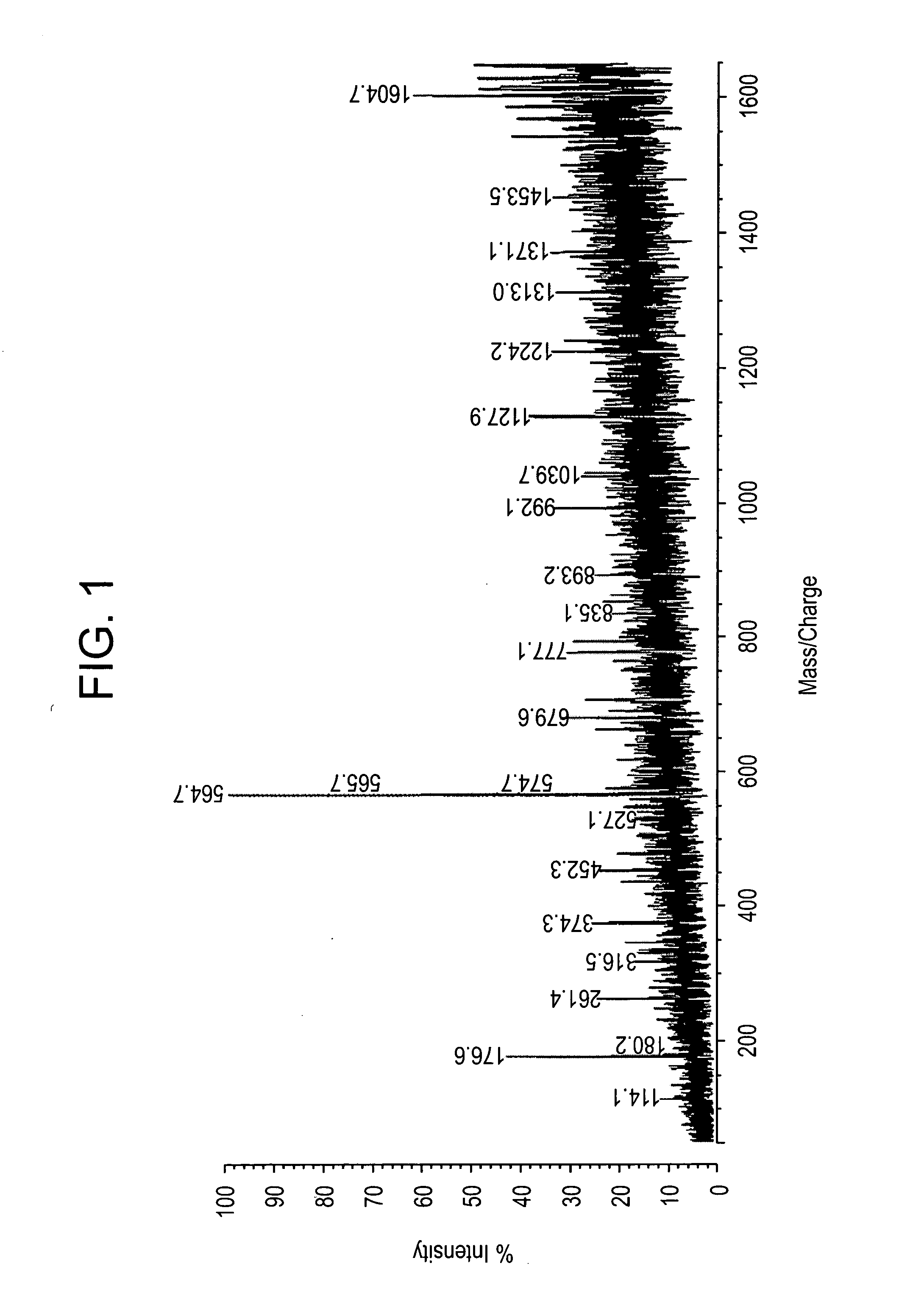
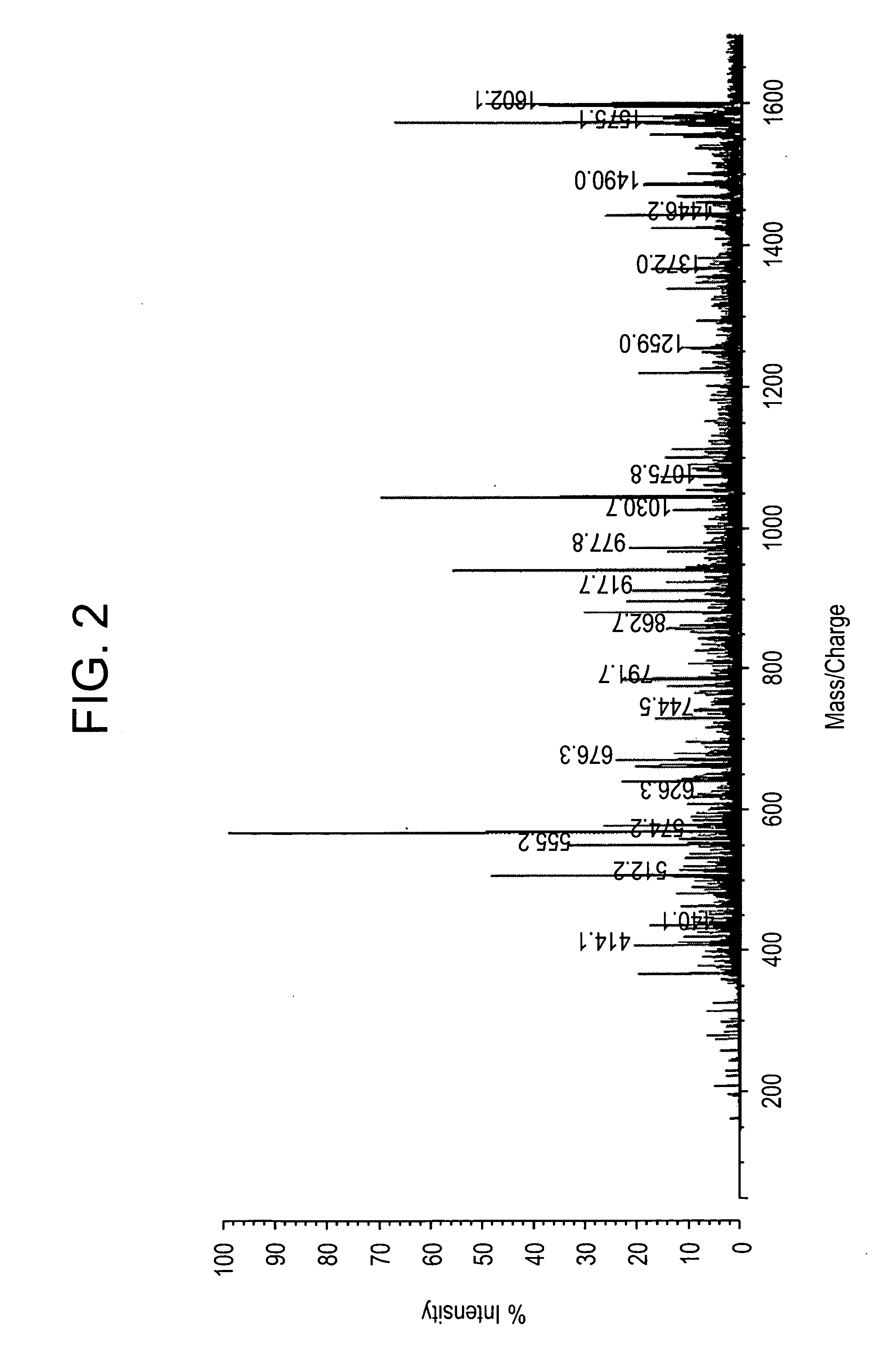
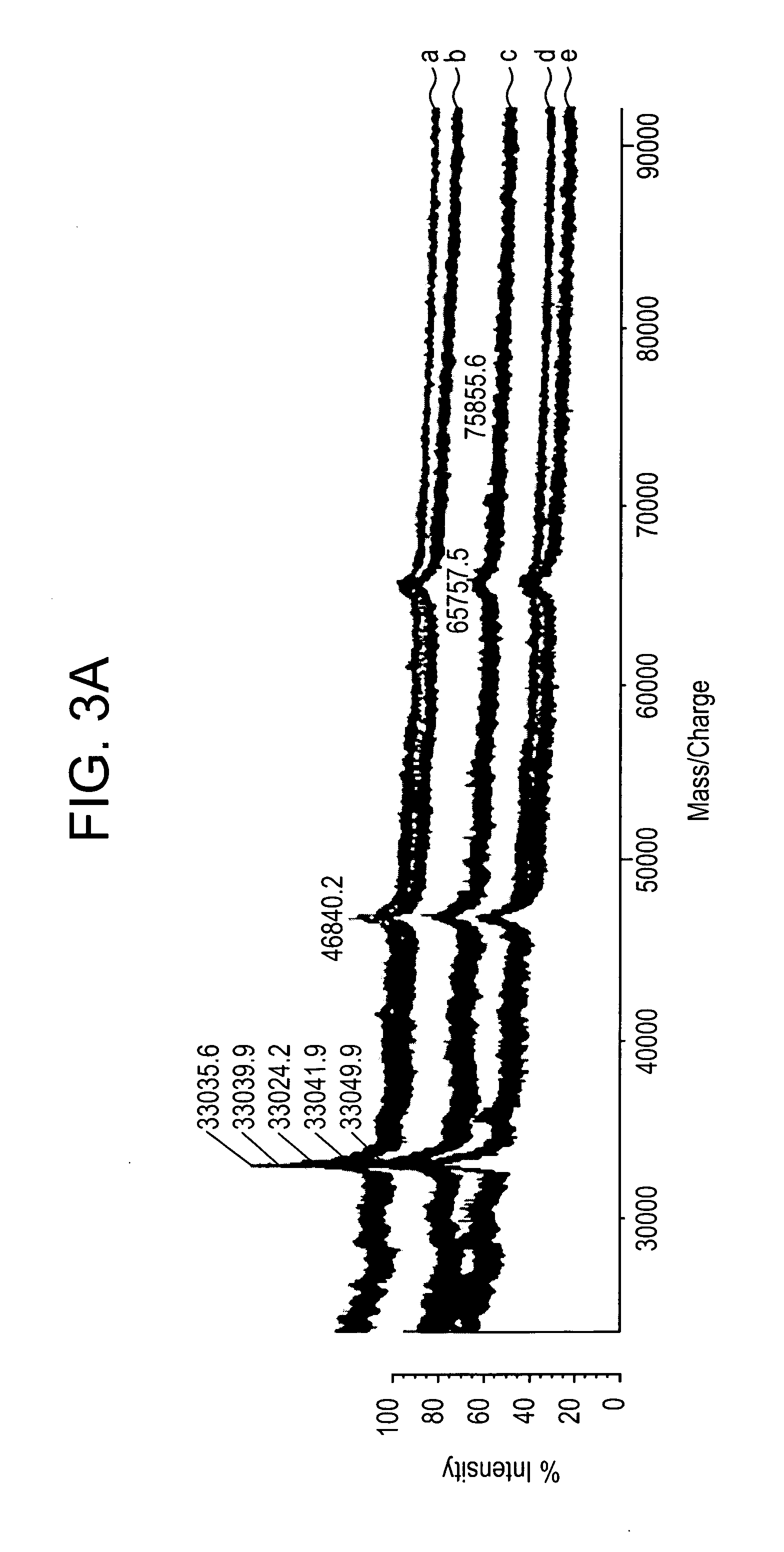
Direct MALDI Cellular and Tissue Identification of Proteins Materials and Methods

[0218]All studies were approved by NYU IRB and IACUC protocols. Chemicals were purchased from: Sigma (St. Louis, Mo., USA), Invitrogen (Carlsbad, Calif., USA), Sakura Finetek (Torrence, Calif., USA), and Scott Specialty Gasses (Plumsteadville, Pa., USA), and used without further purification. All samples were analyzed with an Axima-CFR+(PSD) or QIT (CID) (Shimadzu, Columbia, Md., USA). The instruments were calibrated with four point samples of CHCA (a-cyano-4-hydroxy-cinnamic acid): 190.04 (M+H)—monoisotopic, bradykinin Fragment 1-7: 757.40 (M+H)—monoisotopic, angiotensin II (human): 1046.54 (M+H)—monoisotopic, cytochrome C (equine): 12,361.96 (M+H)—average, aldolase (rabbit muscle): 39,212.28 (M+H)—average, and albumin (Bovine serum): 66,430.09 (M+H)—average. MALDI-PSD and CID were chosen for this study because we could study both larger proteins and significantly larger peptides.

Cells

[0219]Cells—Human...


PUM


Abstract

The present invention relates to the use of post source decay (PSD) or collision induced dissociation (CID) direct tissue (DT) MALDI-TOF or DT-MALDI-TOF-TOF mass spectrographic identification of biological molecules in a tissue or cellular sample without the need for further protein extraction. This method provides for studying cells or tissues by direct tissue MALDI (DT-MALDI), thereby substituting in situ protein release for further protein extraction. Mass / intensity data was processed with Mascot© software interrogation of the NCBI database. These results are proof of principle that DT-MALDI, combined with bioinformatics, can directly identify proteins in cells and tissues from their mass spectra.

Description

CROSS-REFERENCE TO RELATED APPLICATIONS[0001]The present application is a non-provisional application claiming the priority of copending provisional applications U.S. Ser. No. 60 / 722,206, filed Sep. 30, 2005, and U.S. Ser. No. 60 / 784,016, filed Mar. 20, 2006, the disclosures of which are incorporated by reference herein in their entireties. Applicants claim the benefits of these applications under 35 U.S.C. sctn. 119(e).FIELD OF THE INVENTION[0002]The present invention relates generally to methods for the in situ determination of the types and amount of proteins in a tissue sample by direct tissue matrix associated laser desorption ionization imaging time-of-flight (DT-MALDI-TOF) measurement. The methods allow for direct analysis of tissue samples, without the need for protein extraction and analysis of protein content of the tissue sample by other standard methods, e.g. electrophoretic analysis or other cumbersome or time consuming methods. The methods also allow for the rapid and ...

Claims


Application Information

Patent Timeline
Patent Type & Authority Applications(United States)
IPC IPC(8): H01J49/00
CPCG01N33/6851H01J49/164G01N2800/52
Inventor PEVSNER, PAULNAFTOLIN, FREDERICKMILLER, DOUGLAS C.HILLMAN, DEANSTALL, BRIAN K.WISHNIES, STEVEN M.
Owner NEW YORK UNIV
Why Eureka
Try Eureka