Computer-assisted identification and treatment of affected organ tissue

a technology of organ tissue and computer assisted identification, applied in the field of medical imaging technologies and procedures, can solve the problems of high degree of skill required to take the measurements needed to generate the 3d map, insufficient information for determining the amount of therapy to deliver, and physician ordinarily must devote a significant amount of tim

Inactive Publication Date: 2011-04-14
CELL GENETICS
View PDF3 Cites 33 Cited by
  • Summary
  • Abstract
  • Description
  • Claims
  • Application Information

AI Technical Summary

Problems solved by technology

One problem with the above approach is that a high degree of skill is required to take the measurements needed to generate the 3D map.
Another problem is that the map, even if generated by a highly skilled physician, does not accurately reveal the mass of the scar tissue, and thus does not provide sufficient information for determining the amount of therapy to deliver.
Yet another problem is that the physician ordinarily must devote a significant amount of time (typically 45 minutes or more) to generating the map.
In general, existing medical imaging technologies and procedures often do not enable physicians to determine the precise locations and boundaries of the affected organ tissue, or to accurately calculate the volume or mass of such tissue.
Without such information, the physician typically cannot accurately administer therapy, such as stem cell, gene, pharmaceutical, protein, and / or device therapy.
Existing imaging technologies used for catheterization procedures generally do not provide sufficient information for enabling physicians to accurately and reliably deliver therapy to areas of interest.

Method used


Image

  • Computer-assisted identification and treatment of affected organ tissue
  • Computer-assisted identification and treatment of affected organ tissue
  • Computer-assisted identification and treatment of affected organ tissue

Examples


Embodiment Construction

Specific medical imaging technologies and procedures will now be described for identifying, quantifying and treating myocardial infarcts or other damaged or affected organ tissue. Although the following description focuses on detecting and treating damaged tissue of the heart, as will be apparent, aspects of the disclosed methods are also applicable to disorders involving dead or damaged tissue of other organs, such as the kidneys, brain, liver, bladder, spleen, and pancreas.

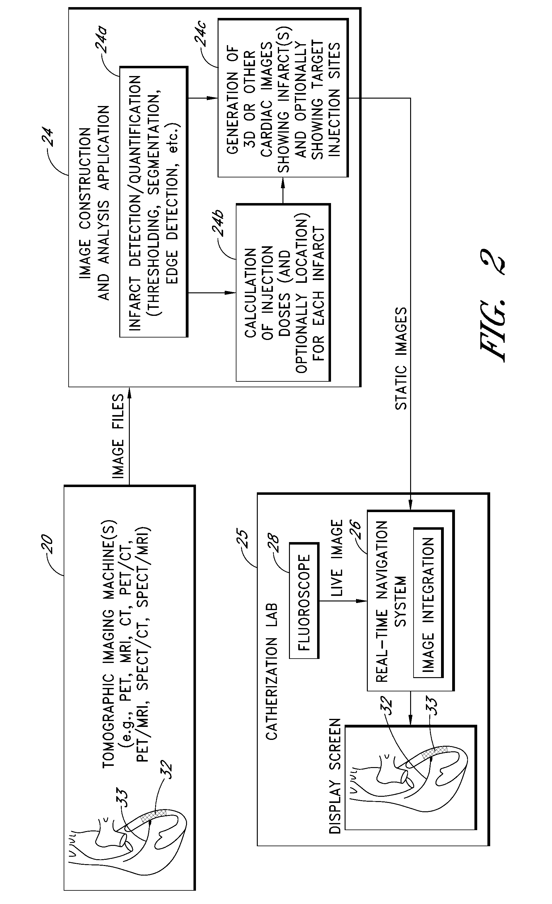
I. OVERVIEW (FIGS. 1 AND 2)

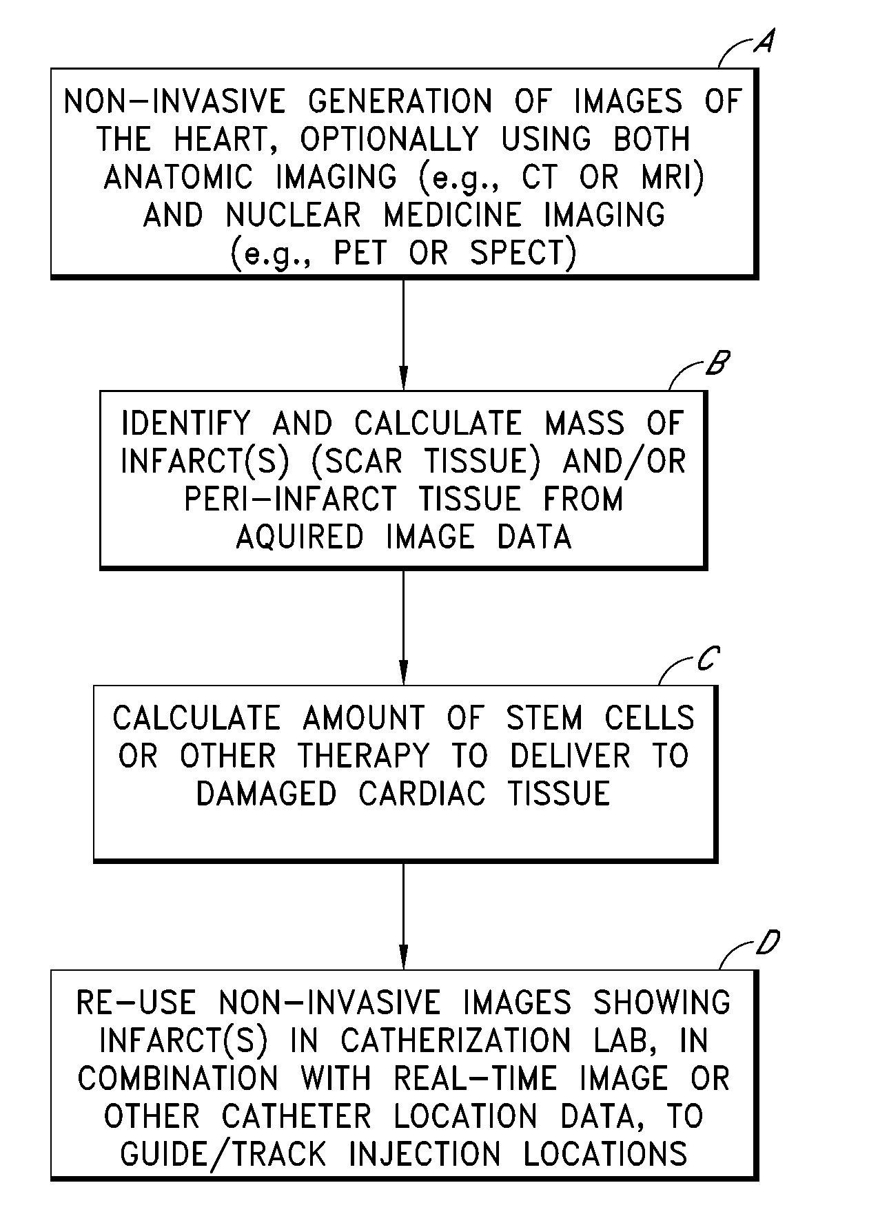
FIG. 1 illustrates an overall process, depicted as four steps or blocks A through D, for identifying, quantifying and treating one or more myocardial infarcts (also referred to as scar tissue), and / or damaged or ischemic tissue surrounding such infarcts (referred to as “peri-infarct tissue”). Example implementation details of these four steps are described in further detail in subsequent sections. As will be apparent, the process shown in FIG. 1 can also be applied to organs other than...


PUM


Abstract

Computer-assisted processes are disclosed for using nuclear medicine image studies (e.g. PET or SPECT), preferably in combination with anatomic image studies (e.g., CT or MRI), to identify and quantify regions of affected organ tissue (e.g., myocardial infarcts), and to calculate doses of stem cells or other therapy to deliver to such regions. The resulting (static) image data showing the affected tissue may also be integrated with live (moving) image data, such as a fluoroscopy image, during a subsequent interventional procedure to generate a hybrid image showing the real time location of an injection catheter relative to the affected tissue.

Description

BACKGROUND OF THE INVENTION1. Field of the InventionThis disclosure relates to medical imaging technologies and procedures for identifying and quantifying myocardial infarcts and / or other areas of affected organ tissue, and for delivering stem cell therapy, gene therapy, protein therapy, pharmaceutical therapy, device therapy, and / or other types of therapy to the affected tissue.2. Description of the Related ArtA myocardial infarct or scar is a localized area of dead or damaged myocardial tissue resulting from a heart attack. A myocardial infarct may be treated by injecting an appropriate therapeutic substance, such as stem cells or a pharmaceutical compound, into the damaged tissue using an injection catheter.A known procedure for identifying and treating myocardial infarcts involves the use of the NOGA™ Cardiac Navigation system to generate a three dimensional (3D) map of the heart. The physician initially uses a special catheter system to generate measurements of electrical activ...

Claims


Application Information

Patent Timeline
Patent Type & Authority Applications(United States)
IPC IPC(8): A61B5/055A61B5/053G16H30/40G16H50/20G16Z99/00
CPCA61B5/053G06T2207/30048A61B6/037A61B6/12A61B6/463A61B6/481A61B6/503A61B6/504A61B6/5217A61B6/5235A61B6/5247A61B6/541A61B2017/00247A61B2018/00392A61B2019/5251A61B2019/5291A61M25/0084A61M2025/0089G06F19/321G06F19/3437G06F19/3468G06T7/0012G06T7/0083G06T7/0097G06T2207/10072G06T2207/10084G06T2207/10121A61B5/415A61B2034/2051A61B2090/365G06T7/12G06T7/174G16H50/50G16H30/40G16H50/20G16Z99/00
Inventor KORN, RONALD L.NATHAN, MARK D.DIB, NABIL
Owner CELL GENETICS
Why Eureka
Try Eureka