

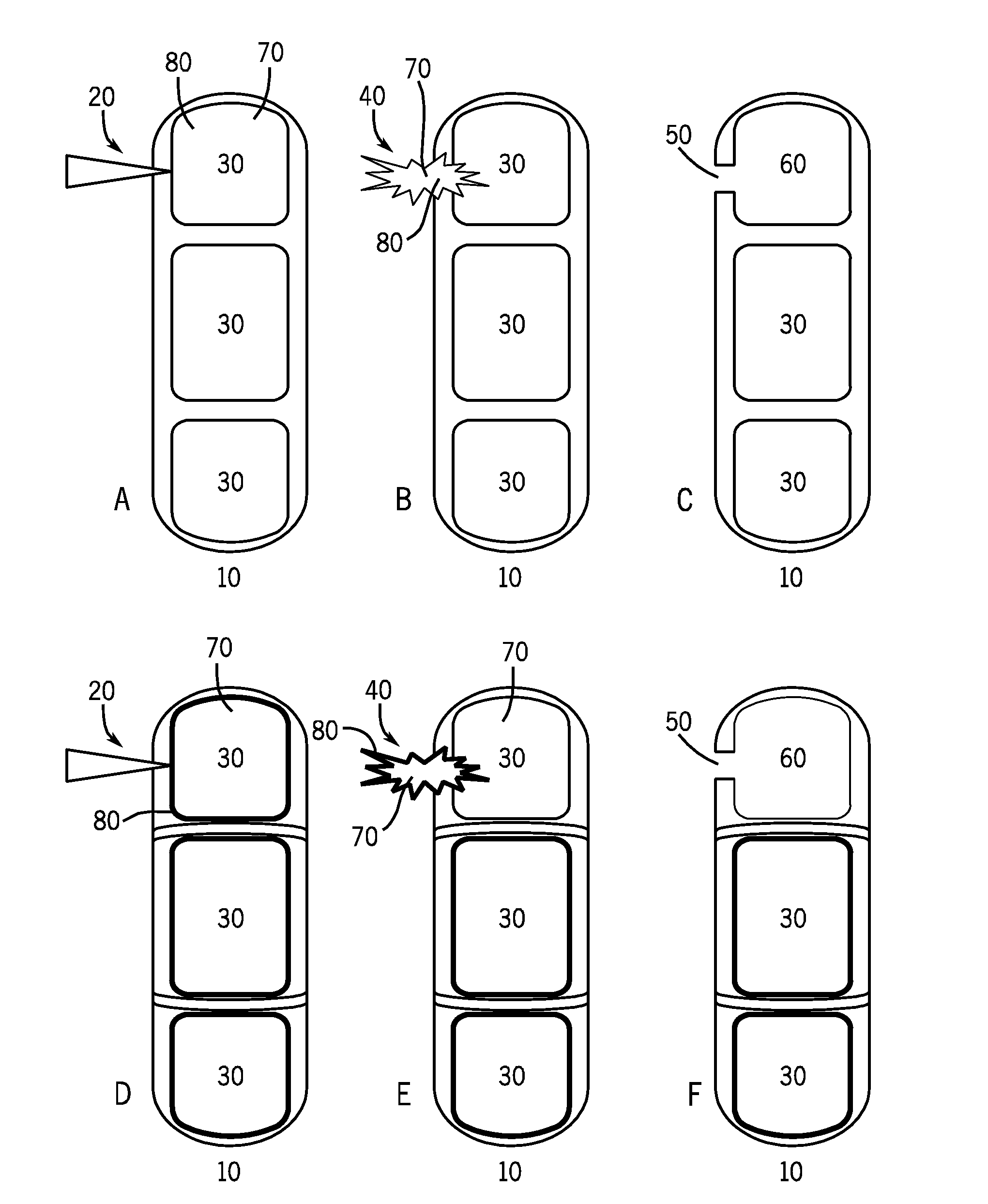
Visual indication of rupture of drug reservoir
a technology of drug reservoir and visual indication, which is applied in the direction of eye treatment, drug composition, prosthesis, etc., can solve the problems of difficult to determine whether the therapeutic agent is being or has been correctly released, and methods may present therapeutic, diagnostic, economic, or simply logistical drawbacks
- Summary
- Abstract
- Description
- Claims
- Application Information
AI Technical Summary
Benefits of technology
Problems solved by technology
Method used
Image

Examples
example 1
Visualization of a Laser-Activated Fluorophore Release
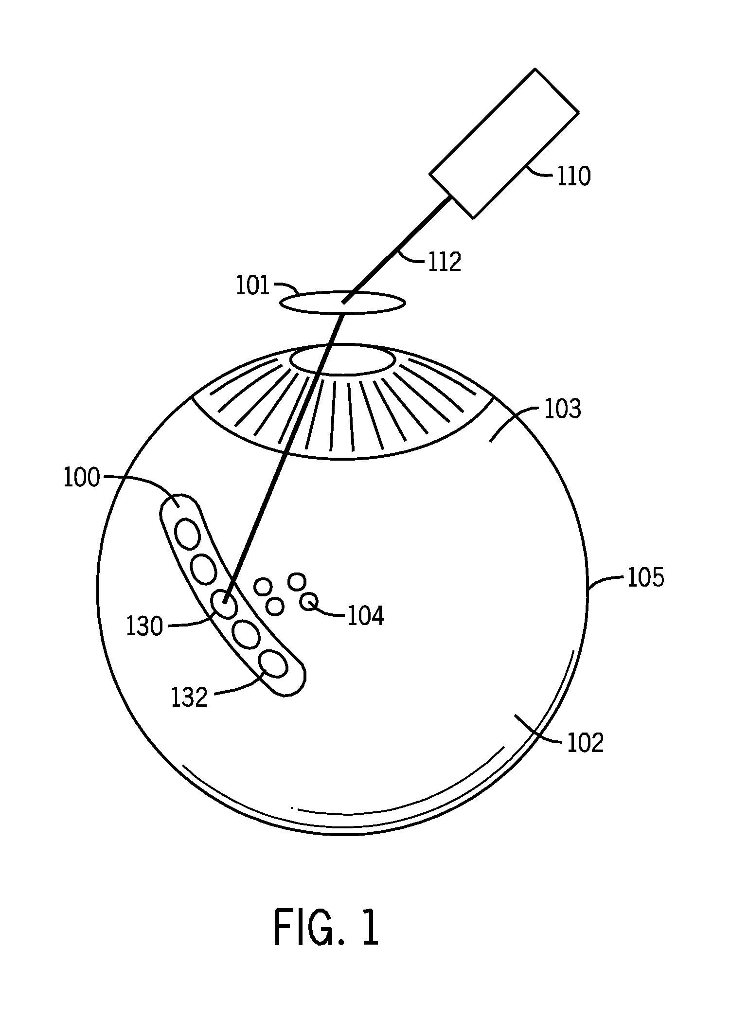
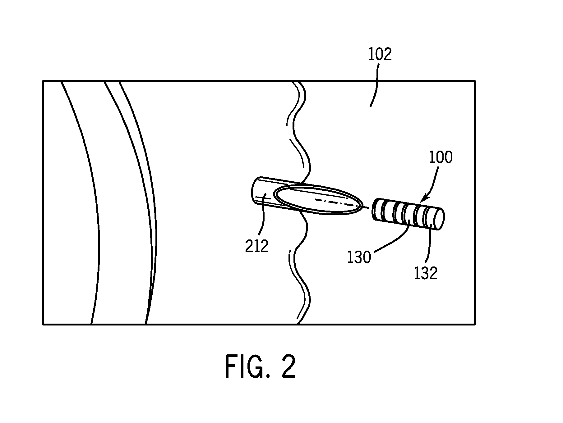
[0059]In a proof of concept demonstration, illustrated in FIG. 6, a drug delivery device 600 was fabricated as a tubular polymeric device. Drug reservoir 610 of the exemplary drug delivery device was filled with a composition comprising a dexamethasone salt and sodium fluorescein. The drug delivery device 600 was placed within an acceptor compartment of a Franz cell. A Franz cell is a specially designed apparatus that allows for the determination of the precise amount of bioactive compound that has penetrated through a membrane. The membrane is positioned between an upper donor chamber and a lower acceptor chamber. A laser was used to irradiate the drug delivery device with a wavelength at 532 nm through a membrane separating the donor and acceptor compartments of the Franz cell. The laser-induced rupture of the drug reservoir was verified by visually observing a fluorescent streams 620 emanating from the drug delivery device int...
example 2
Visualization of a Laser-Activated Reservoir Breach
[0060]In a further proof of concept demonstration, a tubular polymeric drug delivery device (black polyolefin shrink tube) was fabricated. A drug reservoir of the exemplary drug delivery device was filled with a composition comprising a dexamethasone salt and sodium fluorescein. The drug delivery device was placed within the vitreous of an eye of an albino pig using an 18 gauge needle. A laser was used to irradiate the drug delivery device with a wavelength of 532 nm (750-1000 mw, 50 ms, 50 μm laser pulse) to rupture the barrier layer to elute the dexamethasone salt and sodium fluorescein mixture contained within the drug reservoir. A sample of the vitreous containing the activated drug delivery device was removed from the eye and placed in a cuvette for observation of the drug delivery device over time. The laser-induced rupture of the drug reservoir was verified by visually observing formation of a gas bubble at the site upon whic...
example 3
Visualization of a Laser-Activated Exposure of Two Therapeutic Agents
[0061]In a further proof of concept demonstration, illustrated in FIG. 7, a tubular polymeric drug delivery device 700, made from black polyolefin shrink tube, containing two drug reservoirs separated by a divider was fabricated. A first drug reservoir 710 of the exemplary drug delivery device 700 is filled with a composition comprising a dexamethasone salt and a fluorescein salt. A second drug reservoir 720 of the exemplary drug delivery device 700 is filled with a composition comprising a dexamethasone salt and a rhodamine salt. The drug delivery device is placed within the vitreous of an eye of an albino pig using an 18 gauge needle. A laser is used to irradiate the drug delivery device with a wavelength of 532 nm (750-1000 mw, 50 ms, 50 μm laser pulse) to rupture the barrier layer of the first drug reservoir to elute the dexamethasone salt and fluorescein salt mixture contained within the first drug reservoir 7...
PUM
 Abstract
Description
Claims
Application Information