System for Determining Patient Heart related Parameters for use in Heart Imaging

a heart imaging and patient technology, applied in the field of system for determining patient heart related parameters for use in patient heart imaging examination, can solve the problems of inability to consider temporal, burdensome, time-consuming and sensitive to intra and inter observer errors

Inactive Publication Date: 2011-05-05
SIEMENS MEDICAL SOLUTIONS USA INC
View PDF23 Cites 7 Cited by
  • Summary
  • Abstract
  • Description
  • Claims
  • Application Information

AI Technical Summary

Problems solved by technology

The known systems are typically based on the analysis of static medical images and fail to consider temporal information inside an image sequence.
This is burdensome, time consuming and sensitive to intra and inter observer errors.

Method used


Image

  • System for Determining Patient Heart related Parameters for use in Heart Imaging
  • System for Determining Patient Heart related Parameters for use in Heart Imaging
  • System for Determining Patient Heart related Parameters for use in Heart Imaging

Examples


Embodiment Construction

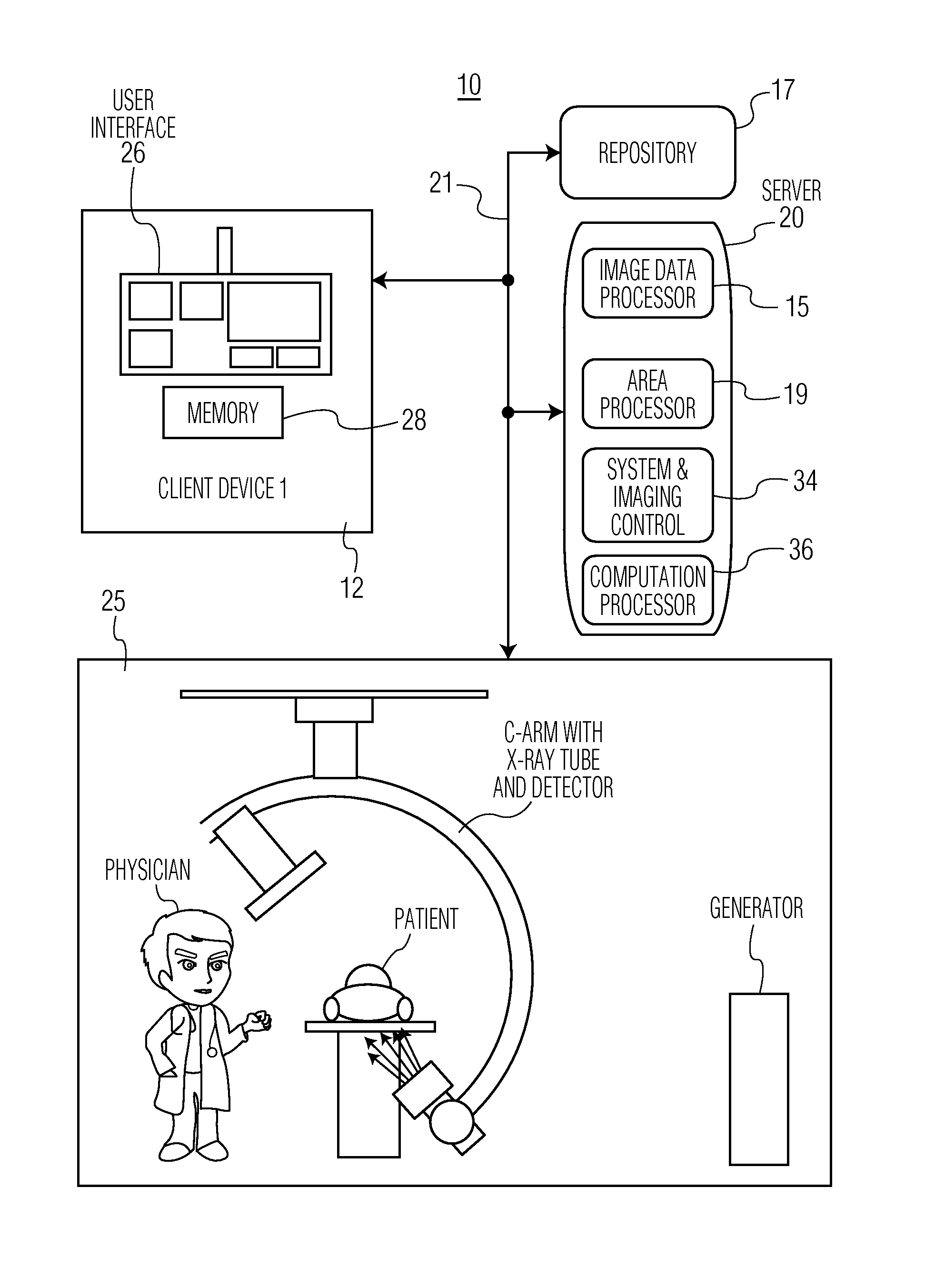
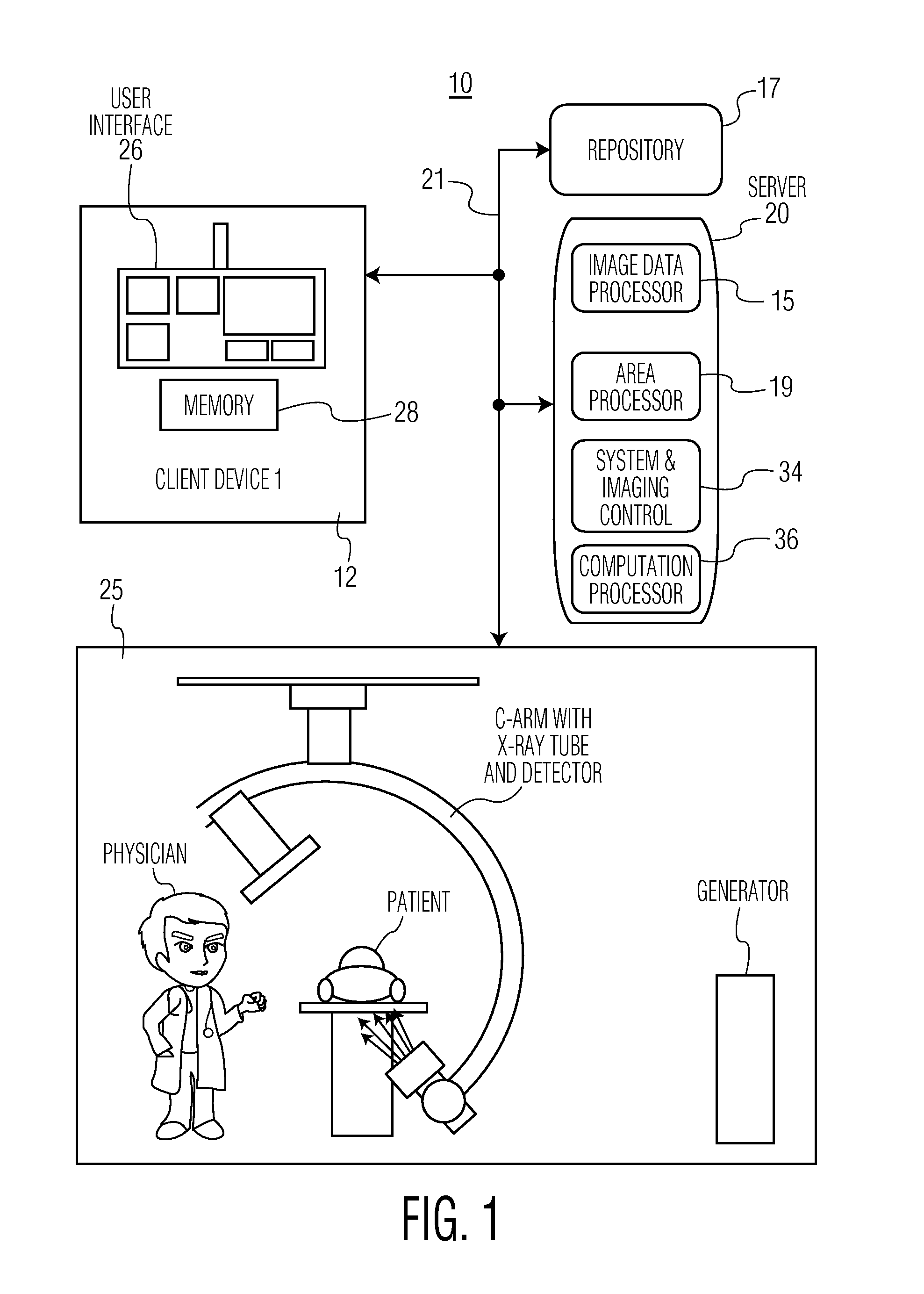
[0012]A system automatically estimates stroke area and volume, two-dimensional ejection fraction and three-dimensional ejection fraction using integrated spatio-temporal analysis and a geometric left ventricle model. The system advantageously uses spatial information within each image frame and temporal information between image frames to provide robust and accurate parameter estimation for use in X-ray angiography, for example. The system exploits the temporal correlation between end-diastolic (ED) and end-systolic (ES) phases, to advantageously eliminate a need to calculate left ventricle volumes for both ED and ES phases. Instead, the system automatically estimates a left ventricle stroke area and derives the relationship between two-dimensional ejection fraction and three-dimensional ejection fraction. The system applies a constraint instead of a known geometric left ventricular model and is readily adapted to accommodate patient heart variation and minimizes need for human inte...


PUM


Abstract

A system uses integrated spatio-temporal analysis in X-ray angiography, for example, by using spatial information within each image frame and temporal information between image frames to provide robust and accurate estimation of stroke area and volume, two and three dimensional ejection fraction and to accommodate patient heart variation. A system determines patient heart related parameters for use in patient heart imaging examination. An image data processor processes data representing multiple cardiac images of a patient over multiple heart beat cycles of the patient to derive data representing a distribution curve of a heart section area over multiple heart beat cycle times and indicating heart section area change over a heart beat cycle. An area processor determines a heart section area in response to user command. Also a computation processor determines a heart function parameter in response to the determined heart section area and the indicated heart section area change.

Description

[0001]This is a non-provisional application of provisional application Ser. No. 60 / 989,215 filed Nov. 20, 2007, by W. Qu et al.FIELD OF THE INVENTION[0002]This invention concerns a system for determining patient heart related parameters for use in patient heart imaging examination by determining a heart function parameter in response to a determined heart section area and a heart section area change.BACKGROUND OF THE INVENTION[0003]X-Ray angiographic cardiac (e.g., left ventricular) analysis is used in a cardiac catheterization laboratory to assess and measure cardiac functions. In response to injection of a contrast medium into a left cardiac chamber, a left ventricular silhouette is viewed using a digital imaging system. The digital imaging system generates one or two image sequences (single plane or biplane) for further quantitative analysis. Further, an Ejection fraction (EF) is a cardiac function used routinely to judge if a patient has heart disease. It comprises,EF=VED-VESVED...

Claims


Application Information

Patent Timeline
Patent Type & Authority Applications(United States)
IPC IPC(8): A61B5/029A61B5/02
CPCA61B6/481A61B6/504A61B6/5211G06T7/0012G06T2207/30104G06T2207/10116G06T2207/20076G06T2207/30048G06T7/208G06T7/277
Inventor QU, WEICHEN, JINGHUAJIA, YUANYUANSINGH, SUKHVEERKELLER, MICHAEL J.
Owner SIEMENS MEDICAL SOLUTIONS USA INC
Why Eureka
Try Eureka