Method and system for modelling a human heart and atria

a heart and atria technology, applied in the field of human heart and atria modelling, can solve the problems of not being able to routinely use 3d techniques, and not being able to accurately model the heart and atria. the effect of convenien

Active Publication Date: 2021-07-20
MEDTRACE AS
View PDF9 Cites 0 Cited by
  • Summary
  • Abstract
  • Description
  • Claims
  • Application Information

AI Technical Summary

Benefits of technology

[0035]In some embodiments, steps C-D are iteratively repeated by dividing one or more of the at least two portions further into sub-portions (step C) and arranging the sub-portions in relation to each other by comparing the arrival times of their first-pass peaks (step D), thereby further segmenting the model of the human heart.
[0062]The steps of the first as well as the second preferred embodiment can conveniently be automated by employing a computing device comprising a computer readable storage medium containing instructions that, when executed by a processor, are configured to cause said computing device to execute or perform said method steps.Definitions

Problems solved by technology

However, blood flow is not the sole factor in heart disease or cardiac diagnostics.
Besides, diseases directly affecting the LA, such as atrial fibrillation or atrial degeneration might lead to dilatation.
Because LA configuration is highly variable, the conversion of area to volume is subject to measurement error and observer bias.
Direct three-dimensional measurements of LA are possible using specialized equipment, but is rarely performed in clinical routine.
Even with CMR the 3D technique is not routinely used, mainly because this requires additional scanning- and labour time.
Left atrial enlargement increases the risk of major adverse cardiac events independent of coronary vasodilator capacity.
The methods employed in this study were however completely manual, and to some degree observer-dependent, complicating efforts to integrate measures of MBF and LA volumes in a standard workflow.
Deriving LA volume from PET data is thus known to be labour intensive and prone to observer variation.
However, at present, no reliable non-invasive methods are available for quantitatively measuring EVLW and preloading.

Method used


Image

  • Method and system for modelling a human heart and atria
  • Method and system for modelling a human heart and atria
  • Method and system for modelling a human heart and atria

Examples


Embodiment Construction

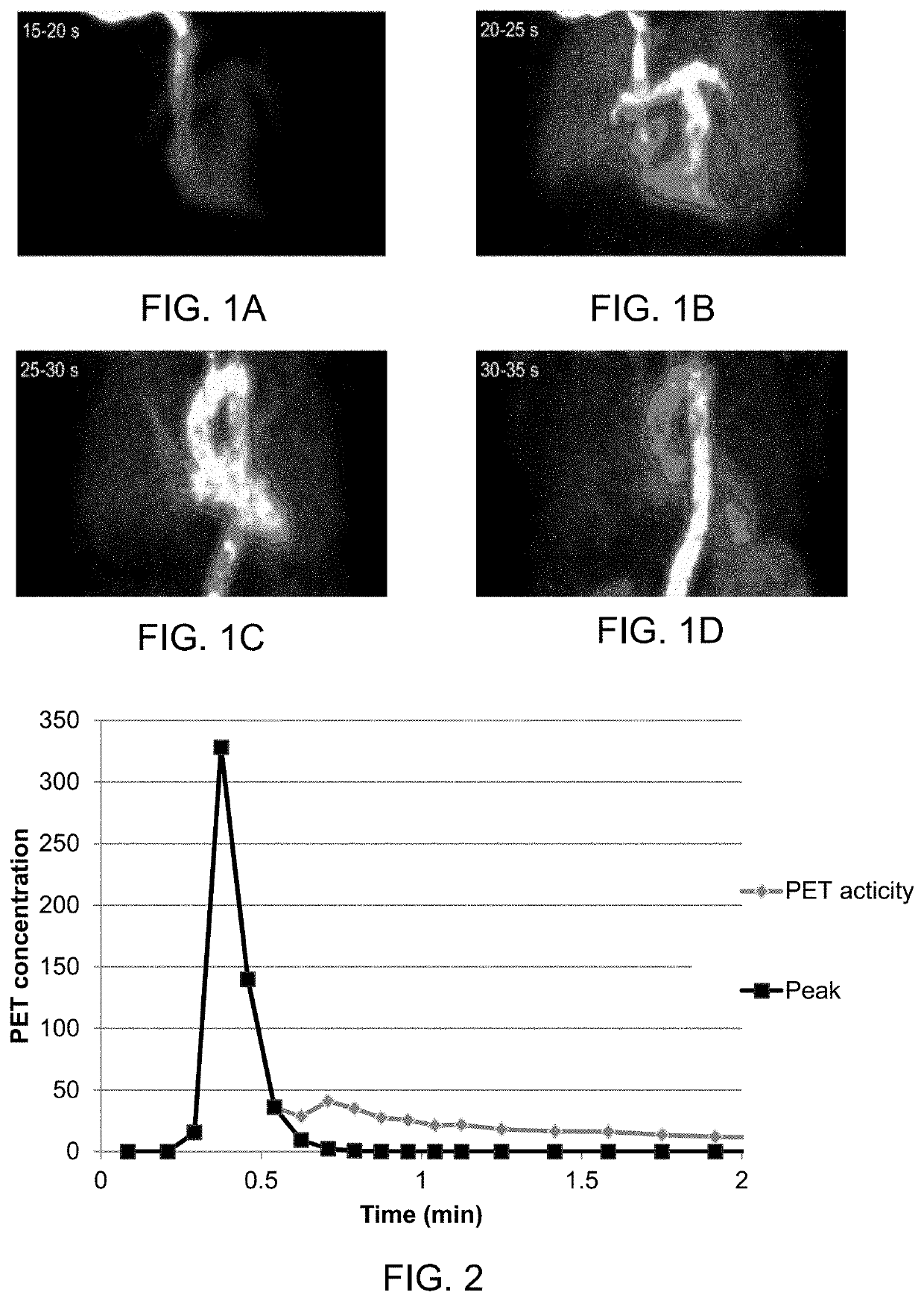
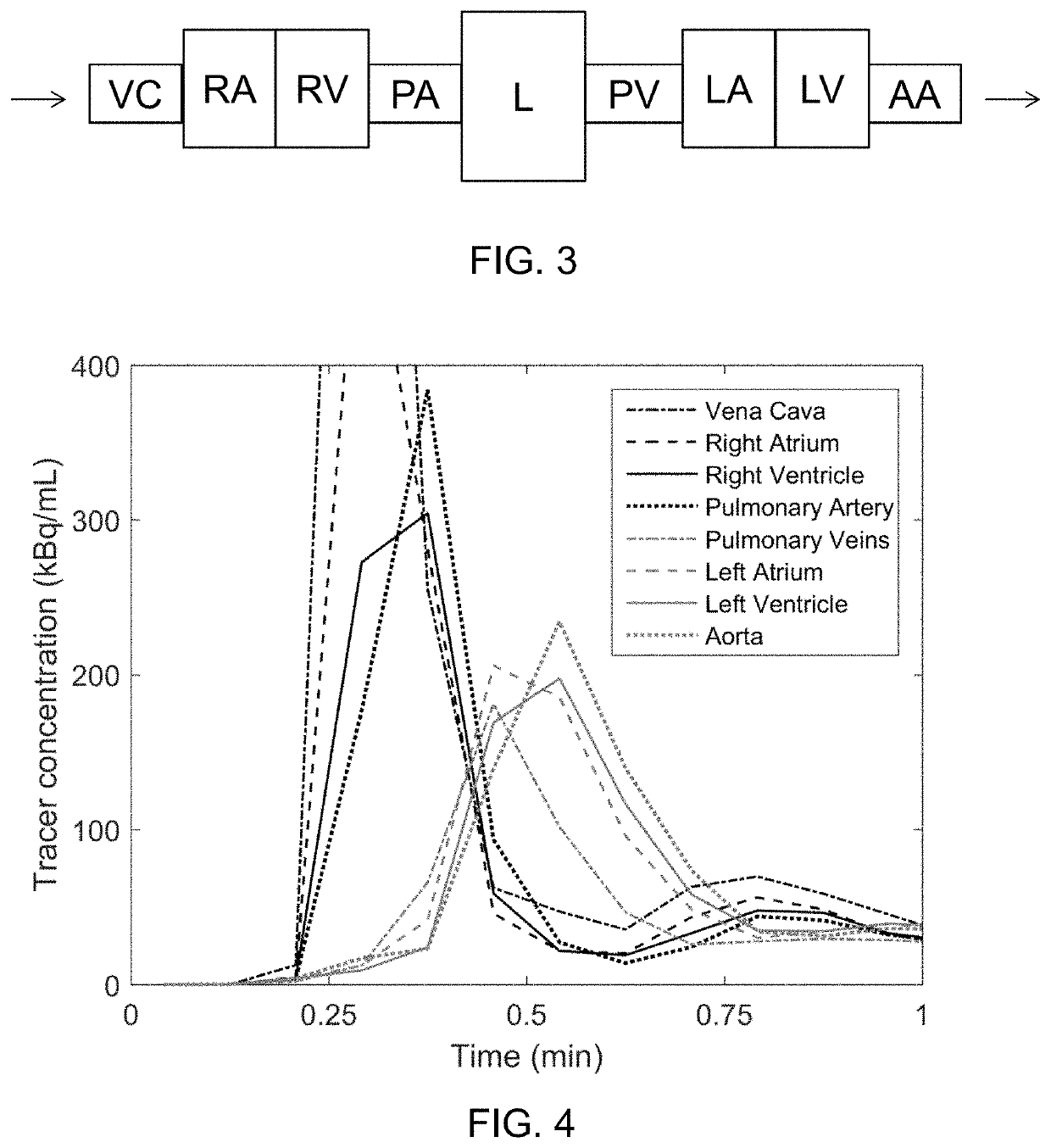
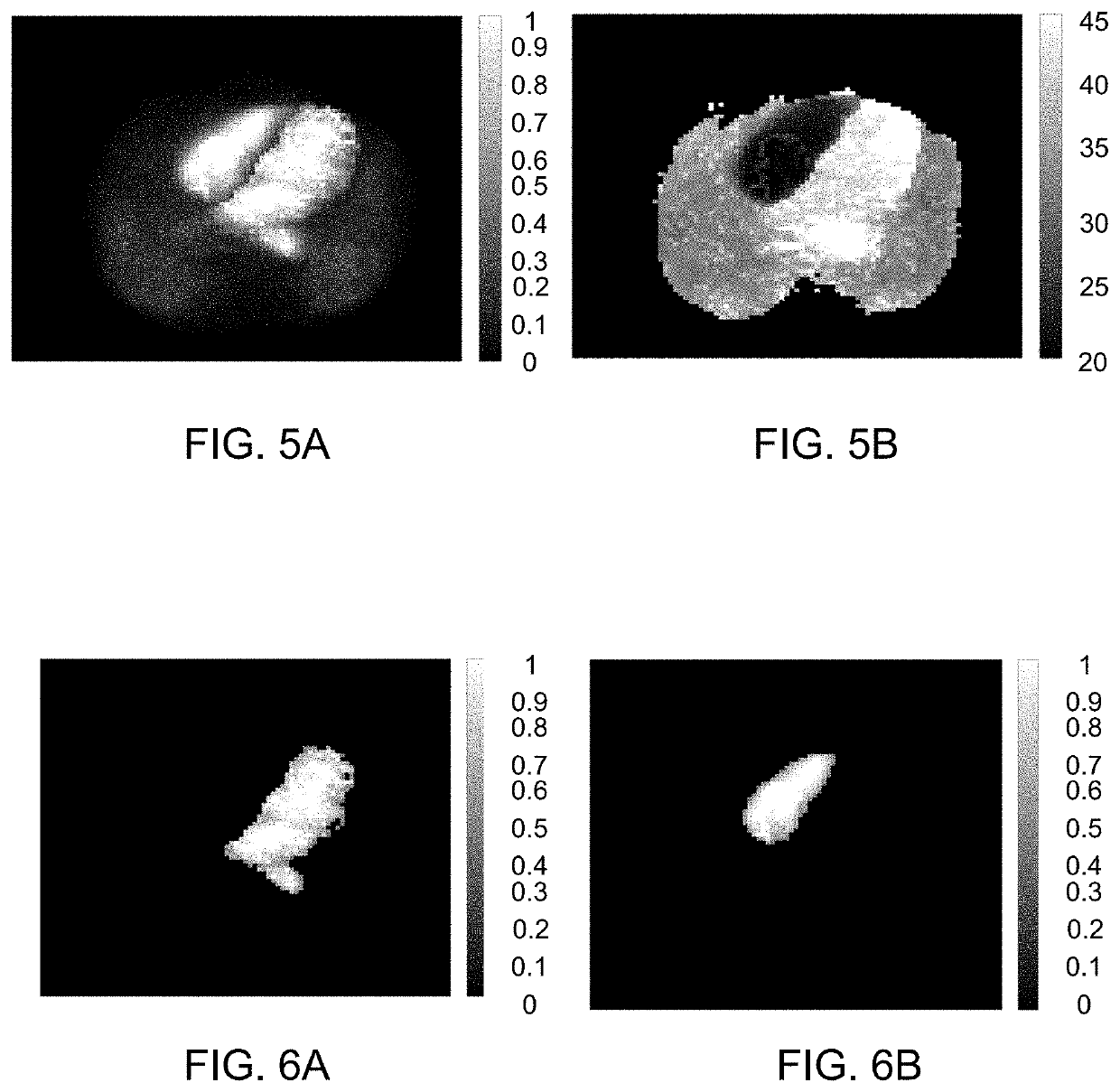
[0082]The present disclosure relates as mentioned in general to a method for modelling a human heart and / or chambers and cavities therein, such as the left and right atrium, or a human central circulation comprising a human heart, based on PET images. By segmenting the various chambers and large vessels in the central circulation, a segmented model is obtained, from which further parameters and information can be obtained.

[0083]Other imaging technologies such as CT, MRI and scintigraphy using gamma-cameras can also be used.

[0084]The present disclosure in particular relates to the inventors' finding that, inter aiia, the left atrium (LA) can be segmented automatically using images of bolus area-under-curve and midpoint time. Using the midpoint time of the left-ventricular (LV) cavity as reference, the LA can be segmented as regions with high first-pass activity and a midpoint time shorter than that of the LV. In analogous fashion, the right atrium (RA) can be segmented, and for both ...


PUM


Abstract

A method and a system for modelling a human heart based on a plurality of emission tomography images representing concentrations of a tracer that has been injected at a specific time is presented. The method comprising: extracting time activity curves of a tracer that has been injected for a plurality of pixels and / or voxels; identifying first-pass peaks of the time activity curves corresponding to an arrival time of the injected tracer at the corresponding pixel / voxel; defining a model comprising at least two portions of the heart; and arranging the at least two portions in relation to each other by comparing the arrival times of the first-pass peaks. The method and model may be used to obtain an estimate of the volume of the left or right atrium of the human heart.

Description

CROSS-REFERENCE TO RELATED APPLICATIONS[0001]This patent application is a national stage filing under 35 U.S.C. 371 of International Application No. PCT / DK2017 / 050367, filed Nov. 10, 2017, which claims the benefit of priority of Danish Patent Application No. PA 2016 70895, filed Nov. 11, 2016, each of which are incorporated herein by reference in their entirety.FIELD OF THE INVENTION[0002]The present disclosure relates to a method and system for modelling a human heart, and / or chambers and cavities therein, such as the left and right atrium, based on emission tomography images. The disclosure further relates to identification of conditions based on the model.BACKGROUND OF INVENTION[0003]Positron emission tomography (PET) is an imaging technique that is used to observe metabolic processes in the body. The system detects gamma rays emitted indirectly by a positron-emitting radionuclide (tracer), which is introduced into the body. Three-dimensional images of tracer concentration within...

Claims


Application Information

Patent Timeline
Patent Type & Authority Patents(United States)
IPC IPC(8): G06T7/00A61B6/03A61B6/00G06T15/08A61B5/029A61B5/0275
CPCG06T7/0016A61B6/037A61B6/481A61B6/486A61B6/503A61B6/507A61B6/5205G06T15/08A61B5/029A61B5/02755G06T2207/10076G06T2207/10104G06T2207/30048G06T2207/30101G06T2210/41G06T7/62G06T2207/10081
Inventor SØRENSEN, JENSHARMS, HENDRIK JOHANNES
Owner MEDTRACE AS
Why Eureka
Try Eureka