Device and method for in vivo imaging

a technology of in vivo imaging and lumen, which is applied in the field of in vivo devices and methods for imaging in vivo lumens, can solve the problems of complex viewing and detection of pathologies in the gi tract, such as tumors, lesions, ulcers, etc., and achieves the effects of high resolution, energy saving and improved imaging

Active Publication Date: 2011-05-26
GIVEN IMAGING LTD
View PDF8 Cites 19 Cited by
  • Summary
  • Abstract
  • Description
  • Claims
  • Application Information

AI Technical Summary

Benefits of technology

[0010]Embodiments of the invention provide a device that may scan a lumen wall in-vivo, typically illuminating in flashes or continuously, and may provide a high resolution image of the wall, thus providing an energy efficient and improved device and method for detecting pathologies on or beneath the surface of the lumen wall.

Problems solved by technology

Viewing and detecting pathologies in the GI tract, such as tumors, lesions, ulcers, polyps and the like may be complicated due to different parameters such as the fact that the item or pathology sought to be viewed may have very similar color and texture as the color and texture of its background tissue or by the fact that a pathology may be too small to visualize using standard in-vivo imaging methods.

Method used


Image

  • Device and method for in vivo imaging
  • Device and method for in vivo imaging
  • Device and method for in vivo imaging

Examples


Embodiment Construction

[0026]In the following description, various aspects of the present invention will be described. For purposes of explanation, specific configurations and details are set forth in order to provide a thorough understanding of the present invention. However, it will also be apparent to one skilled in the art that the present invention may be practiced without the specific details presented herein. Furthermore, well-known features may be omitted or simplified in order not to obscure the present invention.

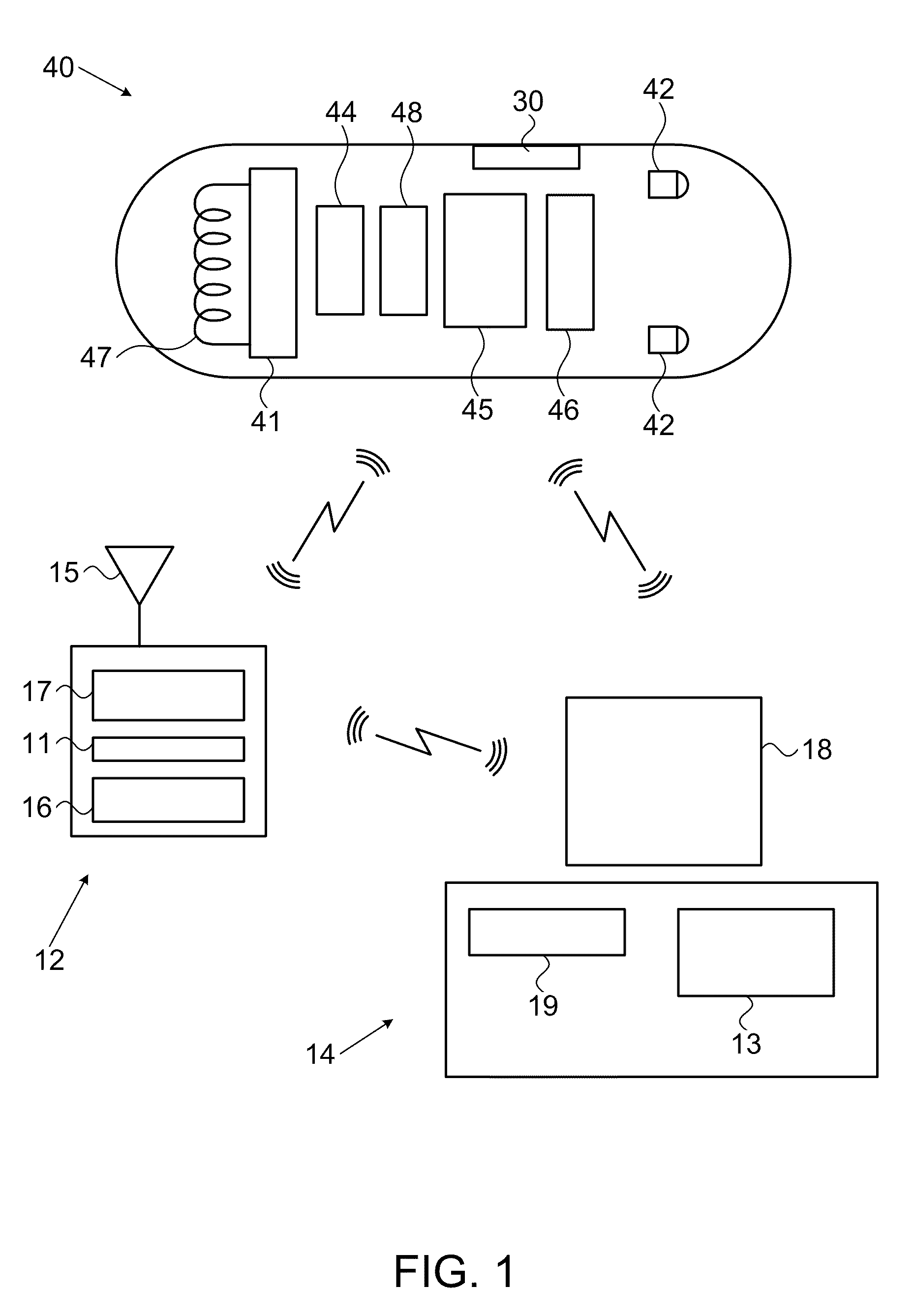
[0027]Reference is made to FIG. 1, which shows a schematic diagram of an in-vivo imaging system according to an embodiment of the present invention. Typically, the in-vivo imaging system may include an in-vivo imaging device 40, an external receiving device and / or recording device 12, e.g., data receiver, and a workstation 14. The in-vivo imaging device 40 may have an imager 46 for capturing image frames or a stream of image frames, an illumination source 42 for illuminating the body lum...


PUM


Abstract

Devices and methods for optically scanning an in-vivo lumen, and for determining contact between an in-vivo device and an in-vivo lumen wall. The device may include an elongated housing having a cylindrical portion. At least one illuminating body may provide illumination at the circumference of the cylindrical portion, and at least one light sensor may sense light that penetrated and was scattered from the tissue of the in vivo lumen. The device may include means for preventing direct illumination from the illuminating body from reaching the light sensor and for decreasing the amount of direct light reflection from the outer surface of the in vivo lumen onto the light sensor.

Description

FIELD OF THE INVENTION[0001]The present invention relates to an in-vivo device and method for imaging an in-vivo lumen. More specifically, the present invention relates to an autonomous in vivo sensing and / or imaging device and method for imaging scattered light and for detecting pathologies of in-vivo lumens, e.g., of the gastrointestinal (GI) tract wall, using scattered light analysis and spectral analysis methods.BACKGROUND OF THE INVENTION[0002]Known devices may be efficient in providing in-vivo sensing, such as imaging or pH sensing. Autonomous in-vivo sensing devices, such as swallowable or ingestible capsules or other devices, may move through a body lumen, sensing as they move along. An autonomous in-vivo sensing device such as an imaging device may include, for example, an imager for obtaining images from inside a body cavity or lumen, such as the GI tract, while the in-vivo imaging device passes through the GI lumen. The imager may, for example, be associated with an optic...

Claims


Application Information

Patent Timeline
Patent Type & Authority Applications(United States)
IPC IPC(8): A61B6/08
CPCA61B5/0062A61B5/0071A61B5/073A61B5/0084A61B5/0075
Inventor BLIT, SHMUELRABINOVITZ, ELISHA
Owner GIVEN IMAGING LTD
Why Eureka
Try Eureka