
Immunomodulation by iap inhibitors
- Summary
- Abstract
- Description
- Claims
- Application Information
AI Technical Summary
Benefits of technology
Problems solved by technology
Method used
Image

Examples
example 1
NKT Cell Development in Fetal Thymic Organ Culture (FTOC) is Blocked by Treatment with IAP Inhibitors
[0185]In order to investigate the role of IAP family members in NKT cell development, pharmacologic mimetics of the endogenous IAP inhibitor second mitochondrial activator of apoptosis (SMAC) were obtained. Three chemically distinct inhibitors were used for most experiments. One such inhibitor, LBW-242, is a potent inhibitor of IAP family members which binds to the XIAP BIR domain at submicromolar concentrations (IC50=280 nM). LBW-242 was used for most experimental replications.
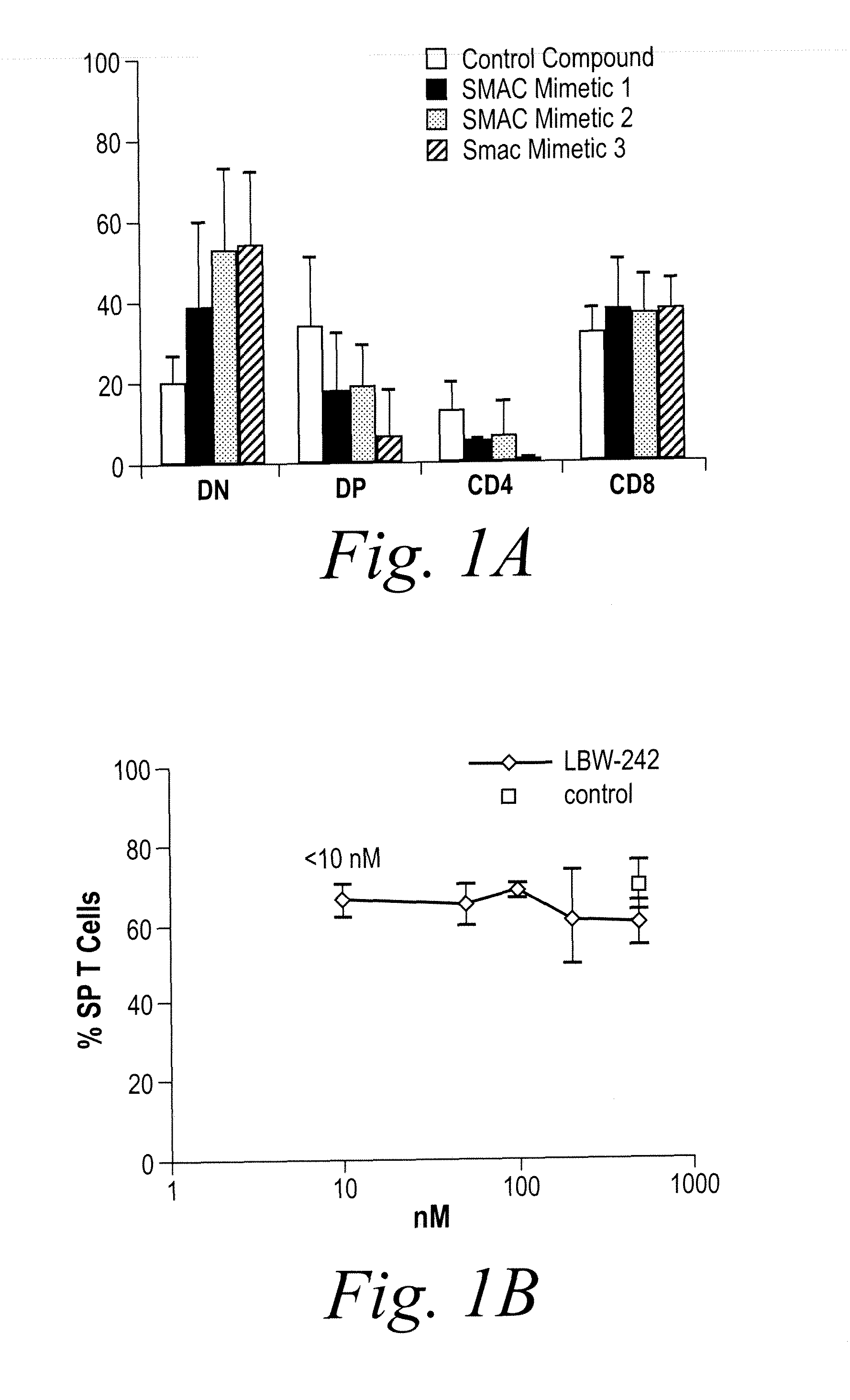
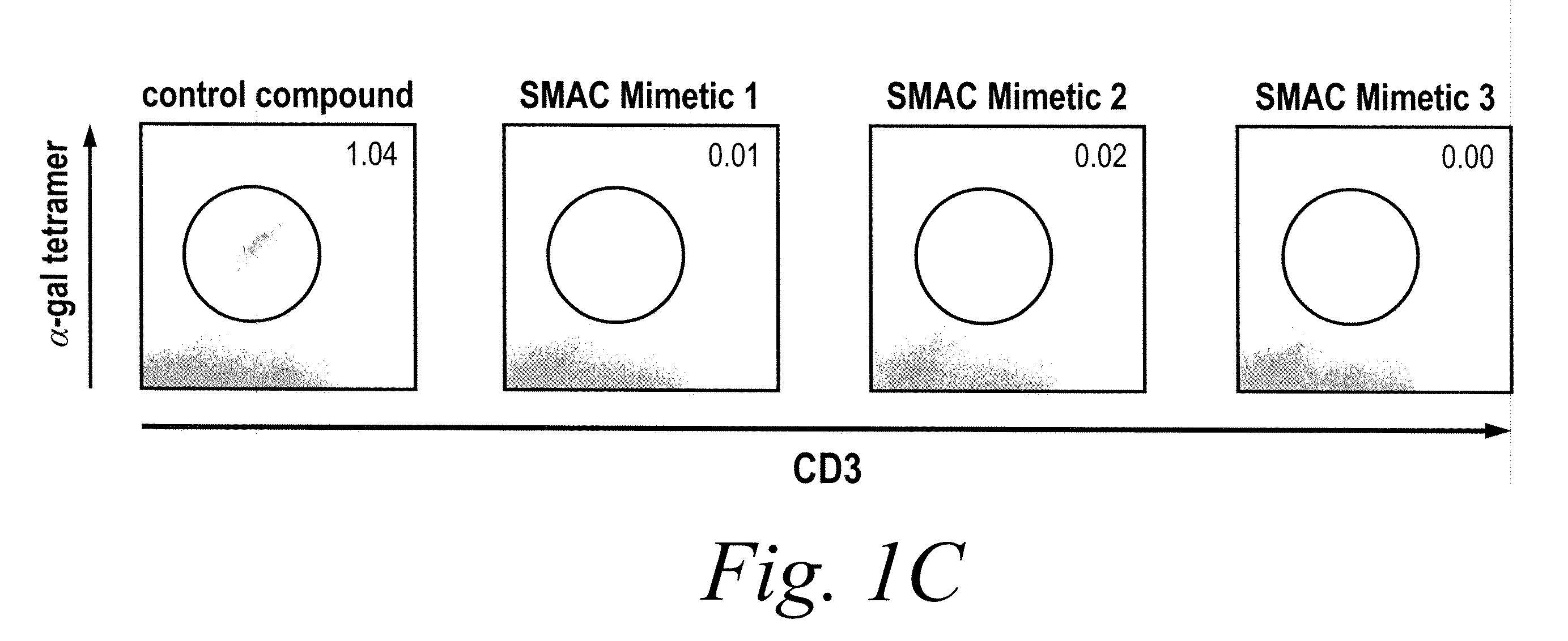
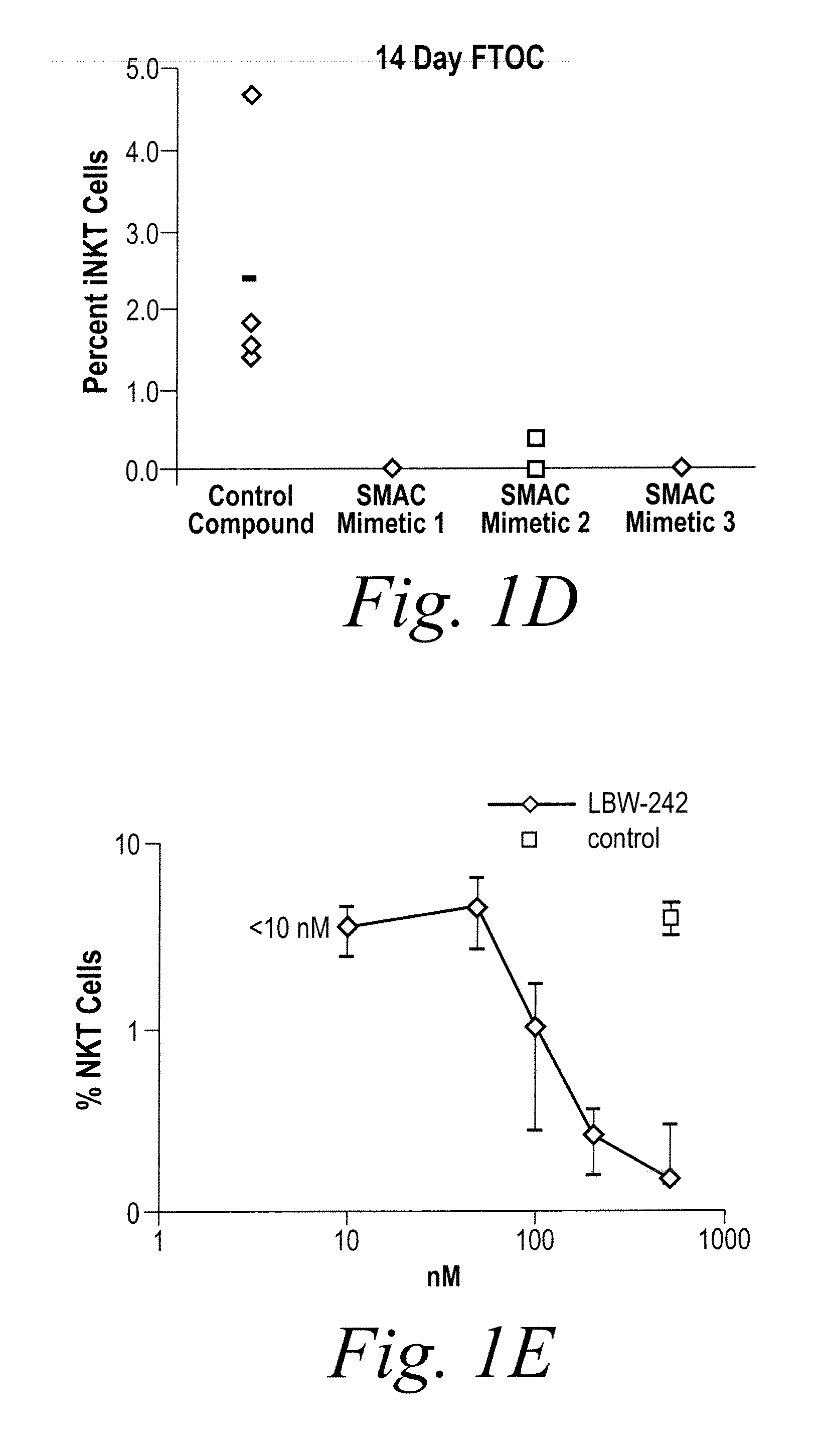
[0186]NKT cell development was studied using fetal thymic organ cultures (FTOCs). Embryonic day 16.5 embryos from C57BL / 6 mice were cultured in the presence of three distinct IAP inhibitors, a control compound, or vehicle (PBS). After 14 days, cultures were harvested and analyzed by flow cytometry. As shown in FIG. 1, NKT cell development in fetal thymic organ culture (FTOC) is blocked by treatment with IAP in...
example 2
Inhibition of IAP Family Members does not Sensitize Mature CD4+ T Cells to Apoptosis
[0187]The role of IAP family members in mature NKT cell survival was assessed. Spleen-derived CD4+ T cells from BALB / c mice were activated with anti-CD3 and anti-CD28 for 24 hours and then analyzed by flow cytometry. As shown in FIG. 2, inhibition of IAP family members does not sensitize mature CD4+ T cells to apoptosis. 2×10⁶ BALB / c splenic CD4+ T cells were stimulated with anti-CD3 and anti-CD28 in RPMI. After 24 hours viable cells were quantified by trypan blue exclusion, and the resulting cell counts are depicted in FIG. 2A. Apoptotic cells were quantified in the cultures from FIG. 2A using flow cytometry with annexin V and 7-AAD (results are depicted in FIG. 2B). Both LBW-242 and the control compound were used at 500 nM. Treatment of CD4+ T cells with IAP inhibitors did not alter the total number of T cells, nor did it contribute to apoptosis, as shown in FIGS. 2A-B. NKT cells were identified in...
example 3
Inhibition of IAP Family Members Enhances Cytokine Secretion from Activated T Cells
[0188]In order to determine the functional consequences of IAP inhibition in NKT cells, spleen cells from BALB / c mice were stimulated with the NKT cell specific agonist α-galactosylceramide (α-galcer). Surprisingly, α-galcer stimulated spleen cells treated with IAP inhibitors showed increased secretion of both IFN-γ (interferon γ) and IL-2 (interleukin-2), as shown in FIGS. 3A-B. 5×10⁵ BALB / c spleen cells from three separate mice were cultured in RPMI with α-galcer in the presence of LBW-242 or a control compound. After 48 hours, cytokine levels were measured in the culture supernatants by ELISA, and the results are depicted in FIG. 3A (IFNγ) and FIG. 3B (IL-2). A similar, dose-dependent effect on IL-2 production was observed in unfractionated BALB / c or C57BL / 6 CD4+ T cells stimulated with anti-CD3 and anti-CD28, as shown in FIGS. 3C-D. 105 splenic CD4+ T cells from BALB / c mice were cultured in RPMI w...
PUM
| Property | Measurement | Unit |
|---|---|---|
| Electrical conductance | aaaaa | aaaaa |
| Electrical conductance | aaaaa | aaaaa |
| Electrical conductance | aaaaa | aaaaa |
Abstract
Description
Claims
Application Information