Detection of xenotropic murine leukemia virus
a technology seroconversion antibodies, which is applied in the field of detection of xenotropic murine leukemia virus related virus, can solve the problems of shortening the life of cfs subjects, not being able to extend the results to detection of seroconversion antibodies against xmrv in humans, and achieving high stringency hybridization conditions
- Summary
- Abstract
- Description
- Claims
- Application Information
AI Technical Summary
Benefits of technology
Problems solved by technology
Method used
Image

Examples
example 1
Blood Preparation and Nucleic Acid Isolation
[0175]Whole blood was drawn from subjects by venipuncture using standardized phlebotomy procedures into 8-mL greencapped Vacutainers containing the anti-coagulant sodium heparin (Becton Dickinson). Plasma was collected by centrifugation, aspirated and stored at −80° C. for later use. The plasma was replaced with PBS and the blood resuspended and further diluted with an equal volume of PBS. PBMCs were isolated by layering the diluted blood onto Ficoll-Paque PLUS (GE Healthcare), centrifuging for 22 min at 800 g, aspirating the PBMC layer and washing it once in PBS. The PBMCs (approximately 2×10⁷ cells) were centrifuged at 500 g for 7 min and either stored as frozen unactivated cells in 90% FBS and 10% DMSO at −80° C. for further culture and analysis or resuspended in TRIzol (Invitrogen) and stored at −80° C. for DNA and RNA extraction and analysis.
[0176]DNA was isolated from TRIzol according the to manufacturer's protocol and also isolated ...
example 2
PCR
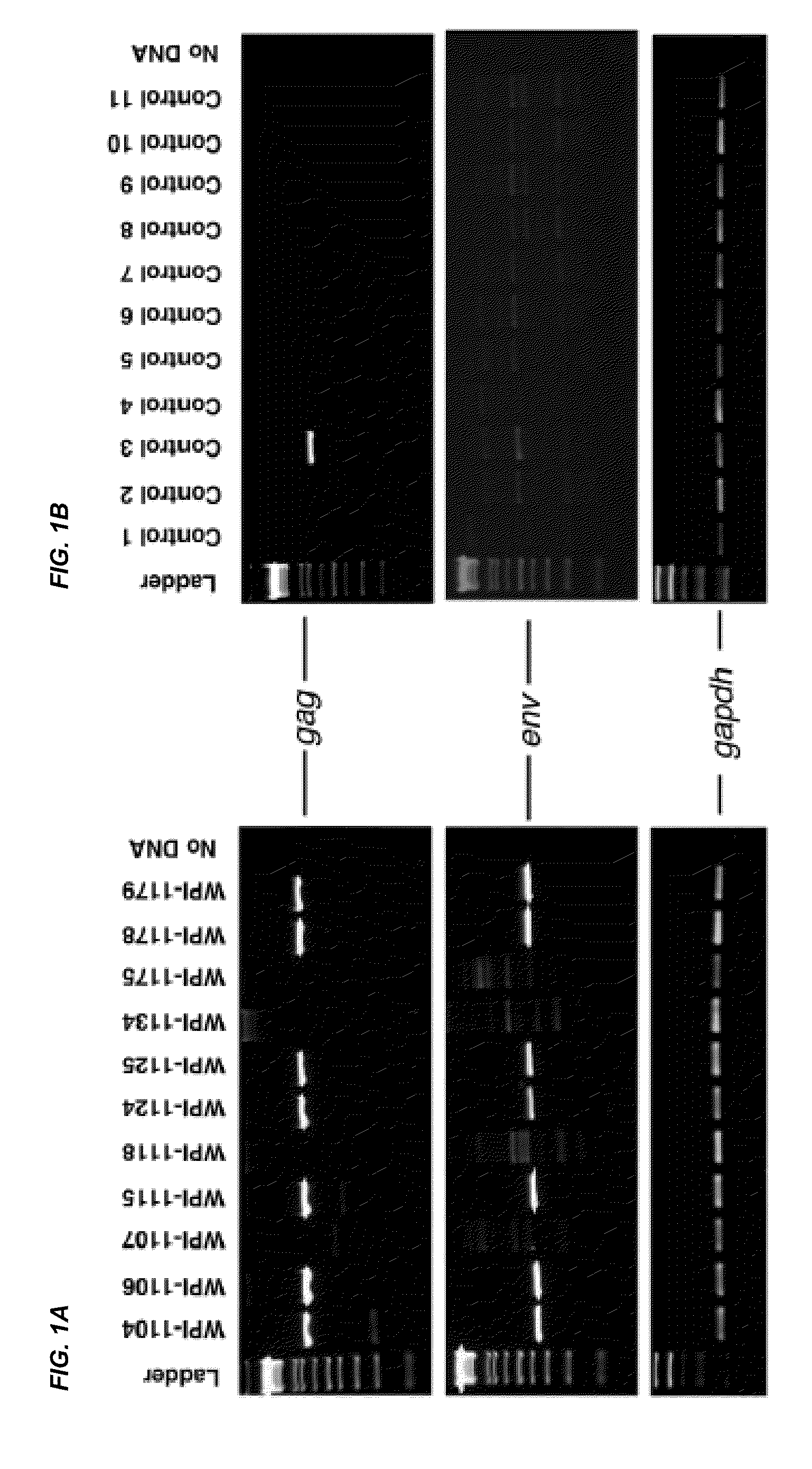
[0177]To avoid potential problems with laboratory DNA contamination, nested PCR was performed with separate reagents in a separate laboratory room designated to be free of high copy amplicon or plasmid DNA. Negative controls in the absence of added DNA were included in every experiment. Identification of XMRV gag and env genes was performed by PCR in separate reactions.
[0178]Reactions were performed as follows: 100 to 250 ng DNA, 2 μL of 25 mM MgCl2, 25 μL of HotStart-IT FideliTaq Master Mix (USB Corporation), 0.75 μL of each of 20 μM forward and reverse oligonucleotide primers in reaction volumes of 50 μL. For identification of gag, 419F (5′-ATCAGTTAACCTACCCGAGTCGGAC-3′) (SEQ ID NO: 5) and 1154R (5′-GCCGCCTCTTCTTCATTGTTCTC-3′) (SEQ ID NO: 6) were used as forward and reverse primers. For env, 5922F (5′-GCTAATGCTACCTCCCTCCTGG-3′) (SEQ ID NO: 7) and 6273R (5′-GGAGCCCACTGAGGAATCAAAACAGG-3′) (SEQ ID NO: 8) were used. For both gag and env PCR, 94° C. for 4 min initial denaturation was...
example 3
Cells
[0180]Isolation, separation and culture of primary cells.
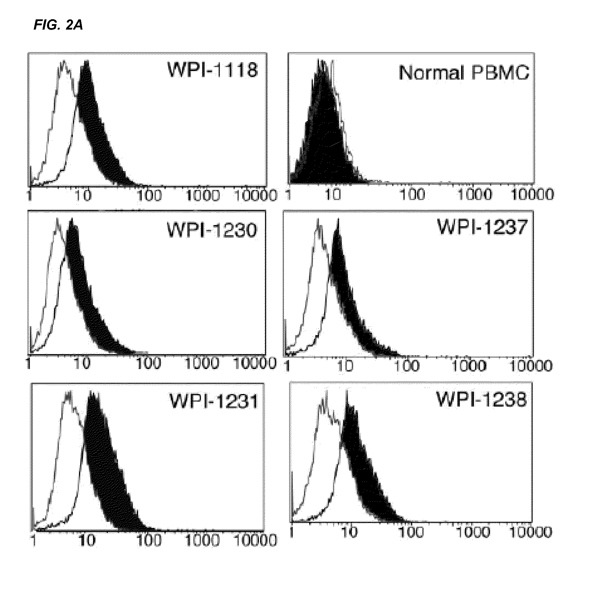
[0181]Leukopaks of peripheral blood from healthy donors were collected according to a NIH approved IRB #99-CC-0168 protocol. Subjects' peripheral blood and plasma samples were from frozen banked samples obtained under NIH exempt status. Mononuclear leukocytes from both normal and subjects' cells were isolated by Ficoll-Hypaque gradient centrifugation. The light density fraction (buffy coat) was collected, washed twice with PBS. PBMC were activated by 1 μg / mL PHA (Abbott Diagnostics) and after 72 hours the cells were cultured with 20 units / mL of IL-2 (Zeptometrix) and subcultured every 3-5 days. For isolation of CD4+ T cells, CD8, CD11b, CD14, CD19, CD33 and CD56 positive cells were removed using magnetic activated cell sorting (MACS) methods according to manufacturer's instructions (Miltenyi Biotec, Inc.). After isolation, the CD3+, CD4 T cells (>95% pure) were cultured in RPMI-1640 medium supplemented with 10% fetal calf...
PUM
 Abstract
Description
Claims
Application Information