
Methods and apparatus for performing an arthroscopic procedure using surgical navigation

a technology of surgical navigation and arthroscopic procedure, applied in the field of surgical methods and equipment, can solve the problems of affecting the function of the hip joint, significant recovery time, and reducing the range of motion of the robotic arm, so as to prevent the robotic arm from damaging the tissue, avoid the risk of surgical damage, and ensure the effect of accurate positioning of the anchor

Inactive Publication Date: 2011-08-04
STRYKER CORP
View PDF20 Cites 32 Cited by
  • Summary
  • Abstract
  • Description
  • Claims
  • Application Information

AI Technical Summary

Benefits of technology

[0017]Registration markers may be placed in the femur and / or acetabulum before any scans are taken. These registration markers may be placed percutaneously so that they reside on or in the bone of the patient, below the surface of the skin, where they may remain in place during the patient scan, and until and during, the patient's arthroscopic procedure. The registration markers may be placed at their desired location using a needle or gun type delivery device or any other delivery device that is appropriate. Preferably, the placement of the registration marker does not leave a significant opening in the patient's skin, so that the opening, if any, may be covered with a bandage, or require only a small amount of stitching to close. The opening may be small enough that it requires no coverage at all. This approach will allow the patient to resume their normal activity after the scan without worrying about a registration marker protruding through their skin or a large wound which may become infected.
[0035]The surgical navigation system may include a fixture to mount the patient's leg. The fixture allows manipulation of the leg and may include registration markers. Additional arthroscopic access to pathology may be enabled by moving the femur. Tracking this movement via the leg fixture allows the surgical navigation system to guide tools to treat pathology (for example, remove bone to relieve CAM impingement) more effectively.

Problems solved by technology

One of many problems that can affect the function of the hip joint is a decreased range of motion.
However, such open surgery requires significant recovery times and causes substantial pain for the patient.
It is also possible to perform the procedure arthroscopically, however, the arthroscopic approach is extremely challenging due to the tight spaces of the hip joint, which restrict visualization of the surgical site and manipulation of surgical instruments about the surgical site.
Among other things, it can be difficult for the surgeon to precisely identify the impinging bone which needs to be removed in the arthroscopic procedure.
Due to these difficulties, surgeons often remove too much tissue, or too little tissue, during an arthroscopic decompression.
However, this procedure is tedious and time-consuming.
Furthermore, it is impractical to do arthroscopically because of the limited access to the interior of the hip joint.
If the reference body is not placed in exactly the same position that it occupied during scanning, the 3-D virtual model will not be properly registered with the patient anatomy and the image generated from the 3-D virtual model will not be in proper registration with the live image generated by the arthroscope during the procedure.
This could result in the surgeon removing too much tissue, not enough tissue, or the wrong tissue during the arthroscopic procedure.
However, the procedure of Monahan et al. requires that a potentially cumbersome mechanical linkage apparatus be connected to the arthroscope and tools during the procedure.
If the pin is not placed in exactly the same position that it occupied during scanning, the 3-D virtual model will not be in proper registration with the anatomy, which could result in the surgeon removing too much tissue, not enough tissue, or the wrong tissue during the arthroscopic procedure.
Because of this, it is not desirable to have a reference pin protruding from the patient's leg during this time, since this can expose the patient to the risk of infection, cause trauma to the tissue if the pin should be bumped, etc.
Furthermore, in practice, it is also extremely difficult, if not impossible, to remove the reference pin after scanning and thereafter replace it, prior to the arthroscopic procedure, in exactly the same location that it occupied during scanning.

Method used


Image

  • Methods and apparatus for performing an arthroscopic procedure using surgical navigation
  • Methods and apparatus for performing an arthroscopic procedure using surgical navigation
  • Methods and apparatus for performing an arthroscopic procedure using surgical navigation

Examples


example 1

[0099]As noted above, hip arthroscopy is becoming increasingly more common in the diagnosis and treatment of various hip pathologies. However, due to the anatomy of the hip joint and the pathologies associated with the same, hip arthroscopy is currently practical for only selected pathologies.

[0100]One procedure which is sometimes attempted arthroscopically relates to femoral debridement for treatment of cam-type femoroacetabular impingement (i.e., cam-type FAI) and / or acetabular debridement for treatment of acetabular femoroacetabular impingement (i.e., pincer-type FAI). More particularly, with cam-type FAI, irregularities in the geometry of the femur can lead to impingement between the femur and the rim of the acetabular cup. Treatment for cam-type FAI typically involves debriding the femoral neck and / or head, using tools such as burrs, to remove the bony deformities causing the impingement. See FIGS. 10 and 11. In this respect it should be appreciated that it is important to debr...

example 2

[0123]In the example discussed above, the system merges the modified 3-D virtual model image with the live scope image so as to provide the surgeon a visual guide to follow during the debridement procedure. However, the modified 3-D virtual model is “blind” to the location of the debridement tool during the debridement procedure, and the virtual guide is provided solely in the form of visual markings placed on the modified 3-D virtual model prior to surgery.

[0124]Alternatively and / or additionally, it is also possible to place a “tracker” on the debridement tool, so that the system can determine the current location of the debridement tool during surgery. Furthermore, the system can integrate information about the current location of the tracked debridement tool into the modified 3-D virtual model, so as to guide the surgeon on how the debridement tool should be advanced relative to the anatomy so as to remove the impinging bone. By way of example but not limitation, in this construc...


PUM


Abstract

The method for performing an arthroscopic procedure on a joint using surgical navigation. A 3-D virtual model of the anatomy is created from a scan of the anatomy. The 3-D virtual model is then used to reproduce motion of the joint and plan the arthroscopic procedure. The 3-D virtual model is placed into registration with the real-world anatomy, so that a virtual image generated by the 3-D virtual model may be placed into registration with an arthroscopic image of the real-world anatomy. Preferably, arthroscopic registration markers, positioned prior to scanning, are used to place the 3-D virtual model in registration with the real-world anatomy. The arthroscopic registration markers may be placed either percutaneously or arthroscopically. At the conclusion of the procedure, the registration markers may be left in place, removed arthroscopically or allowed to biodegrade.

Description

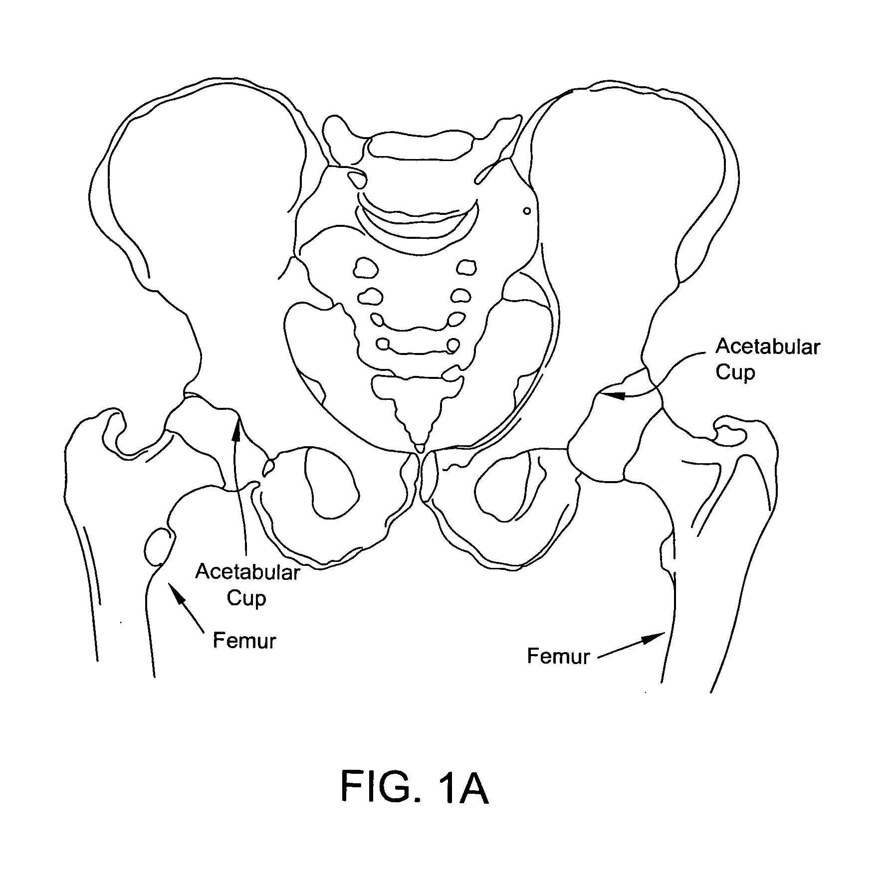
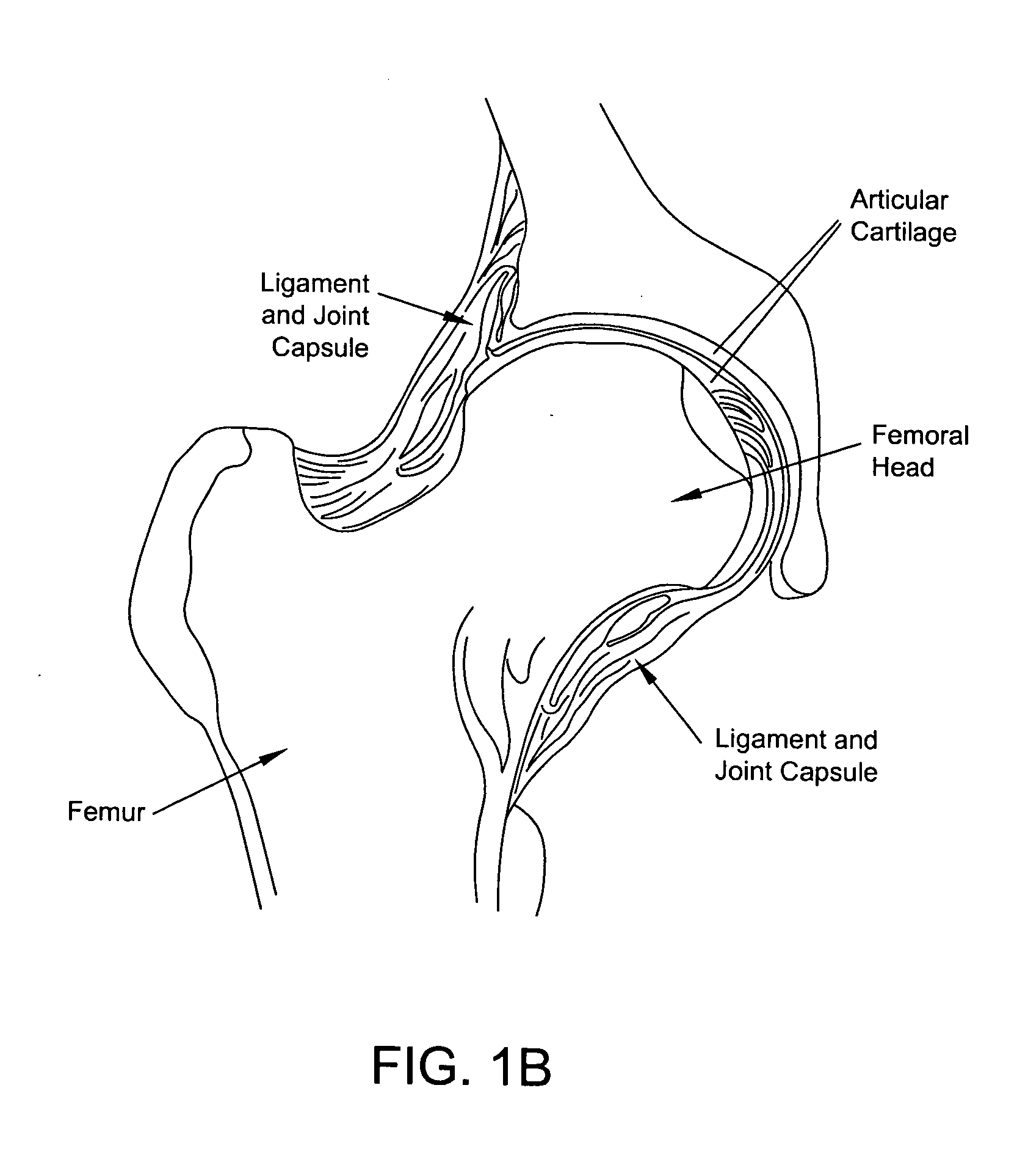
REFERENCE TO PENDING PRIOR PATENT APPLICATIONS[0001]This patent application claims benefit of pending prior U.S. Provisional Patent Application Ser. No. 61 / 262,196, filed Nov. 18, 2009 by Julian Nikoichev et al. for METHOD AND APPARATUS FOR SURGICAL NAVIGATION FOR HIP ARTHROSCOPY (Attorney's Docket No. FIAN-50 PROV), which patent application is hereby incorporated herein by reference.FIELD OF THE INVENTION[0002]This invention relates to surgical methods and apparatus in general, and more particularly to surgical methods and apparatus for arthroscopically treating the hip.BACKGROUND OF THE INVENTION[0003]One of many problems that can affect the function of the hip joint is a decreased range of motion. One common pathology is femoral acetabular impingement (FAT). FAI may occur in two forms. The first form of FAI is called CAM impingement. CAM impingement exists where the head and / or neck of the femur is abnormally shaped, but the acetabular cup is normally shaped. The second form of F...

Claims


Application Information

Patent Timeline
IPC IPC(8): A61B17/56
CPCA61B17/56A61B34/20A61B34/10A61B2034/105A61B34/30A61B90/36A61B2034/2055A61B17/1666A61B2034/2051A61B2090/3983A61B2034/2068
Inventor NIKOLCHEV, JULIANFLOM, JAMES R.
Owner STRYKER CORP
Learn More