Methods and compositions for delivery of exogenous factors to nervious system sites

a technology of exogenous factors and nervious system, which is applied in the direction of drug compositions, peptide sources, peptide/protein ingredients, etc., can solve the problems of limiting functional recovery, lacking the technology to successfully direct stem cell differentiation into the appropriate or desired cell type in vivo, and transplanted neural stem cells (nsc) not differentiate into the appropriate cell type for neuron regeneration

Inactive Publication Date: 2011-09-08
TEMPLE SALLY +2
View PDF16 Cites 2 Cited by
  • Summary
  • Abstract
  • Description
  • Claims
  • Application Information

AI Technical Summary

Problems solved by technology

Spinal cord injury (SCI) causes loss of spinal cord cells, damage to ascending and descending axonal tracts and loss of myelination, resulting in paralysis.
However, at present, the technology to successfully direct stem cell differentiation into the appropriate or desired cell type in vivo is lacking.
Specifically, research studying stem cells in the context of treating SCI has shown that transplanted neural stem cells (NSC) do not differentiate into the appropriate cell types for neuron regeneration, such as oligodendrocytes and neurons.
NSCs instead differentiate into primarily astrocytes in vivo, thereby limiting functional recovery (Enzmann et al.
One reason functional recovery from SCI is limited is because the microenvironment of the adult spinal cord lacks the necessary biological cues for proper differentiation of NSCs into neurons and oligodendrocytes.
Achieving delivery of soluble growth factors to the site of SCI is a challenging problem.
Furthermore, long-term pumps, which have been used in other applications to deliver soluble factors, are difficult to use in SCI patients, as the human spinal cord moves significantly as a result of respiratory variations and the pulse, thus catheters tend to migrate.
The failure to improve function is likely due to rapid clearance of Shh from the central nervous system (CNS) (Bambakidis and Miller 2004).
Spinal cord injuries are not only common, but they are at present difficult to treat, because NSCs do not differentiate on their own into oligodendrocytes and neurons.
While some growth factors, such as Shh, are known to drive this differentiation, it has up to now not been known how to harness this beneficial effect in vivo.

Method used


Image

  • Methods and compositions for delivery of exogenous factors to nervious system sites
  • Methods and compositions for delivery of exogenous factors to nervious system sites
  • Methods and compositions for delivery of exogenous factors to nervious system sites

Examples


example 1

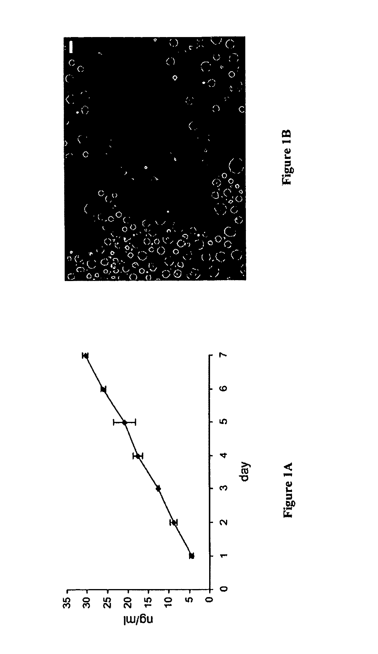
[0184]Shh-Containing Microspheres Release Active Shh Protein In Vitro and are not Toxic to Neural Stem Cells.

[0185]To establish a source of continuous release of Shh, biodegradable microspheres (10-40 μm in diameter) of poly(lactide-co-glycolide) (PLGA) that incorporated 0.5% of Shh as described above (Cat. No. 1314-SH / CF, recombinant human Shh from R&D Systems, Minneapolis, Minn., corresponding to SEQ ID NO:2, the active N-terminal Shh fragment, which corresponds to Cys24-Gly 197 of SEQ ID NO:1) were generated. Microsphere preparation is described above and adapted from the methods of (Fu et al. 2003). To determine the release kinetics 0.1 mg of Shh-containing microspheres or microspheres with PLGA alone (blank sample) were resuspended in 1 ml PBS. 100 μl aliquots were taken out each day and analyzed for the Shh release by ELISA, as shown in FIG. 1A. FIGS. 1A-B show biodegradable PLGA-based microspheres release active Shh in a course of at least 7 days. These microspheres are not t...

example 2

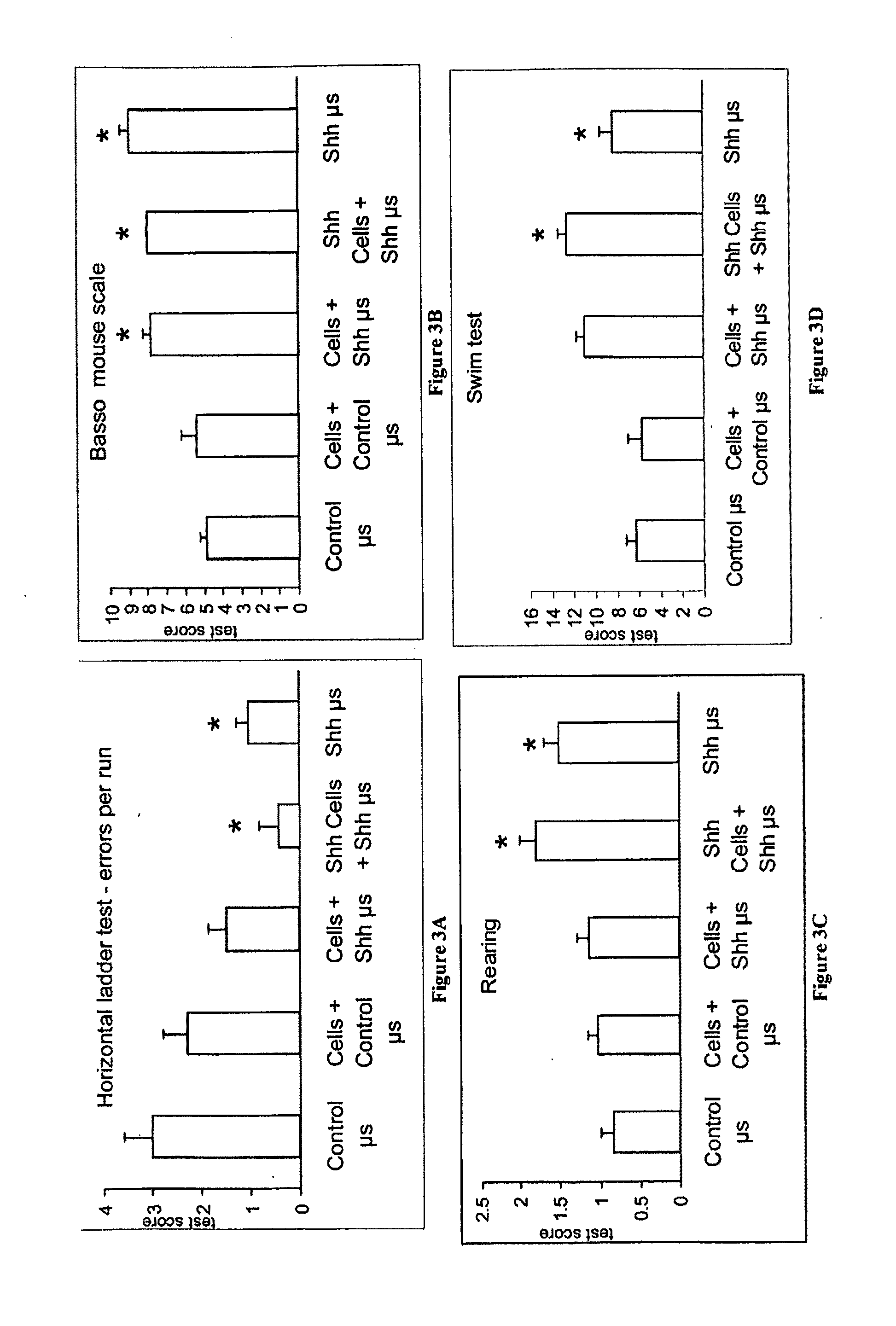
[0189]Transplantation of Shh-Releasing Microspheres or a Combination of Shh-Treated Spinal Cord Neural Stem Cells with Shh-Releasing Microspheres Produced / Resulted in Motor Recovery.

[0190]Shh-releasing microspheres and a combination of Shh-releasing microspheres and endothelial-expanded Shh treated cells were examined as treatments for SCI. To generate endothelial-expanded spinal cord stem cells, E9 mouse spinal cord stem cells were co-cultured with BPAE cells in serum-free medium with or without the addition of 1 μM Shh and 1 μM retinoic acid for 6 days, then removed and injected into adult (10-12 weeks old) mice recipients that had a dorsal over-hemisection spinal cord injury.

[0191]Adult mice were anaesthetized and the dorsal surface of the cord was exposed at T8-9, and the cord was cut down to a depth of 1 mm, representing halfway through the cord from the dorsal surface. This procedure severs the descending corticospinal and ascending sensory spinal axons located in the dorsal c...

example 3

[0194]Injection of Shh-Releasing Microspheres into SCI Resulted in a Reduced Astroglial Scar Formation Compared to Injection of Control Microspheres into SCI.

[0195]After behavioral testing, the mice were sacrificed (four weeks after SCI) and the spinal cords were examined by staining longitudinal sections of the SCI site with GFAP, which is an astrocyte marker (as shown in FIG. 4A). In FIGS. 4A-B, the spinal cord cells appear as bright regions, since they are isolated from GFP transgenic mice and constitutively express green fluorescent protein (GFP). Transplantation of Shh-releasing microspheres resulted in a decrease of the astrocytic scar at the site of injury, as shown in FIG. 4B, demonstrated by reduced staining of GFAP in FIG. 4B (few bright-staining cells on sections treated with Shh-releasing microspheres) compared to FIG. 4A (control microspheres lacking Shh and containing more brighter staining regions). These data illustrate that injection of Shh-releasing microspheres in...


PUM

PropertyMeasurementUnit
timeaaaaaaaaaa
diameteraaaaaaaaaa
sizeaaaaaaaaaa

Abstract

The present invention relates to treatment methods and methods for sustained delivery of one or more exogenous factors to desired nervous system sites. In certain embodiments, the invention relates to the use of biodegradable microspheres to deliver exogenous factors, such as the morphogenic factor, sonic hedgehog (Shh), to the site of spinal cord injury. In certain embodiments, the Shh-releasing microspheres are administered together with stem cells, which may be spinal cord neural stem cells. In certain embodiments, the invention relates to regrowth of neural cells in both the central and peripheral nervous systems.

Description

CROSS REFERENCE TO RELATED APPLICATIONS[0001]The present application claims priority under 35 U.S.C. §119 to U.S. Provisional Application Ser. No. 61 / 034,068, filed Mar. 5, 2008, which is hereby incorporated by reference in its entirety.GOVERNMENT SPONSORED RESEARCH OR DEVELOPMENT[0002]This invention was made in part in the course of research sponsored by the New York State Spinal Cord Injury Research Trust Fund through New York State Department of Health Contract #CO20922, New York State may have certain rights in this invention.FIELD OF THE INVENTION[0003]The present invention relates to treatments and methods for sustained delivery of one or more exogenous factors to predetermined sites in the mammalian nervous system. In certain embodiments, the invention relates to the use of biodegradable microspheres to deliver exogenous factors, such as the morphogenic factor, sonic hedgehog (Shh), to the site of spinal cord injury. In other embodiments, the Shh-releasing microspheres are ad...

Claims


Application Information

Patent Timeline
Patent Type & Authority Applications(United States)
IPC IPC(8): A61K38/20A61K38/18A61K38/30A61K35/12A61K35/30A61P25/00
CPCA61K9/0019A61K9/19C12N5/0623A61K35/30A61K38/1709A61K38/18A61P25/00
Inventor TEMPLE, SALLYLOWRY, NATALIASTERN, JEFFREYGODERIE, SUSAN K.
Owner TEMPLE SALLY
Why Eureka
Try Eureka