Methods and apparatuses for 3D imaging in magnetoencephalography and magnetocardiography

a technology of magnetoencephalography and 3d imaging, which is applied in the field of methods and apparatuses for 3d imaging in magnetoencephalography and magnetocardiography, can solve the problems of not fully exploiting all available information, prior art is unable to and cannot provide a closed-form solution to the problem of 3d image reconstruction. solve the problem of noise n(r2), reduce the effect of noise n(r

Inactive Publication Date: 2011-12-22
SUBBARAO MURALIDHARA
View PDF2 Cites 25 Cited by
  • Summary
  • Abstract
  • Description
  • Claims
  • Application Information

AI Technical Summary

Benefits of technology

[0019]It is an object of the present invention to provide methods and apparatuses for 3D imaging in Magnetoencephalography, Magnetocardiography, and the 3D imaging of electrical activity in neural, muscle, or any biological tissue.
[0020]An advantage of the present invention is that it is information efficient, i.e. it measures and exploits all available information for 3D image reconstruction. Therefore it provides a more accurate 3D image in terms of spatial, temporal, and contrast or intensity resolution and therefore it improves the accuracy of medical diagnosis. Another advantage of the present invention is that, unlike prior art, it does not use heuristic assumptions and it provides a computationally efficient closed-form solution to the problem of 3D image reconstruction.
[0026](d) Solving the vector-matrix equation g(r2)=H(r1,r2) f(r1)+n(r2) and estimating a solution f1(r1) for the 3D image f(r1) using a method that reduces the effect of noise n(r2) so that estimated solution f1(r1) is close to desired solution f(r1).
[0027]The biological tissue in the above method can be that of a mam

Problems solved by technology

Consequently, prior art does not provide a closed-form solution to the 3D image reconstruction problem and it uses heuristic assumptions.
The methods and apparatuses in prior art in MEG and MCG rely solely on data measured on a surface and therefore they do not fully exploit all the available information.
Therefore, prior art is unable to provide a closed-from solution to th

Method used


Image

  • Methods and apparatuses for 3D imaging in magnetoencephalography and magnetocardiography
  • Methods and apparatuses for 3D imaging in magnetoencephalography and magnetocardiography
  • Methods and apparatuses for 3D imaging in magnetoencephalography and magnetocardiography

Examples


Embodiment Construction

[0039]This invention discloses novel methods and apparatuses for 3D imaging in MEG and MCG. A detailed description of the methods and apparatuses are presented in this section.

[0040]The present invention is based on a new theory not found in prior art. It is based on the Field Paradigm. Therefore, the theoretical basis of the present invention is presented with concrete mathematical derivations for 3D imaging in MEG and MCG.

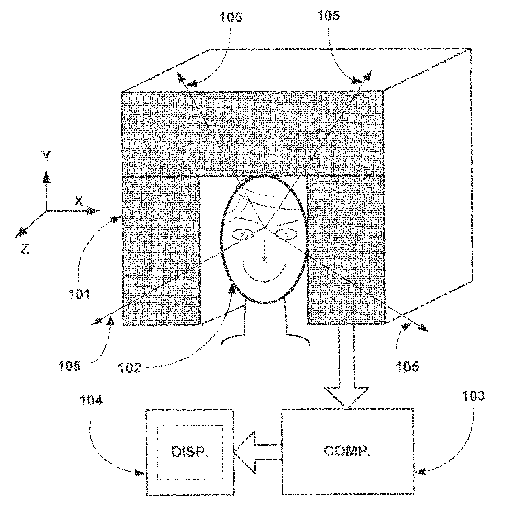
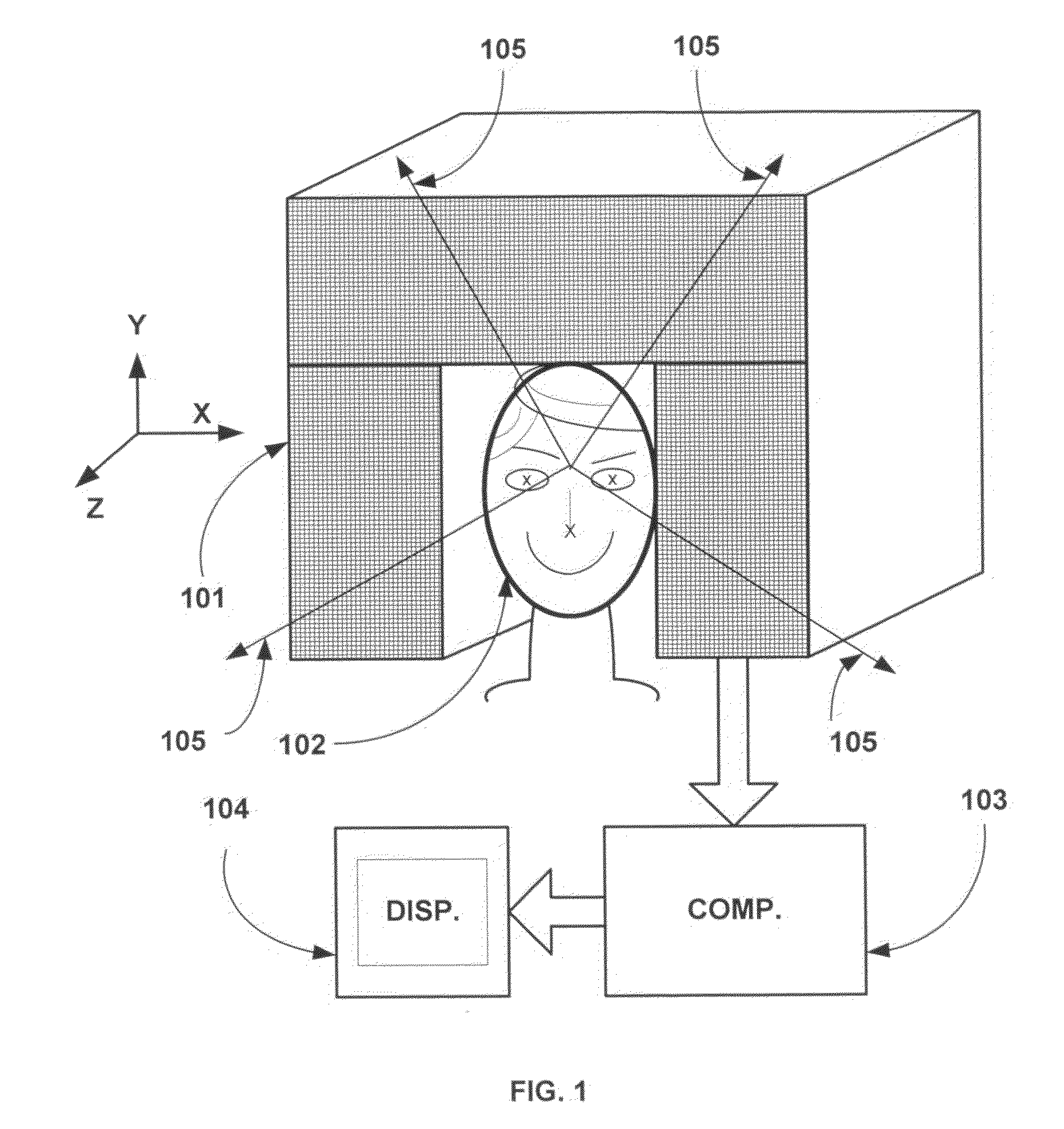
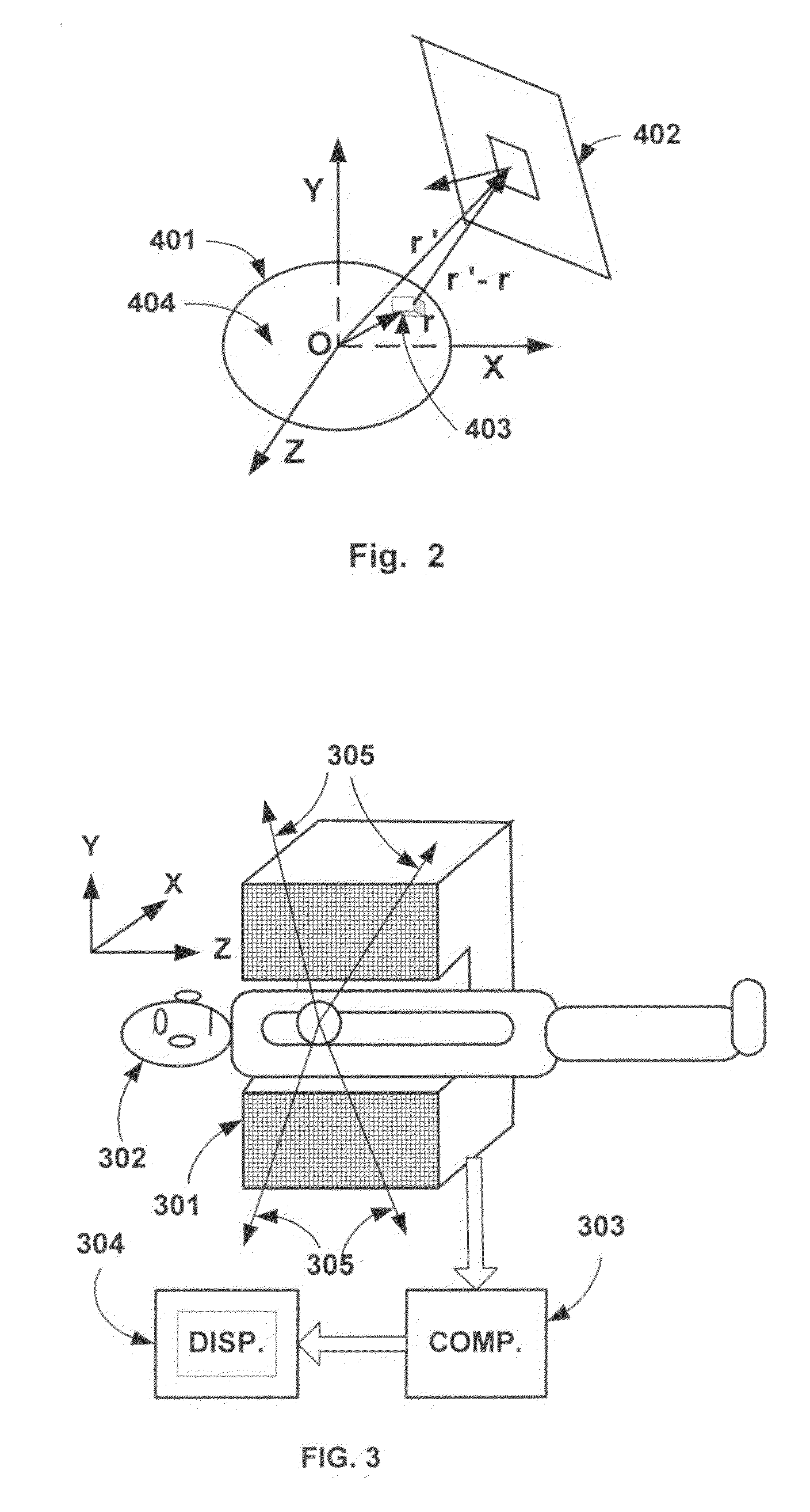
[0041]Consider an object to be imaged with biological tissue in which an electrical current pattern is present. For example, in MEG, this object would be the neural tissue in a brain and in MCG this would be the muscle tissue in a heart. The case of a human head placed in an MEG apparatus is shown in FIG. 1. This object 102 occupies a certain 3D volume space V1 inside a cubical volume, say with an approximate dimension of 200 mm on each side. In FIG. 2, the object is shown as 401 and volume V1 is shown as 404. This cubical volume can be thought of as being made u...


PUM


Abstract

This invention discloses methods and apparatuses for 3D imaging in Magnetoencephalography (MEG), Magnetocardiography (MCG), and electrical activity in any biological tissue such as neural/muscle tissue. This invention is based on Field Paradigm founded on the principle that the field intensity distribution in a 3D volume space uniquely determines the 3D density distribution of the field emission source and vice versa. Electrical neural/muscle activity in any biological tissue results in an electrical current pattern that produces a magnetic field. This magnetic field is measured in a 3D volume space that extends in all directions including substantially along the radial direction from the center of the object being imaged. Further, magnetic field intensity is measured at each point along three mutually perpendicular directions. This measured data captures all the available information and facilitates a computationally efficient closed-form solution to the 3D image reconstruction problem without the use of heuristic assumptions. This is unlike prior art where measurements are made only on a surface at a nearly constant radial distance from the center of the target object, and along a single direction. Therefore necessary, useful, and available data is ignored and not measured in prior art. Consequently, prior art does not provide a closed-form solution to the 3D image reconstruction problem and it uses heuristic assumptions. The methods and apparatuses of the present invention reconstruct a 3D image of the neural/muscle electrical current pattern in MEG, MCG, and related areas, by processing image data in either the original spatial domain or the Fourier domain.

Description

RELATED PROVISIONAL APPLICATION FOR PATENT[0001]This patent application is a continuation of the following US Provisional Application for patent filed by this inventor:[0002]M. Subbarao, “Methods and Apparatuses for Accurate and High-Resolution 3D Tomographic Imaging of Field Emission Sources in SPECT, PET, MRI, Magnetoencephalography, EEG, Inner Density Mapping of Earth, and Related Applications”, Provisional Application for Patent No. 61 / 397,958, filed Jun. 19, 2010.[0003]This patent application provides full details of the description of the above invention related to Magnetoencephalography (MEG) and Magnetocardiography (MCG).FIELD OF INVENTION[0004]This invention discloses novel methods and apparatuses for 3D imaging of electrical activity in human and animal organs such as the brain, the heart, and neural / muscle tissue in other parts of the body. Electrical activity in any neural / muscle tissue results in electrical current patterns that produce magnetic fields according to Biot...

Claims


Application Information

Patent Timeline
IPC IPC(8): A61B5/055
CPCA61B5/04007A61B5/7257A61B5/05A61B5/04008A61B5/243A61B5/245
Inventor SUBBARAO, MURALIDHARA
Owner SUBBARAO MURALIDHARA
Why Eureka
Try Eureka