Intravascular ultrasound detection of blood-flow distribution in an arterial wall

a technology of intravascular ultrasound and blood flow distribution, which is applied in the field of early identification of defects of the vascular system, can solve the problems of intraplaque hemorrhage and rupture, limited understanding of their functional anatomy and specific role in these diseases, and amplifying the chances of hemorrhag

Active Publication Date: 2011-12-29
MAYO FOUND FOR MEDICAL EDUCATION & RES
View PDF1 Cites 15 Cited by
  • Summary
  • Abstract
  • Description
  • Claims
  • Application Information

AI Technical Summary

Benefits of technology

[0010]In addition, the method assigns a density value (representing a change of density of the blood flow within the wall of the blood vessel) to every position of the transducer. The assignment of corresponding density values is made based on comparison among the image indicators which, in one embodiment, may include color-coded image pixels. In this case, analysis of the formed images may include analyzing a numb

Problems solved by technology

It has been speculated that a higher spatial density of vasa vasorum may contribute to the increased propensity towards neovascularization upon exposure to atherosclerotic risk factors, and may thereby subsequently lead to intraplaque hemorrhage and rupture.
While vasa vasorum have been implicated in a number of diseases, including atherosclerosis, understanding their functional anatomy and specific role in these diseases has been limited by the small size of the vasa vasorum and difficulty in imaging them.
The newly created capillaries, being very fragile, can be easily ruptured therefore amplifying the chances of hemorrhage.
Moreover, the plaques with high density of vasa vasorum are more prone to rupture and cause heart attacks and sudden death.
The existing methodologies are not necessarily suitable to accomplish this goal.
Well known

Method used


Image

  • Intravascular ultrasound detection of blood-flow distribution in an arterial wall
  • Intravascular ultrasound detection of blood-flow distribution in an arterial wall
  • Intravascular ultrasound detection of blood-flow distribution in an arterial wall

Examples


Embodiment Construction

[0026]References throughout this specification to “one embodiment,”“an embodiment,”“a related embodiment,” or similar language mean that a particular feature, structure, or characteristic described in connection with the referred to “embodiment” is included in at least one embodiment of the present invention. Thus, appearances of the phrases “in one embodiment,”“in an embodiment,” and similar language throughout this specification may, but do not necessarily, all refer to the same embodiment. It is to be understood that no portion of disclosure, taken on its own and / or in reference to a figure, is intended to provide a complete description of all features of the invention.

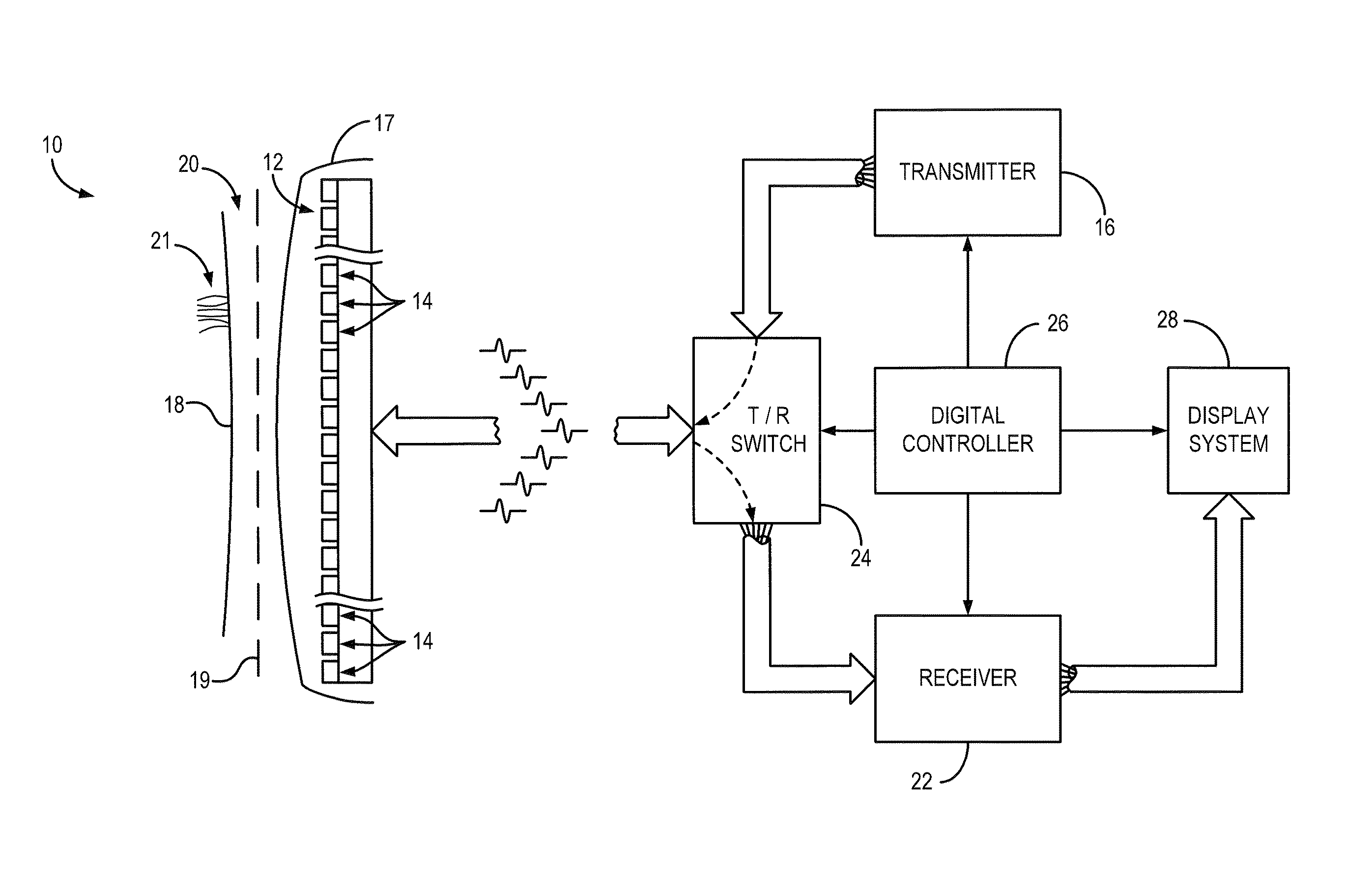
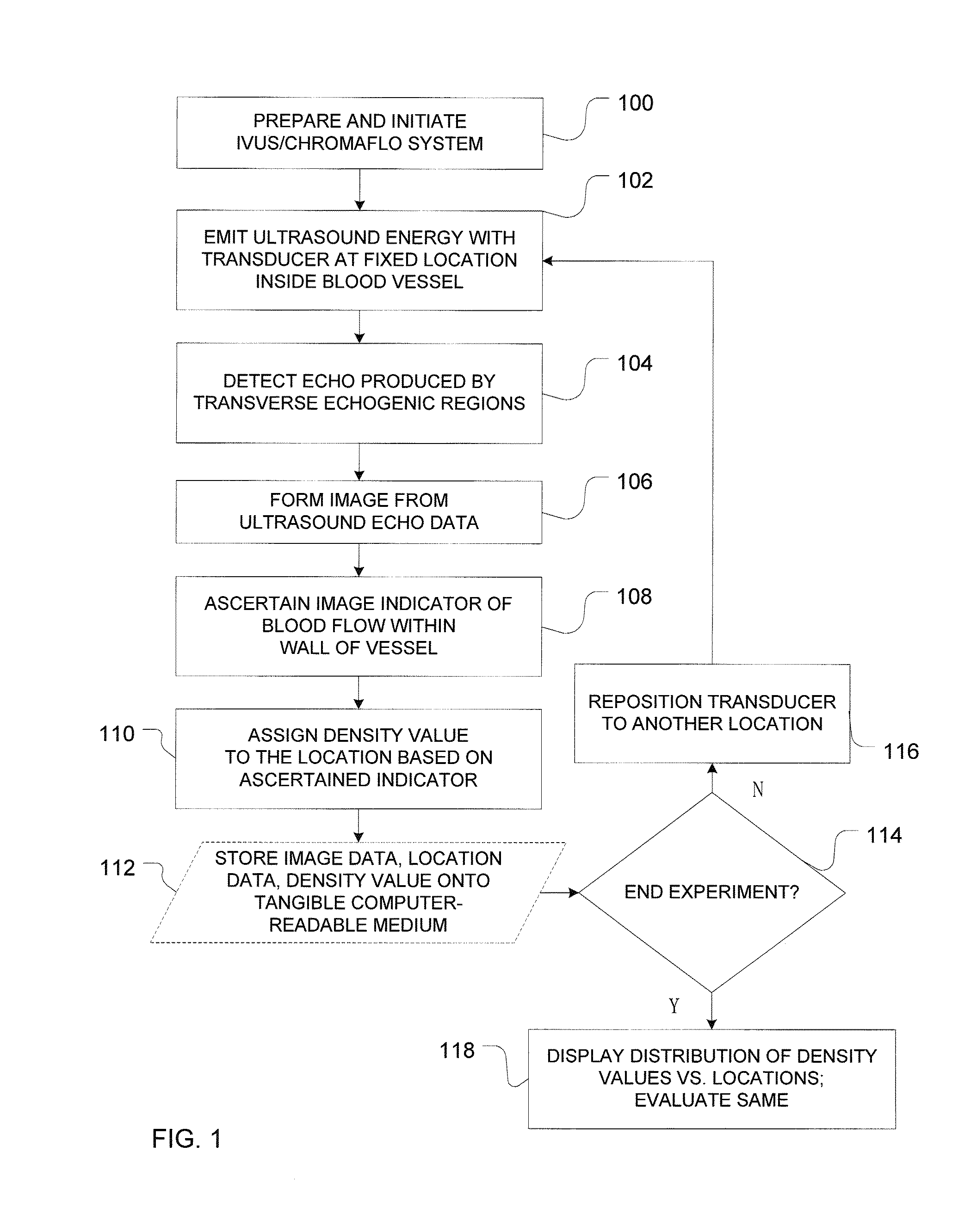
[0027]In addition, in drawings, with reference to which the following disclosure may describe features of the invention, like numbers represent the same or similar elements wherever possible. In the drawings, the depicted structural elements are generally not to scale, and certain components are enlarged relative t...


PUM


Abstract

A system and method for quantitative determination of density of vasa vasorum in an arterial wall that utilizes a detection of temporal and/or spatial displacement of blood-flow with the use of intravascular ultrasound system. Locations of extrema in the spatial distribution can be identified to detect vascular defects. The system and method support a clinically-useful application for early detection of indicators of diseases, such as coronary atherosclerosis.

Description

CROSS-REFERENCE TO RELATED APPLICATIONS[0001]This application claims the benefit of and priority from a U.S. Provisional Patent Application No. 61 / 358,319 filed on Jun. 24, 2010 and titled “Intravascular Ultrasound Detection of Blood-Flow Distribution in an Arterial Wall.” The disclosure of the above-mentioned patent application in incorporated herein by reference in its entirety.STATEMENT REGARDING FEDERALLY SPONSORED RESEARCH OR DEVELOPMENT[0002]This invention was made with government support under Grant Number NIH HL065342 awarded by the NIH National Heart, Lung, and Blood Institute. The government has certain rights in the invention.TECHNICAL FIELD[0003]The present invention relates to methods of early identification of defects of the vascular system and, in particular, to the detection of changes in the density of vasa vasorum using an intravascular ultrasound system.BACKGROUND ART[0004]Earlier experimental studies concluded that changes in the density of vasa vasorum—small blo...

Claims


Application Information

Patent Timeline
IPC IPC(8): A61B8/06
CPCA61B8/06A61B8/12A61B8/0891A61B8/0858
Inventor LERMAN, AMIRRITMAN, ERIK L.KLINE, TIMOTHY L.MORITZ, REGINA
Owner MAYO FOUND FOR MEDICAL EDUCATION & RES
Why Eureka
Try Eureka