

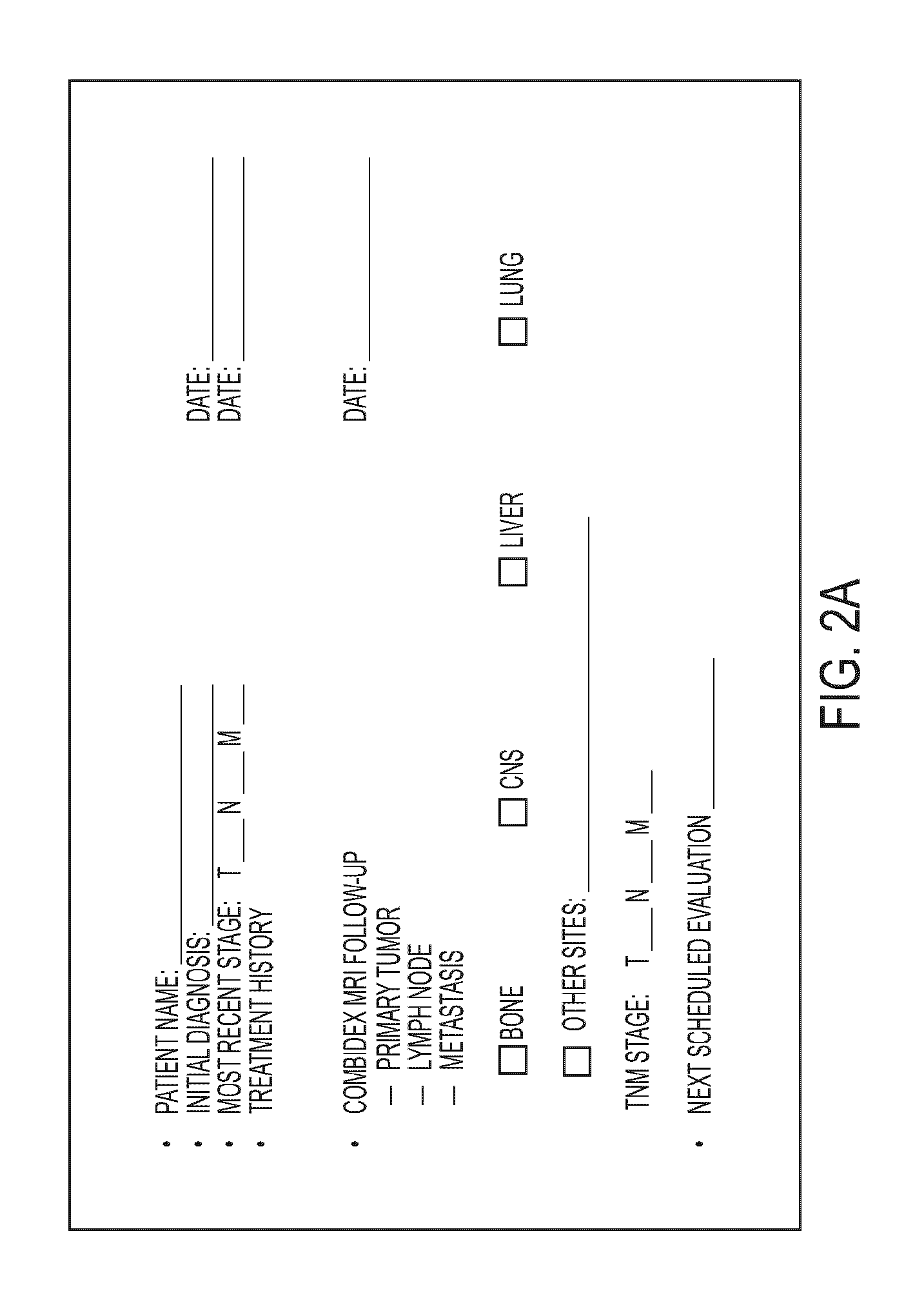
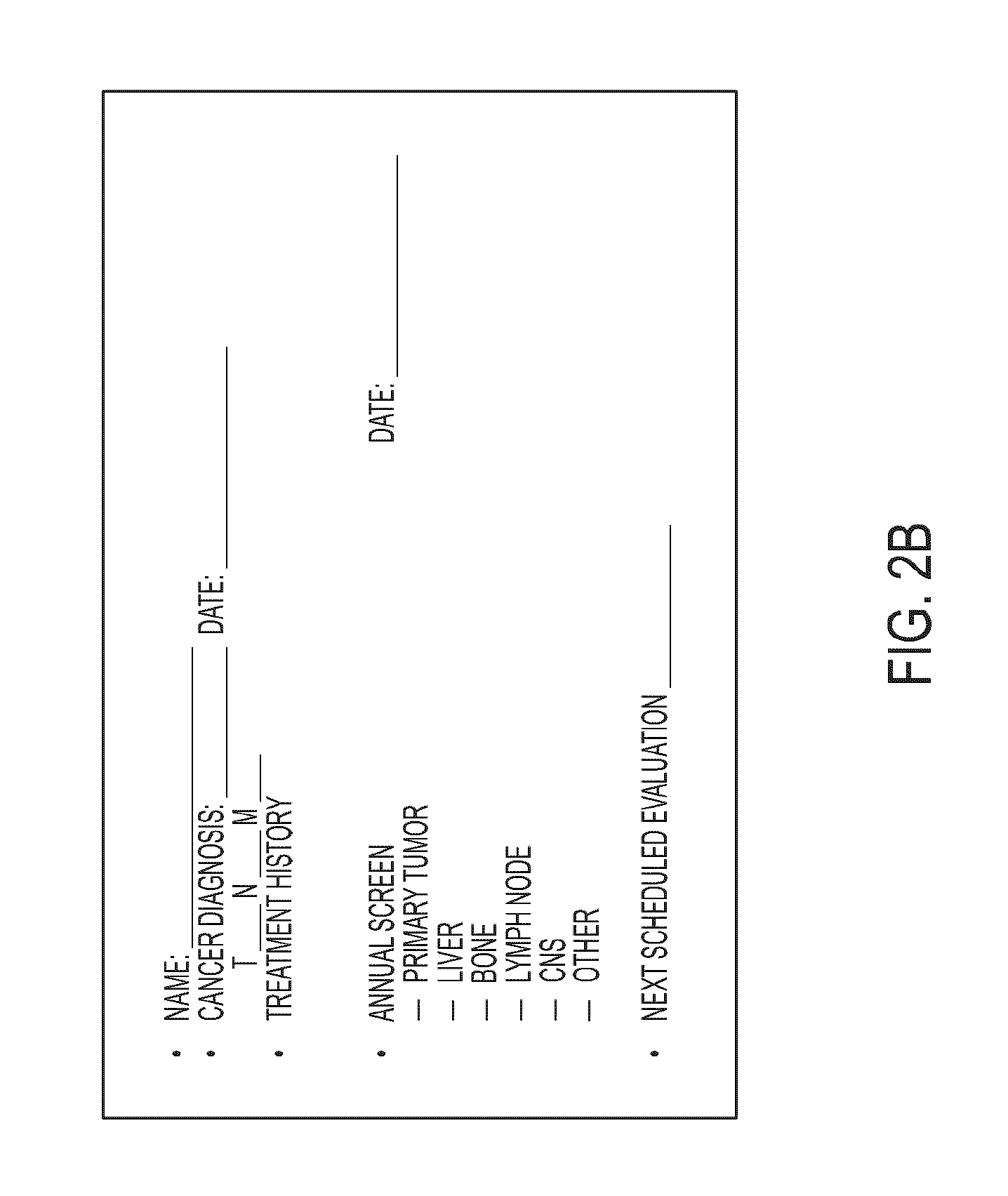
Macrophage-Enhanced MRI (MEMRI) in a Single Imaging Session
a single imaging session and macrophage technology, applied in the field of whole-body mri scanning and cancer staging, can solve the problems of breast cancer remaining a leading cause of death in middle-aged women, breast cancer remains an incurable disease, and mortality rates have decreased
- Summary
- Abstract
- Description
- Claims
- Application Information
AI Technical Summary
Benefits of technology
Problems solved by technology
Method used
Image


Examples
example 1
MEMRI Evaluation of Patient with Suspected Cancer in Single Breast
[0103]This situation involves a patient presenting with known or suspected cancer of one breast but a normal mammogram of the opposite breast. Recently, it has been suggested that such patients should undergo contrast-enhanced breast MRI to rule out other cancer foci (see Lehman, et al (2007) N Eng. J Med 356, 1295-303). In such an evaluation, the contrast agent is usually a gadolinium chelate and abnormal breast tissue is expected to show a focal accumulation of gadolinium in the expanded extracellular space associated with the cancer that was not clinically or mammographically evident. Though sensitive, this procedure is fraught with false positives—the abnormal regions must be biopsied and four of five such regions will not be cancerous—and does not provide information on the possible metastasis to local lymph nodes. In the present invention, the patient at risk is administered the macrophage biomarker and the brea...
example 2
Patient with Breast Cancer and Bone Pain
[0108]When metastatic breast cancer is suspected, it is important to rule out the most common sites of metastasis, as well as recurrence or new cancer in the breast. If whole-body MRI is performed during macrophage enhancement, in other words, if a whole-body MEMRI is performed, identification of soft tissues where there is excessive macrophage density will identify where metastasis may be present. In addition, bone MRI is the best examination for bone metastasis, and the identification of macrophage dense areas by MEMRI would increase diagnostic accuracy in bone. Finally, displacement of normal macrophages in liver, spleen, or lymph nodes would suggest the presence of metastasis in these sites. One additional use of the macrophage imaging technique MEMRI is to identify regions where a tissue biopsy may be obtained for pathological information.
example 3
Patient with Bladder Cancer and / or Bone Pain
[0109]This example is similar to the above examples with breast cancer, with the additional advantage that regional nodes can be reexamined along with liver and lung, or other sites of potential metastasis.
[0110]FIGS. 5A and 5B show a patient with bladder cancer with macrophages around the primary tumors. The bladder is indicated generally with an arrow in FIGS. 5A and 5B as the large central light area in the center of the pelvis region. FIG. 5A is the MRI without contrast agent and FIG. 5B is the image with contrast agent of the invention. In FIG. 5A, the tumor's presence is only hinted at by the “crease” in the bladder (shown with an arrow) that seems to be an indication of pressure on or displacement of the bladder along this juncture. FIG. 5B, with contrast agent, clearly shows the line of demarcation for the tumor along that “crease”, with the massive tumor showing as a dark mass directly to the left of this line, continuing down to ...
PUM
| Property | Measurement | Unit |
|---|---|---|
| time | aaaaa | aaaaa |
| time | aaaaa | aaaaa |
| time | aaaaa | aaaaa |
Abstract
Description
Claims
Application Information