
Methods And Marker Combinations For Screening For Predisposition To Lung Cancer

a marker combination and lung cancer technology, applied in the field of methods and marker combinations for screening for predisposition to lung cancer, can solve the problems of limiting the sensitivity of chest radiographs, affecting the normal screening of asymptomatic individuals, and procedures not having sufficient accuracy to be routinely used as screening tests for asymptomatic individuals

Inactive Publication Date: 2012-03-22
ABBOTT MOLECULAR INC
View PDF3 Cites 22 Cited by
  • Summary
  • Abstract
  • Description
  • Claims
  • Application Information

AI Technical Summary

Benefits of technology

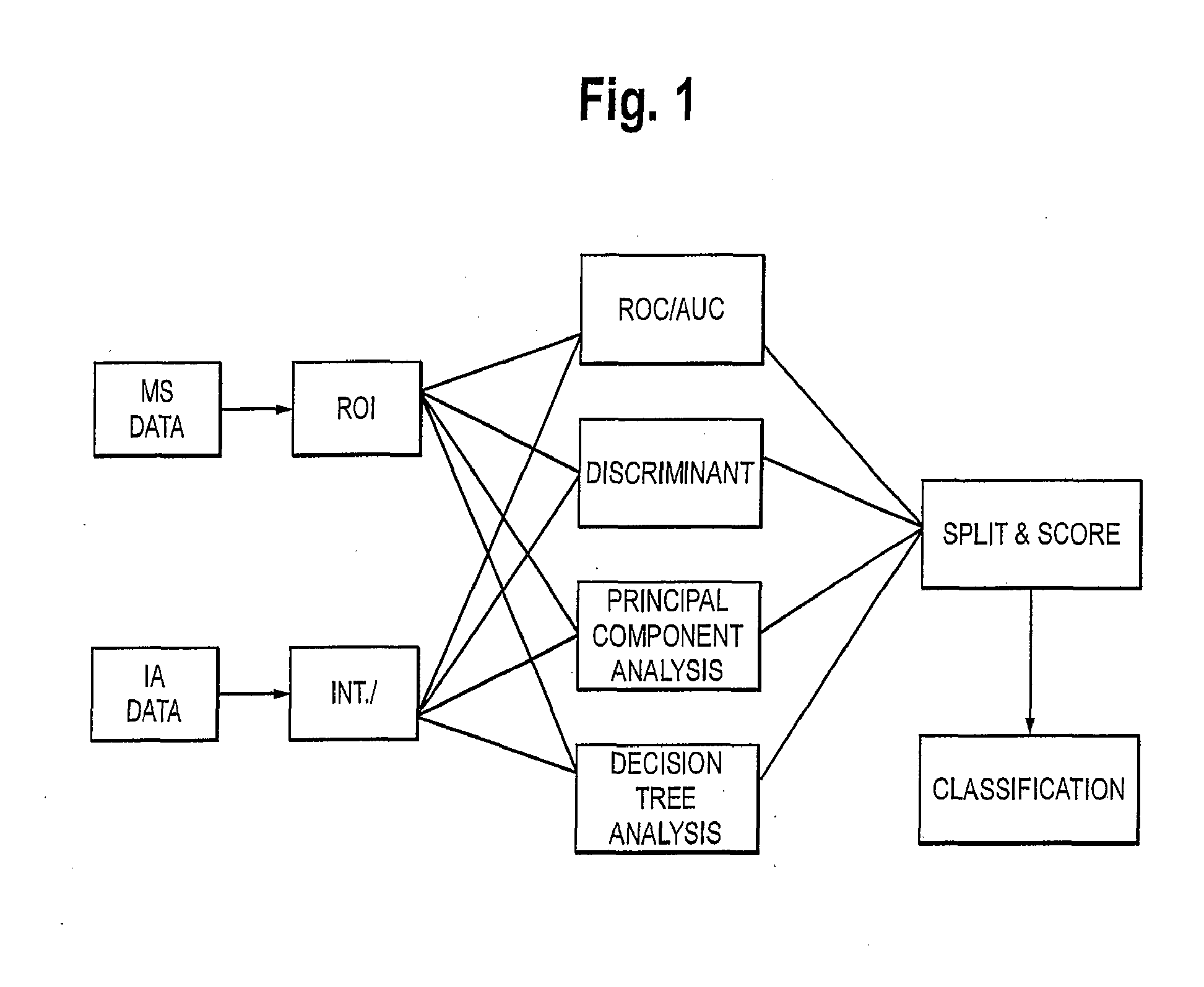
The patent describes a method for detecting lung cancer using a combination of biomarkers and biometric parameters. The method involves quantifying the amount of biomarkers in a sample from a subject and comparing it to a predetermined cutoff for each biomarker. The method can also involve obtaining a value for each biometric parameter and assigning a score to each biomarker based on its comparison to a predetermined cutoff. By combining the scores for each biomarker and biometric parameter, a total score is obtained, which is then compared to a predetermined total score to determine if the subject has a risk of lung cancer. The method is sensitive and specific, with a low detection limit for lung cancer.

Problems solved by technology

Lung cancer is also a major health problem in other areas of the world.
Early stage lung cancer can be detected by chest radiograph and the sputum cytological examination, however these procedures do not have sufficient accuracy to be routinely used as screening tests for asymptomatic individuals.
The potential technical problems that can limit the sensitivity of chest radiograph include suboptimal technique, insufficient exposure, and positioning and cooperation of the patient (See, T. G. Tape, et al., Ann. Intern. Med. 104: 663-670 (1986)).
False-negative interpretations are the cause of most errors and inconclusive results require follow-up testing for clarification (See, T. G. Tape et al., supra).
These statistics show that current procedures are failing to detect lung cancer at an early, treatable stage of the disease and that improved methods of detection and treatment are needed to reduce mortality.
Detecting recurrence by regular monitoring, however, does not greatly affect the mode of treatment and the overall survival time leading to the conclusion that current monitoring methods are not cost effective (See, K. S. Naunheim et al., Ann. Thorac. Surg. 60:1612-1616 (1995); G. L. Walsh et al., Ann. Thorac. Surg. 60: 1563-1572 (1995)).
However, even if implemented in clinical practice, the cost of CT screening would be high and the number of false positives leading to additional testing would also be high.
However, the lack of sensitivity that was characteristic of individual markers still prevents panels of tumor markers from being useful for early detection of lung cancer.
These markers have been found to be useful in staging, classifying, predicting outcomes for, and monitoring of lung cancer patients after their diagnosis has been made; however, these markers have not been found to be useful, either alone or in panels, for the early detection of the disease (See, S. Ando, et al., Anticancer Res. 21: 3085-3092 (2001); U.S. Preventive Services Task Force, Annals of Internal Medicine 140:738-739 (2004)).
The sensitivity of this technique, however, is limited by the degree of protein resolution of the two electrophoretic steps and by the detection step that depends on staining protein in gels.
Also, polypeptide instability will generate artifacts in the two-dimensional pattern.
However, these markers, kits and methods have not been adopted for use in routine practice as these markers and methods have not been duplicated in any laboratory.
To date none of these approaches have yielded novel autoantibodies useful for the early detection of lung cancer.
However, the number of cases and controls are limited (<200 total subjects) and the method needs to be validated on a much larger population.
Other environmental exposures such as asbestos, particulates, etc., can increase the risk of developing lung cancer as well.
Unfortunately, this algorithm is neither sensitive nor specific enough to be useful for the detection of early stage lung cancer.
Indeed, based on the cited algorithm, an individual with a significant smoking history will have a relative risk of 1 / 500 to 1 / 100 for developing lung cancer.

Method used


Image

  • Methods And Marker Combinations For Screening For Predisposition To Lung Cancer
  • Methods And Marker Combinations For Screening For Predisposition To Lung Cancer
  • Methods And Marker Combinations For Screening For Predisposition To Lung Cancer

Examples


example 1

Clinical Specimens

[0277]Clinical samples of patient serum were collected under an Institutional Review Board approved protocol. All subjects who contributed a specimen gave informed consent for the specimen to be collected and used in this project. Serum samples were drawn into a serum separator tube and allowed to clot for 15 minutes at room temperature. The clot was spun down and the sample poured off into 2 mL aliquots. Within 24 hours the samples were frozen at −80° C. and maintained at that temperature until further processing was undertaken. Upon receipt, the samples were thawed and realiquoted into smaller volumes for convenience and refrozen. The samples were then thawed a final time immediately before analysis. Therefore, every sample in the set was frozen and thawed twice before analysis.

[0278]A total of 751 specimens were collected and analyzed. The group was composed of 250 biopsy confirmed lung cancer patients, 274 biopsy confirmed benign lung disease patients, and 227 ...

example 2

Immunoassay Detection of Biomarkers

[0279]A. Abbott Laboratories (Abbott Park, Ill., Hereinafter “Abbott”) Architect™ Assays

[0280]Architect™ kits were acquired for the following antigens: CEA, CA125, SCC, CA19-9 and CA15-3. All assays were run according to the manufacturer's instructions. The concentrations of the analytes in the samples were provided by the Architect™ instrument. These concentrations were used to generate the AUC data shown below in Table 1.

TABLE 1Clinical performance (AUC) of CA125, CEA, CA15-3,CA19-9, and SCC in the small and large cohorts.large cohortsmall cohortMarker#obsAUC#obsAUCCa19-95480.5482560.559CEA5490.6882570.664Ca15-35490.6042570.569Ca1255490.6932570.665SCC5490.6152570.639The #obs refers to the total number of individuals or clinical samples in each group.

[0281]B. Roche Elecsys™ Assay

[0282]Cyfra 21-1 (Cytokeratin 19, CK-19) measurements were made on the Elecsys™ 2010 system (Roche Diagnostics GmbH, Mannheim, Germany) according to the manufacturer's ins...

example 3

Autoantibody Bead Array

[0285]A. Commercially available human proteins (See, Table 4, below) were attached to Luminex™ SeroMap™ beads (Austin, Tex.) and the individual beadsets were combined to prepare the reagent. Portions of the reagent were exposed to the human serum samples under conditions that allow any antibodies present to bind to the proteins. The unbound material was washed off and the beads were then exposed to a fluorescent conjugate of R-phycoerythrin linked to an antibody that specifically binds to human IgG. After washing, the beads were passed through a Luminex™ 100 instrument, which identified each bead according to its internal dyes, and measured the fluorescence bound to the bead, corresponding to the quantity of antibody bound to the bead. In this way, the immune responses of 772 samples (251 lung cancer, 244 normal, 277 benign) against 21 human proteins, as well as several non-human proteins for controls (bovine serum albumin (BSA) and tetanus toxin), were assess...


PUM

PropertyMeasurementUnit
sizeaaaaaaaaaa
chest computed tomographyaaaaaaaaaa
CTaaaaaaaaaa

Abstract

The present invention relates to rapid, sensitive methods for determining whether a subject has or is at risk of developing lung cancer based on certain combinations of biomarkers, or biomarkers and biometric parameters. The methods consist of: a) quantifying in a test sample obtained from a subject, the amount of two or more biomarkers in a panel, the panel comprising at least one antibody and at least one antigen: b) comparing the amount of each biomarker quantified in the panel to a predetermined cutoff for said biomarker and assigning a score for each biomarker based on said comparison: c) combining the assigned score for each biomarker quantified in step b to obtain a total score for said subject: d) comparing the total score in step c with a predetermined total score and e) determining whether said subject has a risk of lung cancer based on the comparison in step d.

Description

RELATED APPLICATION INFORMATION[0001]This application claims priority to U.S. application Ser. No. 11 / 771,727, filed Jun. 29, 2007, which is a continuation-in-part of U.S. application Ser. No. 11 / 644,365 filed on Dec. 21, 2006, which claims priority to U.S. Patent Application No. 60 / 753,331 filed on Dec. 22, 2005, the contents of all are herein incorporated by reference.BACKGROUND OF THE INVENTION[0002]Lung cancer is the second most common cancer for both men and women in the United States, with an estimated 172,500 new cases projected to be diagnosed during 2005 (American Cancer Society statistics). It is the most common cause of cancer death for both sexes, with over 163,000 lung cancer related deaths expected in 2005. Lung cancer is also a major health problem in other areas of the world. In the European Union, approximately 135,000 new cases occur each year (Genesis Report, February 1995). Also, incidence is rapidly increasing in Central and Eastern Europe where men have the wor...

Claims


Application Information

Patent Timeline
Patent Type & Authority Applications(United States)
IPC IPC(8): C40B30/04G01N33/566G01N33/53C07K14/435C07K7/08
CPCG01N33/57423C07K14/4738G01N2333/47G01N2333/4703G01N2333/4725G01N2333/4739G01N2333/4742G01N2333/4748G01N2333/705G01N2333/70596G01N2333/775G01N2333/8125G01N2333/91205
Inventor COLPITTS, TRACEYRUSSELL, ERIC L.FROST, STEPHENRAMIREZ, JAVIERSINGH, BHAWANIRUSSELL, JOHN C.
Owner ABBOTT MOLECULAR INC
Learn More