
Method and apparatus for generating medical images

a medical image and apparatus technology, applied in the field of imaging systems, can solve the problems of image quality degradation, image quality degradation, image quality errors during image review, etc., and achieve the effect of improving the quality of medical images

Inactive Publication Date: 2012-03-29
GENERAL ELECTRIC CO
View PDF4 Cites 48 Cited by
  • Summary
  • Abstract
  • Description
  • Claims
  • Application Information

AI Technical Summary

Benefits of technology

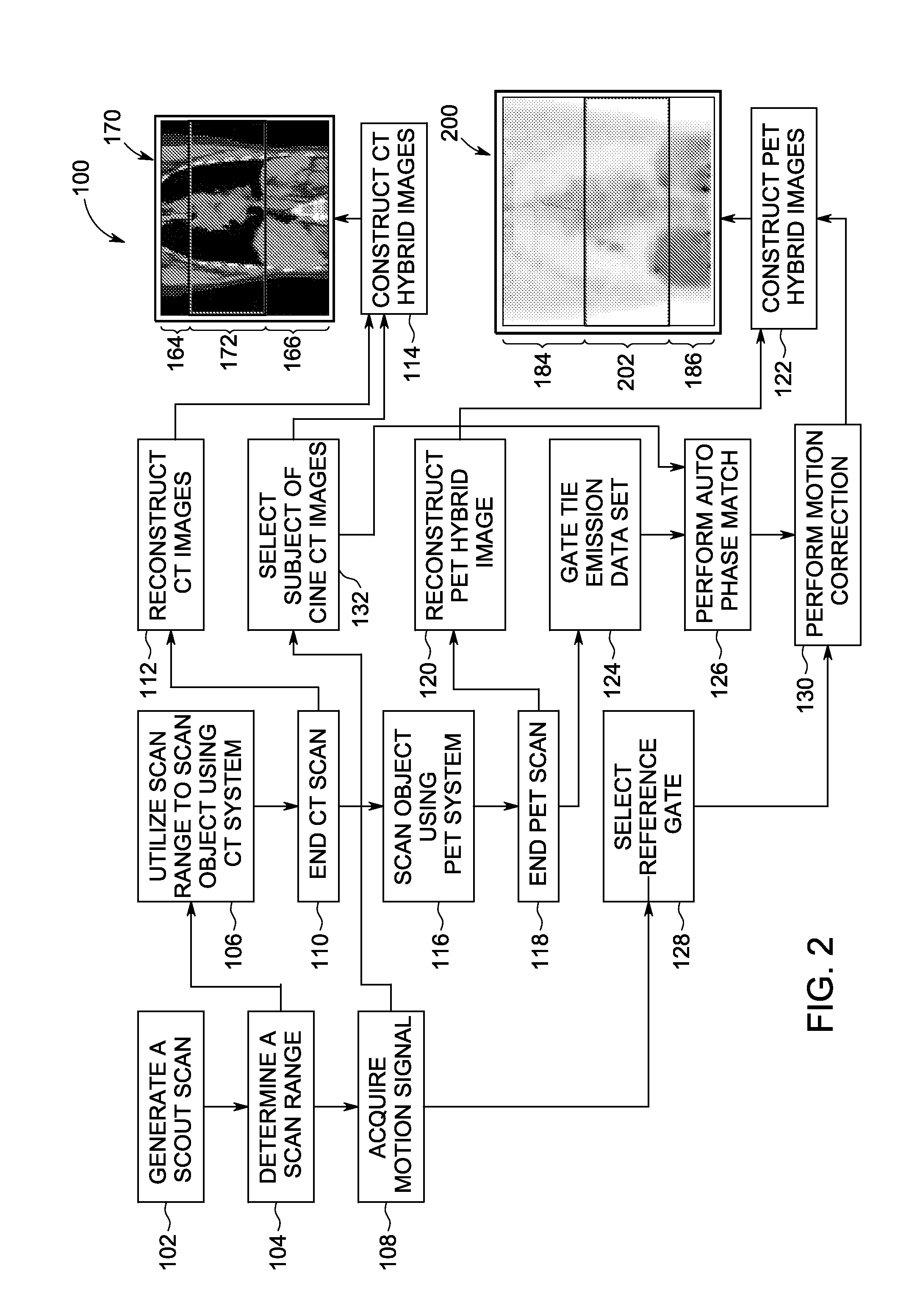
[0005]In another embodiment, a method of improving the quality of a medical image is provided. The method includes generating a plurality of gated Positron Emission Tomography (PET) images, motion correcting the gated PET images using a PET reference gate to generate a hybrid PET series of images, selecting at least one Computed Tomography (CT) image having the same respiratory phase as the gated PET images stored in the PET reference bin, and constructing at least one PET image volume using the hybrid PET series of images.

Problems solved by technology

During operation, the image quality of conventional imaging systems may be affected by the motion of the object being imaged.
In particular, motion of the imaged object can degrade the image quality.
The respiratory motion may lead to errors during image review, such as when a physician is determining the size of a lesion, determining the location of the lesion, or quantifying the lesion.
However, the quantity of data produced by utilizing the various motion correction techniques is typically relatively large.

Method used


Image

  • Method and apparatus for generating medical images
  • Method and apparatus for generating medical images
  • Method and apparatus for generating medical images

Examples


Embodiment Construction

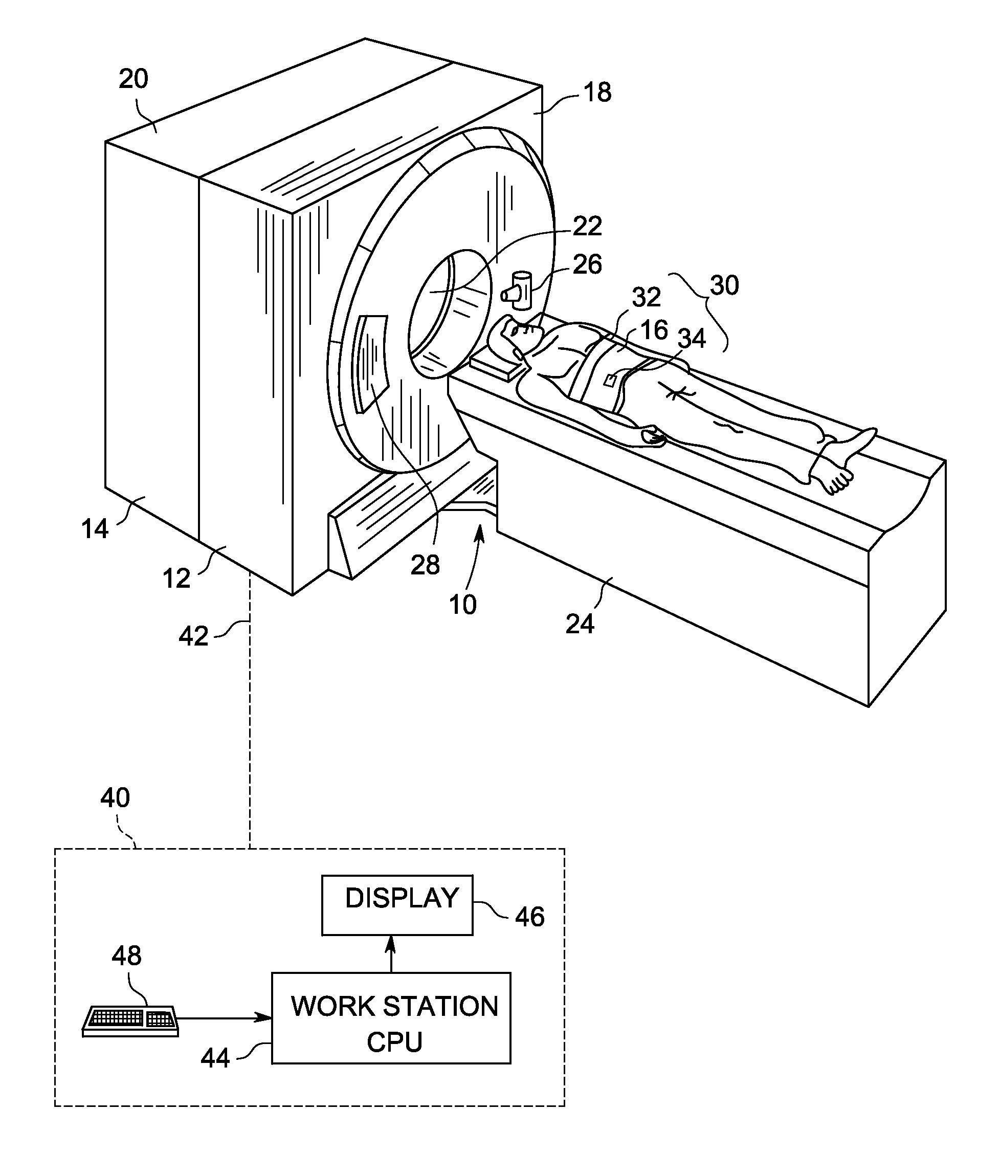
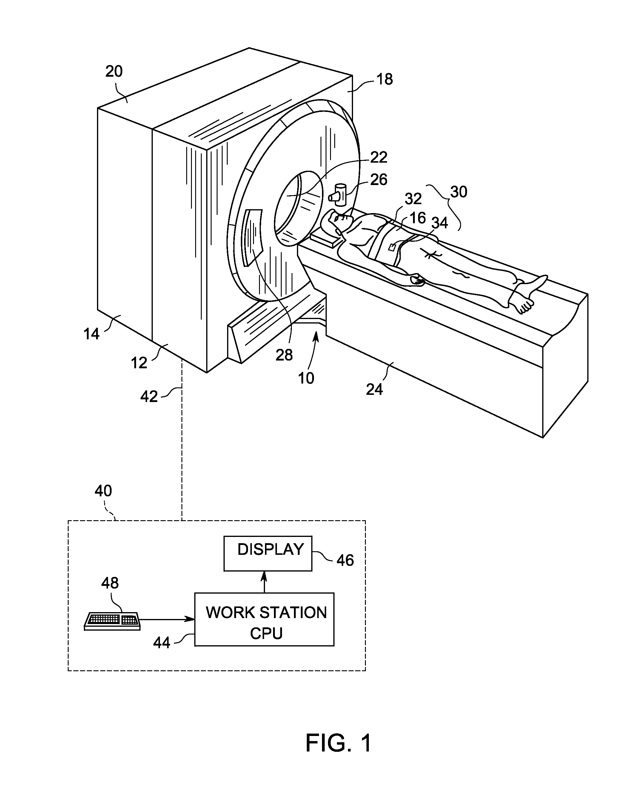
[0014]The foregoing summary, as well as the following detailed description of various embodiments, will be better understood when read in conjunction with the appended drawings. To the extent that the figures illustrate diagrams of the functional blocks of the various embodiments, the functional blocks are not necessarily indicative of the division between hardware circuitry. Thus, for example, one or more of the functional blocks (e.g., processors or memories) may be implemented in a single piece of hardware (e.g., a general purpose signal processor or a block of random access memory, hard disk, or the like) or multiple pieces of hardware. Similarly, the programs may be stand alone programs, may be incorporated as subroutines in an operating system, may be functions in an installed software package, and the like. It should be understood that the various embodiments are not limited to the arrangements and instrumentality shown in the drawings.

[0015]As used herein, an element or step...


PUM


Abstract

A method for generating a hybrid imaging volume includes acquiring a Positron Emission Tomography (PET) imaging dataset of an object using a PET imaging system, the PET imaging dataset including at least one motion affected portion and at least one non-motion affected portion, identifying a motion affected portion of the PET imaging dataset, motion correcting the identified portion of the PET imaging dataset to generate a hybrid portion, and constructing a hybrid PET image volume using the hybrid portion and the at least one non-motion affected portion. A system for implementing the method is also described herein.

Description

BACKGROUND OF THE INVENTION[0001]The subject matter disclosed herein relates generally to imaging systems, and more particularly, to an apparatus and method for generating medical images.[0002]Multi-modality imaging systems exist that scan using different modalities, for example, Computed Tomography (CT), Magnetic Resonance Imaging (MRI), Positron Emission Tomography (PET), and Single Photon Emission Computed Tomography (SPECT). During operation, the image quality of conventional imaging systems may be affected by the motion of the object being imaged. In particular, motion of the imaged object can degrade the image quality. More specifically, image artifacts are produced by movement of the object during image acquisition. Respiratory motion is a common source of involuntary motion in mammals (e.g., people and animals) encountered in medical imaging systems. The respiratory motion may lead to errors during image review, such as when a physician is determining the size of a lesion, d...

Claims


Application Information

Patent Timeline
IPC IPC(8): A61B5/05G01T1/164
CPCA61B5/1113A61B6/5258A61B6/5235A61B6/032A61B6/037
Inventor WOLLENWEBER, SCOTT DAVIDGANIN, ALEXANDER
Owner GENERAL ELECTRIC CO
Learn More