Surgical simulation model generating method, surgical simulation method, and surgical simulator

a simulation model and simulation model technology, applied in the field of surgical simulation model generating method, surgical simulation method, surgical simulator, can solve the problems of membrane tissue surrounding the organ to be operated on that cannot be captured by such means, model that does not incorporate membrane tissue is unsuitable for preoperative simulation, and model that does not incorporate membrane tissue is unsuitable for us

Active Publication Date: 2012-04-05
MITSUBISHI PRECISION
View PDF3 Cites 3 Cited by
  • Summary
  • Abstract
  • Description
  • Claims
  • Application Information

AI Technical Summary

Benefits of technology

[0019]According to the simulation model generating method, surgical simulation method, and surgical simulator of the invention described above, the position achieved by the motion of the simulated organ can be computed in real time.
[0020]The object and advantages of the invention will be realized and attained by means of the elements and combinations particularly pointed out in the claims.
[0021]It is to be understood that both the foregoing general description and the following detailed description are exemplary and explanatory and are not restrictive of the invention, as claimed.

Problems solved by technology

To acquire information of each individual patient, it is common to use medical image data such as CT or MRI data, but images of the membrane tissues surrounding the organ to be operated on cannot be captured by such means.
Because of the inability to recognize such membrane tissues, there arises the problem that the membrane tissues cannot be modeled.
A model that does not incorporate membrane tissues is unsuitable for use in a preoperative simulation.
However, in this case, the deformation of the organ model would greatly differ from the actual deformation, rendering such a model unsatisfactory for use in a preoperative simulation.
However, it is not common to compute the reaction of the simulated organ and supply the computed reaction to the force sensing device, while at the same time, computing in real time the position achieved by the motion of the simulated organ.
However, since it takes a finite time to complete the numerical computation of the motion equation that uses such a stiffness matrix, it has not been possible to compute the motion of the simulated organ in real time.
Furthermore, the computation using the prior art stiffness matrix has had the problem that the computation may diverge.

Method used


Image

  • Surgical simulation model generating method, surgical simulation method, and surgical simulator
  • Surgical simulation model generating method, surgical simulation method, and surgical simulator
  • Surgical simulation model generating method, surgical simulation method, and surgical simulator

Examples


embodiment 1

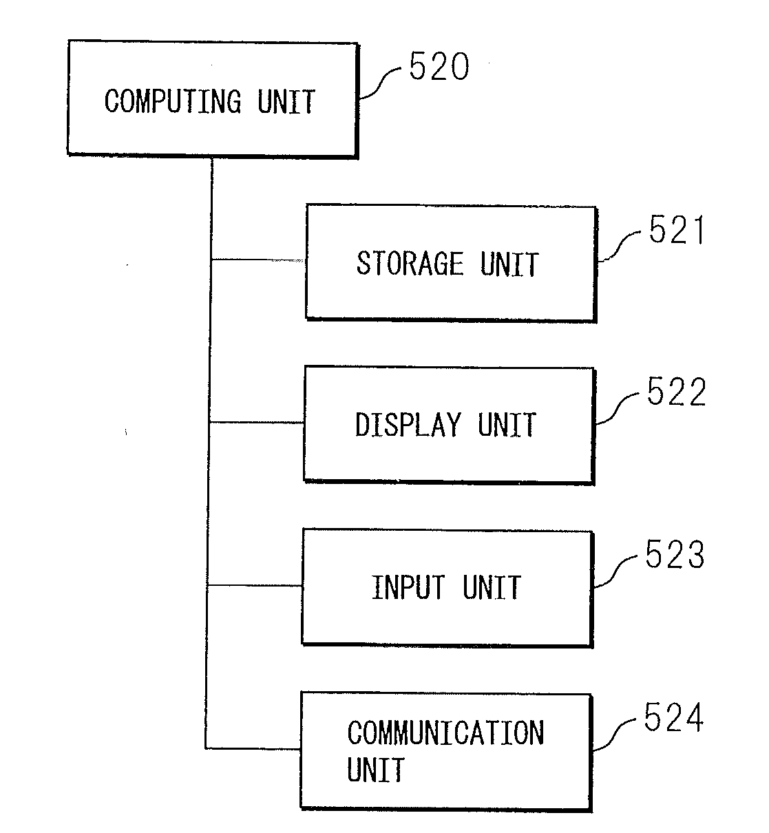
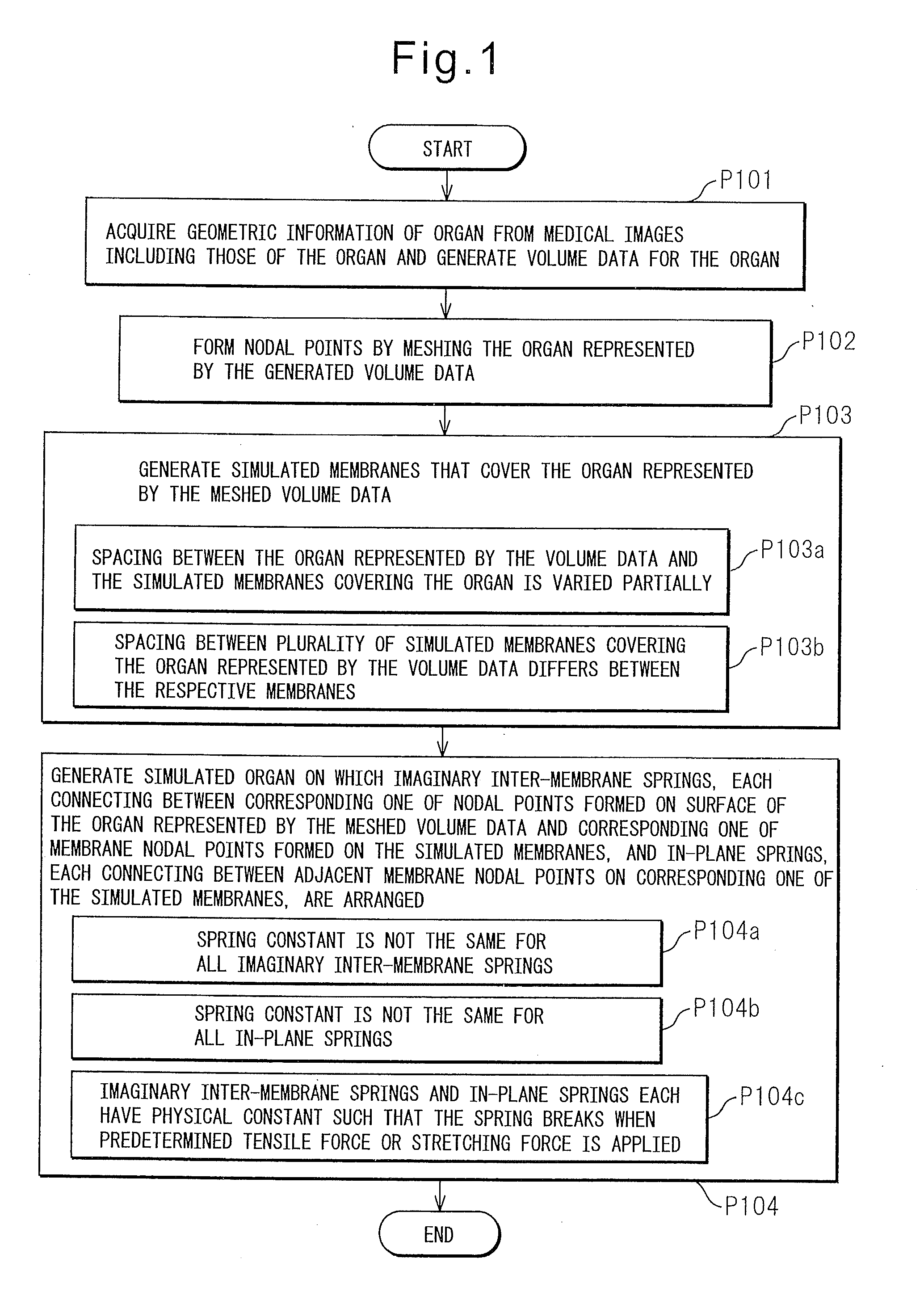
[0035]The first embodiment of the surgical simulation model generating method will be described below. The surgical simulation model generation according to the first embodiment is performed using the surgical simulator shown in FIG. 5A.

[0036]The medical image data storage unit 501 stores the source data of medical images including, for example, those of the organ to be operated on. The source data of medical images is obtained, for example, by CT imaging or MRI imaging.

[0037]The image generating unit 503 generates images, including those of organs, by using the medical image data stored in the medical image data storage unit 501. The images, each representing a cross section of the patient to be operated on, are obtained by scanning the patient's body in thin slices in a prescribed direction. The volume data constructing unit 502 acquires geometrical information of each organ from the medical image data obtained by capturing the images of body parts, including those of the organ, w...

embodiment 2

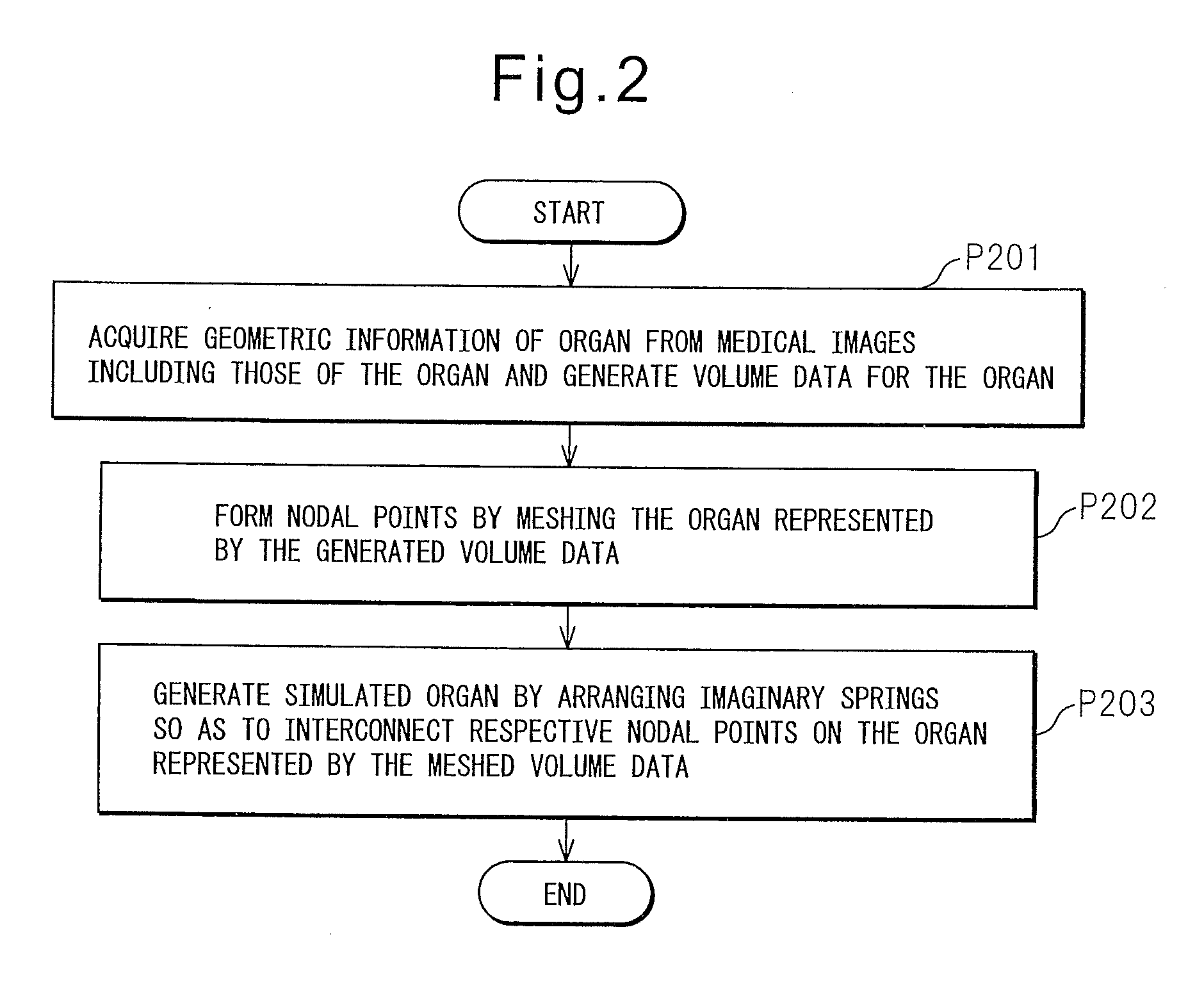
[0053]Next, a method for generating a surgical simulation model intended to simulate a large deformation of a designated organ with high accuracy will be described below with reference to FIG. 2. The surgical simulation model generation according to the second embodiment also is performed using the surgical simulator shown in FIG. 5A.

[0054]In FIG. 2, processes P201 and P202 are the same as the corresponding processes P101 and P102 shown in FIG. 1.

[0055]Next, in process P203, the volume data constructing unit 502 generates a simulated organ by arranging imaginary springs 701a, 701b, 701c, . . . so as to interconnect the respective nodal points on the finite-element model of the organ represented by the volume data meshed with tetrahedrons (FIG. 7). Each imaginary spring is arranged between adjacent nodal points on the organ represented by the volume data.

[0056]The imaginary springs 701a, 701b, 701c, . . . are each formed using a spring model. The imaginary springs are arranged betwee...

embodiment 3

[0061]Next, a description will be given of a surgical simulation that uses the surgical simulation model data generated according to the first embodiment and an apparatus that is used to perform the simulation. The surgical simulation according to the third embodiment is performed using the surgical simulator shown in FIG. 5A.

[0062]The image generating unit 503 retrieves from the surgical simulation model data unit 505 the surgical simulation model data for the simulated organ having the organ represented by the meshed volume data and the simulated membranes covering the organ represented by the meshed volume data. Then, the image generating unit 503 causes the image display device 504 to display the simulated organ having the simulated organ and the plurality of simulated membranes arranged around the organ. In this simulated organ, the imaginary inter-membrane springs and the in-plane springs are arranged as earlier described, defining the dynamic properties of the simulated organ...


PUM


Abstract

A surgical simulation model generating method includes: a first process in which a computing unit acquires geometrical information of an organ from a medical image stored in a storage unit, including an image of the organ, and generates volume data for the organ; a second process in which, after the first process, the computing unit forms nodal points by meshing the organ represented by the generated volume data; a third process in which the computing unit generates a simulated membrane that covers the organ represented by the volume data meshed in the second process; and a fourth process in which the computing unit generates a simulated organ by drawing an imaginary line so as to extend from each nodal point formed on a surface of the organ represented by the volume data meshed in the second process in a direction that intersects the simulated membrane and thereby forming a membrane nodal point at a point where the imaginary line intersects the simulated membrane generated in the third process, and by arranging on each imaginary line an imaginary inter-membrane spring that connects between the nodal point formed on the surface of the organ and the membrane nodal point, while also arranging an in-plane spring that connects between adjacent membrane nodal points on the simulated membrane.

Description

TECHNICAL FIELD[0001]The present invention relates to a method for generating a surgical simulation model used when conducting a surgical simulation before performing a surgical operation using an endoscope, and also relates to a surgical simulation method and a surgical simulator.BACKGROUND ART[0002]With advances in medical technology and medical instruments, many abdominal surgical operations are being performed using a laparoscope. Since laparoscopic surgery is performed by viewing a three-dimensional object displayed on a two-dimensional image display device, training is indispensable for acquiring of the required skill. In actual laparoscopic surgery, the surgery must be planned so as to match each individual patient because the number of blood vessels, the positions of the blood vessels, and the positional relationship of organs, for example, the position and size of a tumor, differ from patient to patient.[0003]For this purpose, it may be appropriate to perform, prior to surg...

Claims


Application Information

Patent Timeline
Patent Type & Authority Applications(United States)
IPC IPC(8): G06T15/00A61B6/03A61B19/00G06T17/20G09B9/00
CPCA61B2019/505G09B23/285G06T17/20A61B2034/105G06T7/74G06T7/248G06V10/42G06F18/22G06T7/0014G06T13/80G06T2207/30004
Inventor KUBOTA, YOSHINOBUMAKIYAMA, KAZUHIDEKIKUKAWA, TAKAAKINAGASAKA, MANABUSAKAMOTO, HIDEOOGATA, MASATO
Owner MITSUBISHI PRECISION
Why Eureka
Try Eureka