Diagnosis apparatus

a diagnostic device and antenna technology, applied in medical science, surgery, vaccination/ovulation diagnostics, etc., can solve the problems of difficult to keep the distance between the antenna and the skin constant, the electromagnetic wave cannot be perfectly removed, and the pain of a person under diagnostic tests is severe, so as to achieve high contrast, low diagnosis cost, and high resolution

Inactive Publication Date: 2012-04-05
NAT UNIV CORP SHIZUOKA UNIV
View PDF3 Cites 31 Cited by
  • Summary
  • Abstract
  • Description
  • Claims
  • Application Information

AI Technical Summary

Benefits of technology

[0030]According to the present invention, it is possible to provide a diagnosis apparatus for detecting abnormal cells via the microwave imaging, which has a high co...

Problems solved by technology

The reflection of the electromagnetic wave from the skin cannot be perfectly removed even by using the matching medium.
As for the shape of the breast, since the ...

Method used


Image

  • Diagnosis apparatus
  • Diagnosis apparatus
  • Diagnosis apparatus

Examples


first embodiment

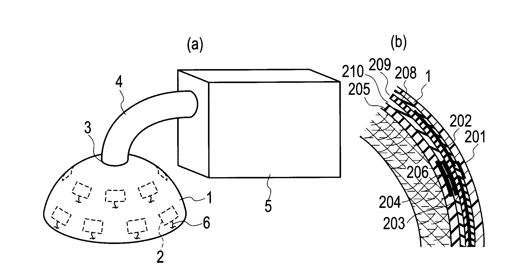
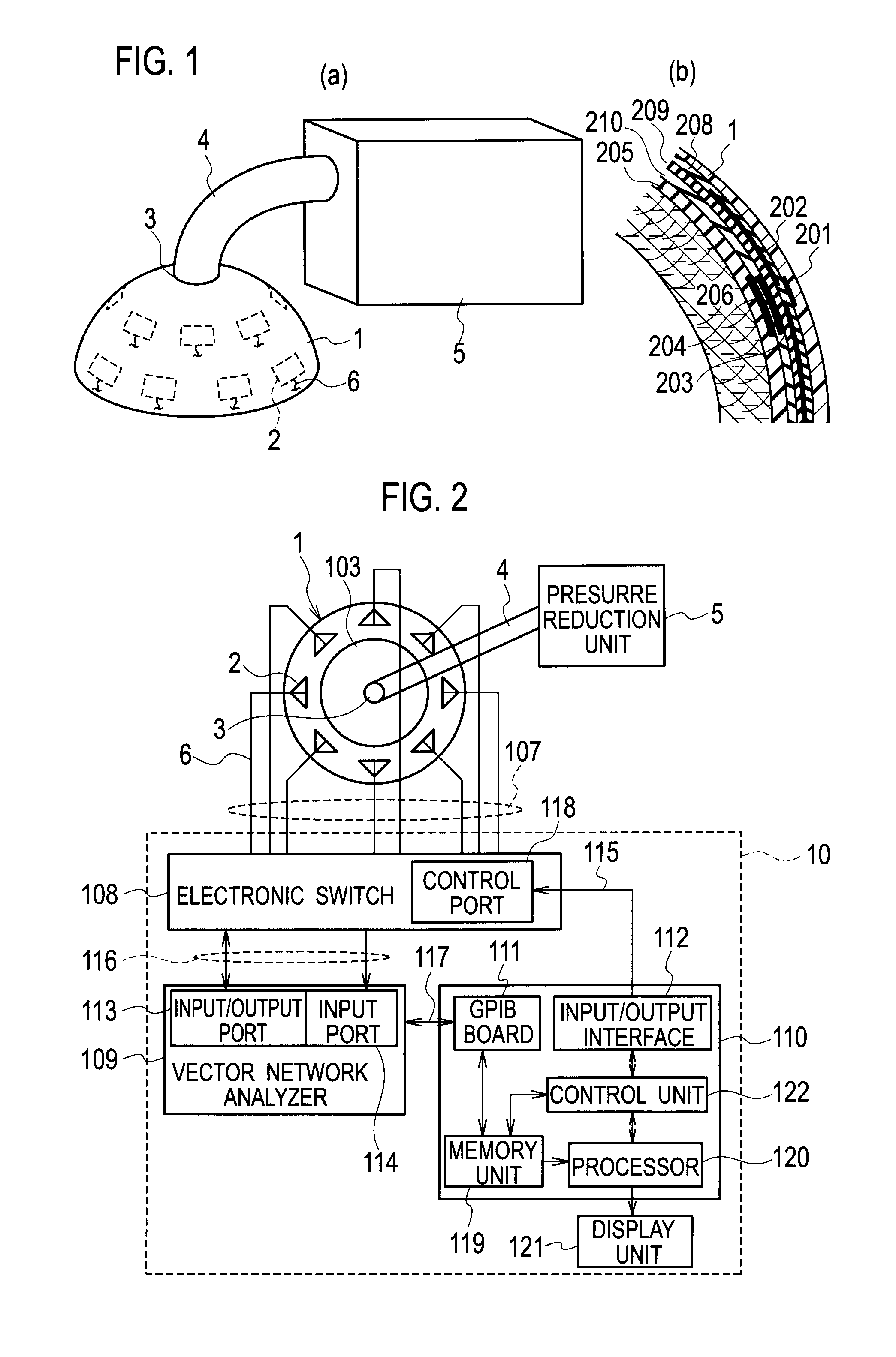
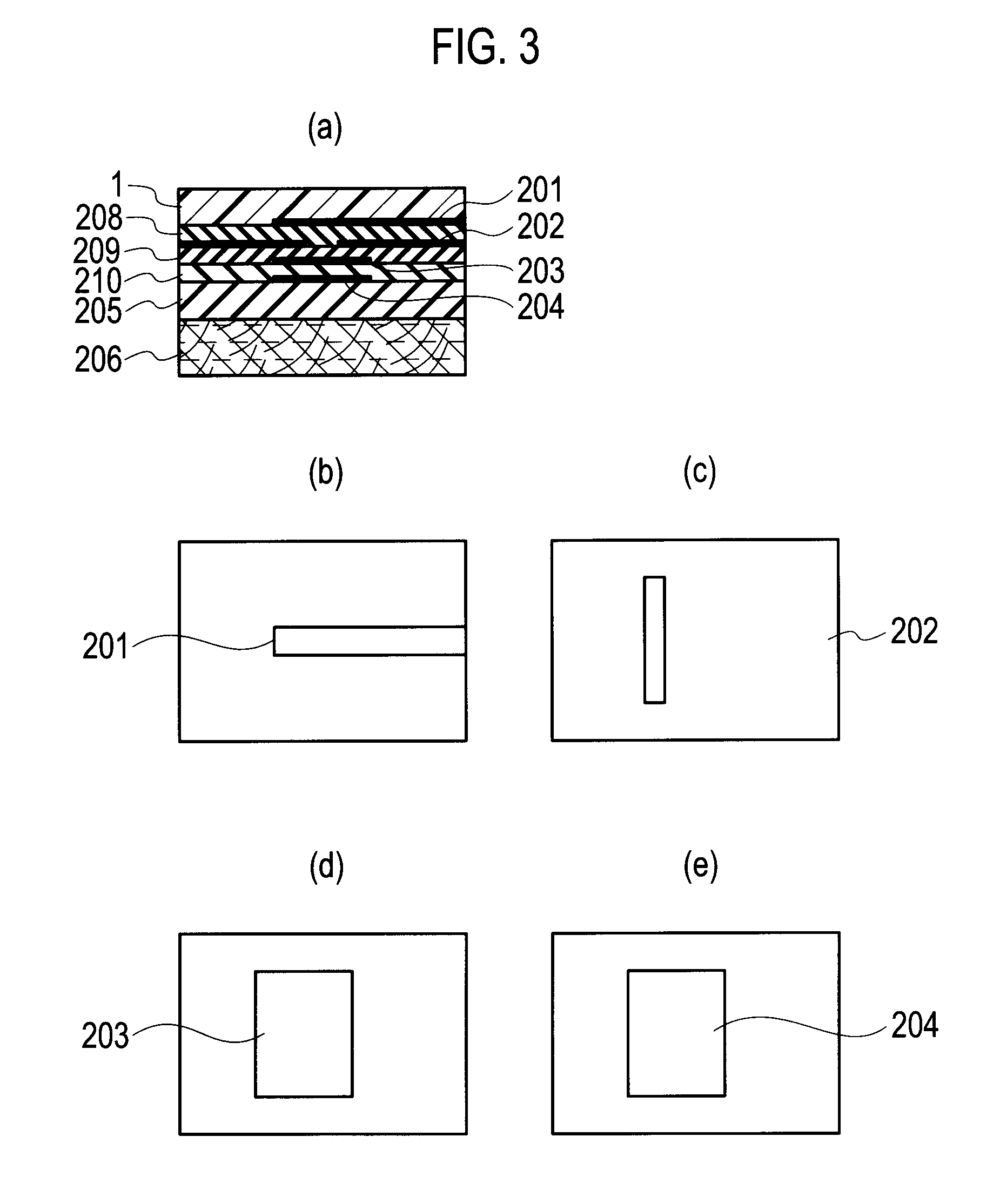
[0065]As illustrated in FIGS. 1 (a) and 2, a diagnosis apparatus pertaining to a first embodiment of the present invention encompasses a probe array (1, 2) made of material having electromagnetic characteristic identical to the electromagnetic characteristic of a target object to be measured, the probe array (1, 2) embraces a receptacle 1 having a semispherical inner wall surface, and a plurality of probes 2, arranged along the inner wall surface, the plurality of probes 2 carry out electrical measurement of a target region in the target object, a fixing mechanism(3, 4 and 5) configured to cover whole of the target region with the probe array (1, 2), bringing skin of the target region into close contact with the inner wall surface, so as to fix a relative position between the target region and the probe array (1, 2), and a measuring, controlling and analyzing mechanism 10 configured to control the plurality of probes 2, to execute the electrical measurement, and to analyze a data ba...

second embodiment

[0103]The probe array is not limited to the flat or conformal antenna of the multilevel structure, as described in the first embodiment. A probe array (301, 302) pertaining to a second embodiment of the present invention is molded with semispherical resin layer, similarly to the first embodiment. However, the second embodiment differs from the first embodiment in that a plurality of probes (antennas) 302 are installed in a hollow portion surrounded by inner and outer wall surfaces of a receptacle 301, and matching medium 305, which has the same dielectric constant and conductivity as the fatty layer of the breast, which is assigned as the target region 304 for measurement, is filled.

[0104]As illustrated in FIG. 15, the probe array (301, 302) includes the receptacle 301 having a semispherical inner wall, a plurality of antennas (probes) 302 arranged in the hollow portion surrounded by the inner and outer wall, and the matching medium 305 filled in the hollow portion.

[0105]Although no...

third embodiment

[0108]In the first and second embodiments, the technique that specifies a lesion (or a pathologically changed portion) by using the frequency-space beamforming via the multi-static radar is described. However, a diagnosis apparatus pertaining to a third embodiment of the present invention further includes a tomographic mechanism for performing tomography (or non-invasive imaging) selectively on the periphery of the abnormal cell portion (lesion), after the detection of the abnormal cell (lesion) in the target region, and the hybrid imaging can be carried out in accordance with the hybrid imaging algorism.

[0109]According to the hybrid imaging that uses the diagnosis apparatus pertaining to the third embodiment, the complex dielectric constant (the dielectric constant and conductivity) distribution of the target region can be estimated at a higher precision. However, as for the tomographic mechanism in the diagnosis apparatus pertaining to the third embodiment, it is possible to use t...


PUM


Abstract

A diagnosis apparatus encompasses a probe array (1, 2) made of material having electromagnetic characteristic identical to the electromagnetic characteristic of a target object, the probe array (1, 2) embraces a receptacle 1 having a semispherical inner wall surface, and a plurality of antennas 2, arranged along the inner wall surface, the plurality of antennas 2 carry out electrical measurement of a target region in the target object, a fixing mechanism configured to cover whole of the target region with the probe array (1, 2), bringing skin of the target region into close contact with the inner wall surface, so as to fix a relative position between the target region and the probe array (1, 2), and a measuring, controlling and analyzing mechanism configured to control the plurality of antennas 2, to execute the electrical measurement, and to analyze a data based on the electrical measurement so as to detect an abnormal cell in the target region. The diagnosis apparatus has a high contrast and a high resolution and does not suffer from any X-ray exposure and is low in diagnosis cost and safe, sure, comfortable and high in speed and high in reliability.

Description

TECHNICAL FIELD[0001]The present invention pertains to a diagnosis apparatus used to diagnose abnormal cells such as early-stage breast-cancers and the like.Background Art[0002]In recent years, a research for detecting early-stage breast-cancers via a microwave imaging has been eagerly performed. Principle for detecting cancers is based upon remarkable differences of electromagnetic parameters (dielectric constant and conductivity) between peripheral (fatty) tissues and a cancer tissue. That is, the principle for detecting cancers is based upon a fact that the backscattered wave from the cancer tissue is larger than the backscattered wave from normal tissues. As examples of the imaging scheme based on the microwave imaging, there are an ultra-wideband (UWB) radar approach (see non-patent literatures (NPLs) 1 and 2) and a tomography approach (see NPL 3).[0003]In the scheme disclosed in NPL 1, by using a monostatic radar, wide band pulses are emitted to a breast from many directions a...

Claims


Application Information

Patent Timeline
IPC IPC(8): A61B5/05
CPCA61B5/0507A61B5/0536A61B2562/143A61B2017/306A61B2019/205A61B10/0041A61B90/17
Inventor KUWABARA, YOSHIHIKO
Owner NAT UNIV CORP SHIZUOKA UNIV
Why Eureka
Try Eureka