Visualizing surgical trajectories

Active Publication Date: 2012-04-26
KONINKLIJKE PHILIPS ELECTRONICS NV
View PDF5 Cites 12 Cited by
  • Summary
  • Abstract
  • Description
  • Claims
  • Application Information

AI Technical Summary

Benefits of technology

[0006]It is an object of the invention to provide a clearer way of displaying sufficiently clear information for the

Problems solved by technology

From the MR images alone it is very difficult or even impossible to visually distinguish small different neurological structures such as the subthalamic nucleus (STN) which is often targeted during deep brain stimulation procedures.
However, registration of the atlas to pre-operative MR images has limited precision due to registration errors inherent to any registration method.
However, also this technique has inherent limited accuracies.
Further, during surgery an implanted probe may unintentionally deviate from a planned trajectory (e.g. due to an imperfection such as a slight bending of the probe) or the anatomy may slightly shift due to the surgical procedure itself (e.g. the implantation of the probe in the tissue generates a small force field that pushes aside the tissue

Method used


Image

  • Visualizing surgical trajectories
  • Visualizing surgical trajectories
  • Visualizing surgical trajectories

Examples


Embodiment Construction

[0025]In the following, the invention will be described by an exemplary embodiment related to neurosurgery using electrophysiological probes. The invention is however not limited to use with a neuro-EP system. Other surgical interventions where knowledge about anatomic information along a surgical trajectory (planned and / or navigated) is useful for clinical and / or diagnostic purposes may benefit as well from this invention. The invention may, e.g., be suitable for optical-needle guided interventions.

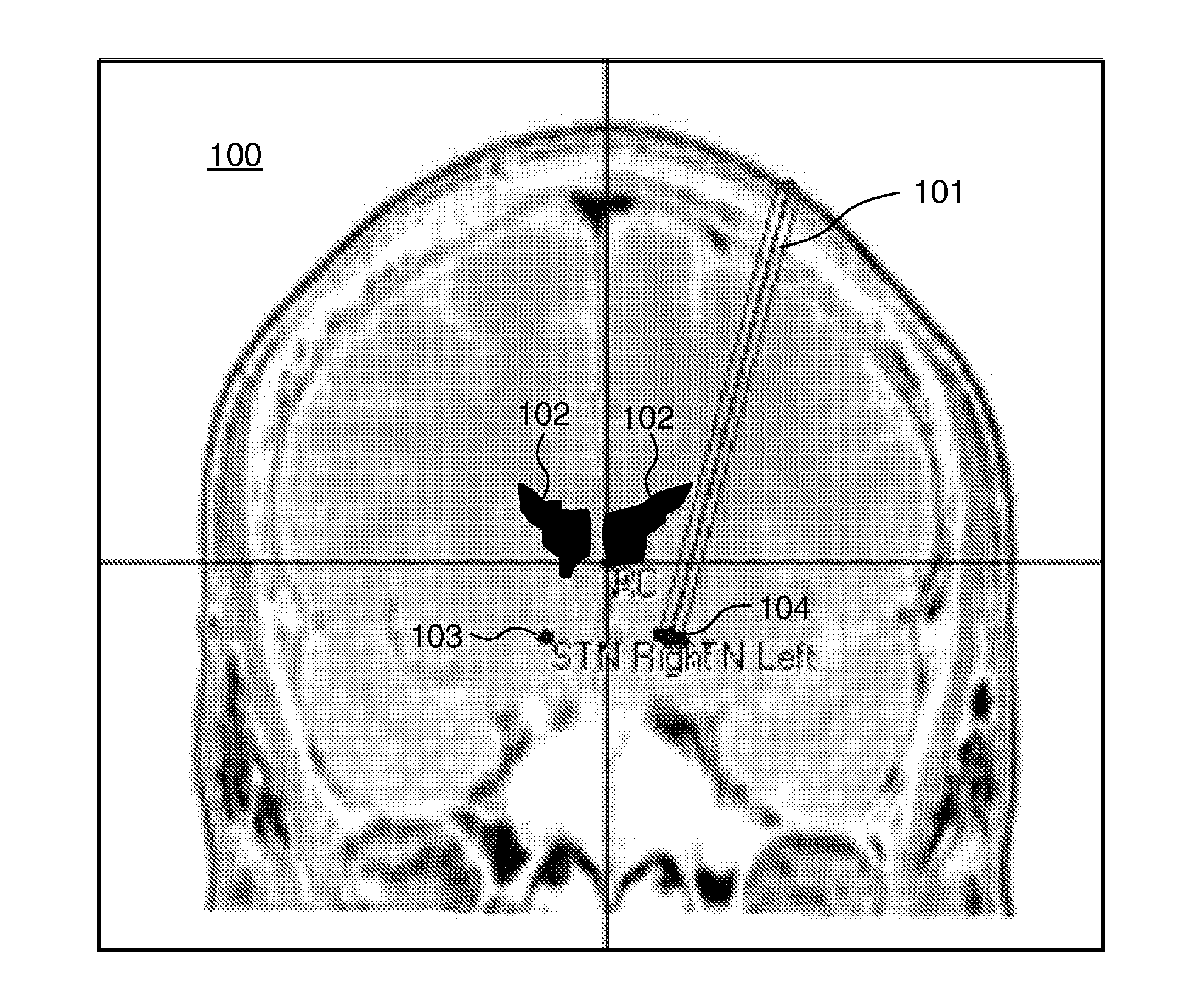
[0026]FIG. 1 shows an image 100 comprising data from an MRI, an atlas and three surgical trajectories 101. It is known to use such an image 100 for pre-operative planning of a surgical trajectory In this image atlas information is used for indicating specific structures 102, 103, 104 in the region of the brain which is shown in the MRI image. The image 100 shows an MRI image of a cross section of a human brain. The surgical trajectory 101 runs through the brain tissue towards the target ...


PUM


Abstract

A method is provided for visualizing a surgical trajectory (32, 101, 42, 46, 47). The method comprises steps of receiving (71) 3D imaging information (31) of a region to undergo surgery and combining (72) the received 3D imaging information (31) with data from a digitized anatomical atlas. As a result, a combined map of the region to undergo surgery is obtained. The combined map comprises expected positions of anatomical structures (102, 103, 104) in the region to undergo surgery. The method further comprises steps of receiving (73) the surgical trajectory (32, 101, 42, 46, 47) for the surgery, determining (74) positions of intersections (43, 44) of the surgical trajectory (32, 101, 42, 46, 47) with the anatomical structures (102, 103, 104) and providing (75) the positions of the intersections (43, 44) in a coordinate system aligned with the surgical trajectory (32, 101, 42, 46, 47).

Description

FIELD OF THE INVENTION[0001]This invention relates to a method for visualizing a surgical trajectory. The method combines 3D imaging information with a digitized anatomic model for segmenting anatomical structures in the 3D imaging information. A planned surgical trajectory is projected onto the combined images for visualizing the surgical trajectory.[0002]This invention further relates to a system and a computer program product for performing said method.BACKGROUND OF THE INVENTION[0003]Such a method is, e.g., known from Guo et al., ‘Development and Application of Functional Databases for Planning Deep-Brain Neurosurgical Procedures’. From Guo et al. it is known to fuse pre-operative MR images of individual patients with digitized anatomical atlases. From the MR images alone it is very difficult or even impossible to visually distinguish small different neurological structures such as the subthalamic nucleus (STN) which is often targeted during deep brain stimulation procedures. Wh...

Claims


Application Information

Patent Timeline
IPC IPC(8): G06K9/00
CPCA61B19/50A61B19/5244A61B2019/5289A61B2019/507A61B2019/524A61B2019/505A61B34/10A61B34/20A61B2034/105A61B2034/107A61B2090/364A61B2090/3762
Inventor CAGNAN, HAYRIYEMARTENS, HUBERT CECILE FRANCOISDOLAN, KEVIN THOMAS
Owner KONINKLIJKE PHILIPS ELECTRONICS NV
Why Eureka
Try Eureka