Ultrasound diagnostic apparatus and method for tracing movement of tissue

a technology of ultrasound and diagnostic equipment, applied in the field of ultrasound diagnostic equipment, to achieve the effect of accurately measuring the movement of tissue in the target obj

Active Publication Date: 2012-05-03
GE MEDICAL SYST GLOBAL TECH CO LLC
View PDF0 Cites 20 Cited by
  • Summary
  • Abstract
  • Description
  • Claims
  • Application Information

AI Technical Summary

Benefits of technology

[0012]In a fifth aspect of the ultrasound diagnostic apparatus, the region of interest setting unit displays a horizontal line in the long axis direction including the first region of interest on the display unit when the first region of interest is set on the vertical straight line, and displays a vertical line normal to the horizontal line including the first region of interest on the display unit when angle of the horizontal line is set. Then an operator can easily set the region of interest.
[0019]The ultrasound diagnostic apparatus described herein can trace a movement of a target object within the predetermined region, including a blood vessel, by using a gradient method and accurately measure the movement of tissue in the target object.

Problems solved by technology

Unfortunately, the correlation of the brightness of pixel as described in Japanese unexamined publication 2002-238903A may vary the diameter of the blood vessel or the blood vessel wall depending on the image data processing.

Method used


Image

  • Ultrasound diagnostic apparatus and method for tracing movement of tissue
  • Ultrasound diagnostic apparatus and method for tracing movement of tissue
  • Ultrasound diagnostic apparatus and method for tracing movement of tissue

Examples


Embodiment Construction

[0035]100>

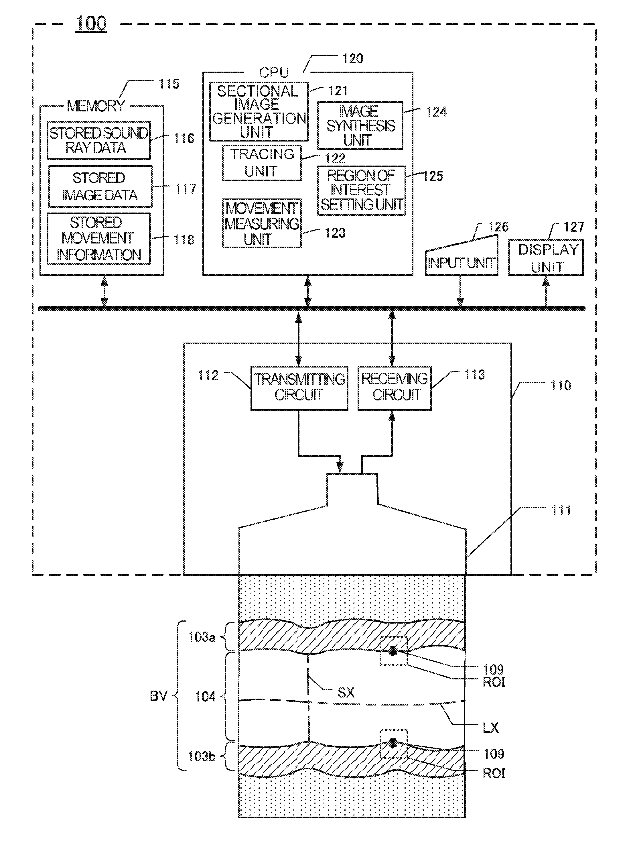
[0036]FIG. 1 is a block diagram showing the configuration of the ultrasound diagnostic apparatus 100. The ultrasound diagnostic apparatus 100 includes a transmitting and receiving unit 110 connected to a parallel bus, a memory 115, a CPU (central processing unit) 120, an input unit 126 for inputting through a mouse or a keyboard and display unit 127 having an LCD unit.

[0037]The transmitting and receiving unit 110 includes an ultrasound probe 111, a transmitting circuit 112 and a receiving circuit 113. The ultrasound probe 111 includes a plurality of ultrasound transducers in a 1-dimensional or a 2-dimensional transducer array. The ultrasound transducers transmit ultrasound waves based on applied driving signal to the target object, receive ultrasound echo reflected from the target object, and output a receiving signal.

[0038]The transmitting circuit 112 includes a plurality of channels, and generates a plurality of driving signals applied to each plurality of ultrasound tra...


PUM


Abstract

An ultrasound diagnostic apparatus includes a transmitting and receiving unit that transmits an ultrasound wave to a target object and receives the ultrasound wave as ultrasound data reflected from the target object including a long axis direction blood vessel. An image generation unit generates an ultrasound image as a sectional image of the blood vessel. A region of interest (ROI) setting unit sets a first ROI on a vertical straight line at a right angle to the long axis direction and a second ROI on a wall of the blood vessel displayed at a designated time. A tracing unit traces movement of tissue in the target object corresponding to the first and second ROIs from the designated time to sequentially following thereafter by a gradient method using a spatial brightness gradient. A second memory unit stores information of the movement of the tissue for a predetermined duration.

Description

CROSS REFERENCE TO RELATED APPLICATIONS[0001]This application claims the benefit of Japanese Patent Application No. 2010-241316 filed Oct. 27, 2010, which is hereby incorporated by reference in its entirety.BACKGROUND OF THE INVENTION[0002]The embodiments described herein relate to an ultrasound diagnostic apparatus for diagnosing blood vessel using ultrasound.[0003]In recent years, number of patients diagnosed with circulatory condition, such as cerebral infarction and cardiac infarction, are on the rise. To prevent from such disease, it is important to detect a symptom of arteriosclerosis in its early stage and to improve a lifestyle.[0004]To diagnose an arterial problem, Japanese unexamined publication 2002-238903A discloses an ultrasound diagnostic apparatus that measures the diameter of blood vessel using B (brightness)-mode image. Japanese unexamined publication 2002-238903A further discloses an ultrasound diagnostic apparatus for tracing the diameter of blood vessel and blood...

Claims


Application Information

Patent Timeline
Patent Type & Authority Applications(United States)
IPC IPC(8): A61B8/14
CPCA61B8/06A61B8/469A61B8/08A61B8/14
Inventor MIYAMA, KOJIOGASAWARA, MASAFUMI
Owner GE MEDICAL SYST GLOBAL TECH CO LLC
Why Eureka
Try Eureka