Method for preparing biological tissue

a biological tissue and tissue technology, applied in the field of biological tissue preparation, can solve the problems of difficult handling, high cell density of biological tissue, and existing methods for preparing biological tissue with high cell density

Active Publication Date: 2016-09-13
DAI NIPPON PRINTING CO LTD
View PDF39 Cites 0 Cited by
  • Summary
  • Abstract
  • Description
  • Claims
  • Application Information

AI Technical Summary

Benefits of technology

[0029]According to the method for preparing tissue of the present invention, biological tissue having a three-dimensional structure of interest can be easily obtained.

Problems solved by technology

Accordingly, such biological tissue is not suitable as transplant tissue for medical purposes.
However, existing methods for preparing biological tissue with high cell density have several drawbacks.
Since a cell sheet is very thin and difficult to handle, superposition of a plurality of such cell sheets is laborious rather than easy.
Thus, nutrition supply to cells that are present inside the tissue and discharge of wastes from such cells were insufficient, and cellular necrosis was likely to take place.
However, such technique has drawbacks such that, for example, it takes several days to prepare tissue, target cells are limited to bone cells or the like, tissue cannot be easily detached from a culture vessel, and a conformation cannot be regulated as intended.

Method used


Image

  • Method for preparing biological tissue
  • Method for preparing biological tissue
  • Method for preparing biological tissue

Examples


reference example 1

1. Preparation of Cell Sheet with Centrifugal Force

1-1. Preparation and Collection of Cell Sheet with Centrifugal Force

[0087]Polyethylene glycol was chemically applied to the entire inner surface of a glass vessel having a bottom with a diameter of 2 cm to render the entire inner surface non-cell-adhesive, 2 ml of a cell suspension containing 4×10⁶ mouse fibroblasts suspended in 10% fetal bovine serum-containing DMEM medium was sowed in the vessel, and culture was conducted at 37° C. in the presence of 5% CO2 for 3 hours while applying centrifugal force of 720 G toward the bottom (FIG. 7A). After culture, centrifugation was terminated, and a cell sheet was easily and rapidly detached and collected from the substrate without destruction of the tissue structure by a physical force, such as via pipetting (FIG. 7B). As a contrast, culture was conducted in the same manner, except that no centrifugal force was applied. As a result, a cell sheet could not be prepared, cells were detached f...

reference example 2

2. Addition of Extracellular Matrix

2-1. Preparation of Extracellular Matrix-Containing Solution

[0089]DMEM medium containing 10% fetal bovine serum was added to a 10-cm polystyrene petri dish to culture mouse fibroblasts, and the cells were grown to reach confluence in order to have the cells to produce an extracellular matrix. Subsequently, multiplied cells were ground using a cell scraper, the cells were completely fragmented via ultrasound treatment, the fragmented product was centrifuged, and the supernatant was collected. The supernatant was used below as an extracellular matrix-containing solution.

2-2. Preparation and Collection of Cell Sheet with Centrifugal Force

[0090]Polyethylene glycol was chemically applied to the entire inner surface of a glass vessel having a bottom with a diameter of 2 cm to render the entire inner surface non-cell-adhesive, 2 ml of a cell suspension containing 4×10⁶ mouse fibroblasts suspended in the extracellular matrix-containing solution prepared in...

example 1

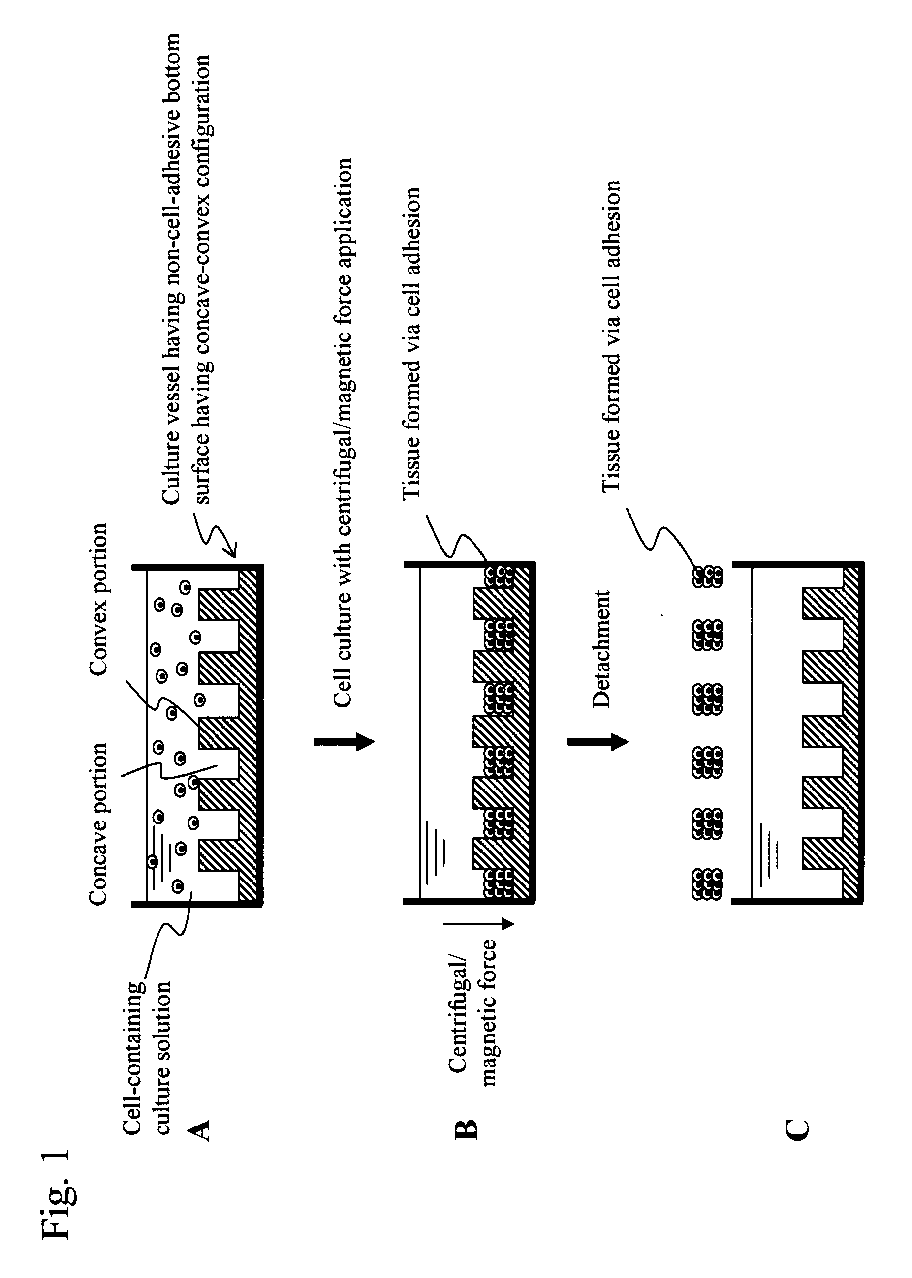
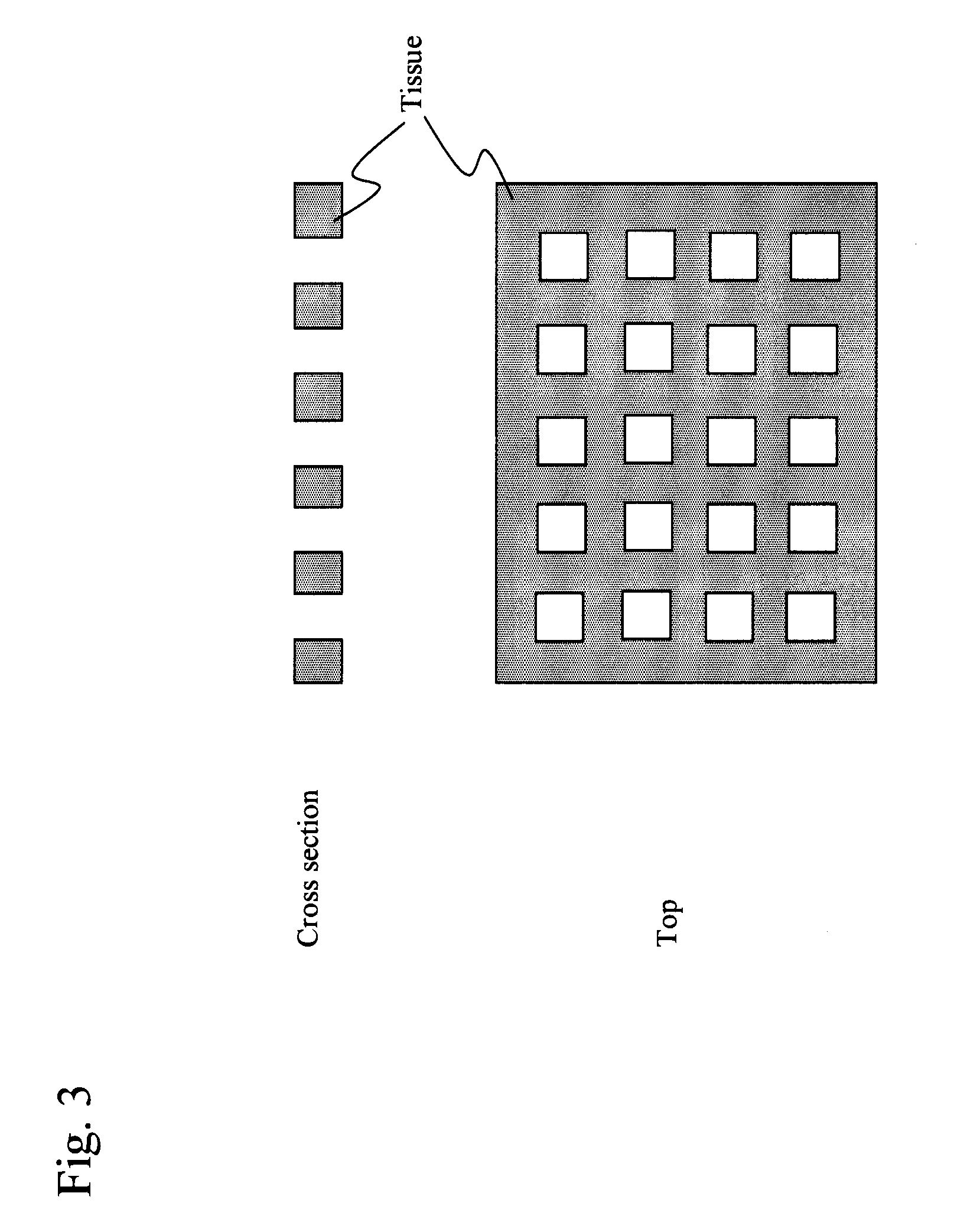
3. Preparation of Cell Sheet Having Holes

3-1. Culture Vessel Having Non-Cell-Adhesive Bottom Surface with Concave-Convex Configuration

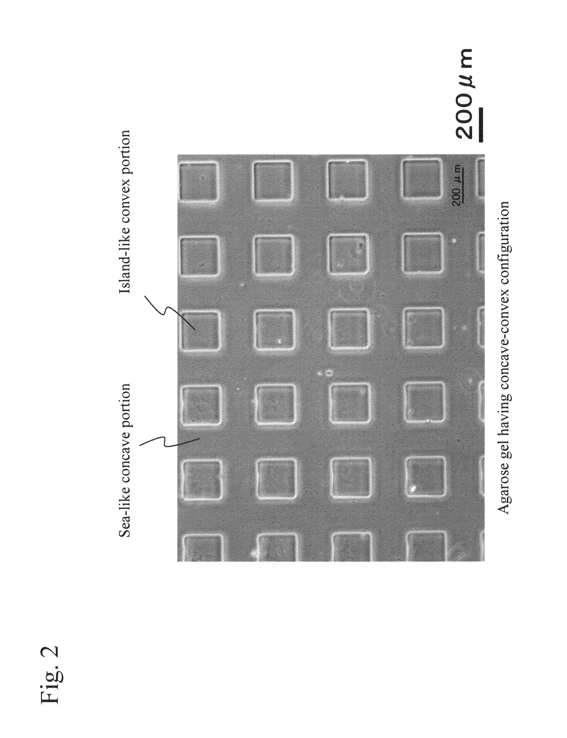
[0091]A silicon wafer was coated with resist (tradename: SU-8; manufacturer: MicroChem) to a thickness of 200 μm, a concave-convex template was prepared via optical lithography, a 3% agarose solution was introduced onto the template for gelation, and the resulting gel was detached from the template. Thus, agarose gel having a concave-convex pattern (200 μm×200 μm squares raised at heights of 200 μm at intervals of 200 μm) was prepared. FIG. 2 shows the concave-convex pattern of the prepared agarose gel. The resulting gel was positioned at the bottom of the vessel, with the entire inner bottom surface being made non-cell-adhesive in 1-1, so that the concave-convex surface of the gel would face upward. Thus, a culture vessel having a non-cell-adhesive and concave-convex bottom surface was prepared (FIG. 11).

3-2. Preparation of Extracellular Matrix-Conta...


PUM

PropertyMeasurementUnit
water contact angleaaaaaaaaaa
static water contact angleaaaaaaaaaa
thicknessaaaaaaaaaa

Abstract

This invention provides a method for obtaining biological tissue having a three-dimensional structure of interest. The method of the invention comprises: adding a cell-containing culture solution to a culture vessel having an inner bottom surface, which is non-cell adhesive and comprises a concave-convex pattern provided thereon; performing cell culture under conditions in which intercellular adhesion takes place while centrifugal or magnetic force toward the inner bottom surface is applied to the cells that were added to the culture vessel to form tissue via intercellular adhesion; and detaching and collecting the resulting tissue from the inner bottom surface at the end in order to obtain tissue having a three-dimensional configuration using a concave-convex pattern as a template.

Description

TECHNICAL FIELD[0001]The present invention relates to a method for preparing biological tissue that is suitable for preparation of biological tissue having a multilayer structure of a plurality of superposed cells in the through-thickness direction.BACKGROUND ART[0002]Biological tissue that is prepared via cell culture on a culture support such as collagen has low cell density. Accordingly, such biological tissue is not suitable as transplant tissue for medical purposes. A technique for preparing tissue suitable for transplantation, such as a cell sheet with high cell density, can be said to be an important technique in the tissue engineering field. However, existing methods for preparing biological tissue with high cell density have several drawbacks.[0003]In general, sowing of cells on a culture support to form a cell sheet is extensively carried out. In order to facilitate detachment of a cell sheet, a technique of providing a layer of a temperature-responsive polymer compound on...

Claims


Application Information

Patent Timeline
Patent Type & Authority Patents(United States)
IPC IPC(8): A61L27/38C12N5/00A61K35/12A61L27/00A61L27/36A61P43/00C12N5/07
CPCA61L27/3895C12N5/0062A61K35/12C12N2525/00C12N2535/10A61P43/00
Inventor HASE, MASAHIKO
Owner DAI NIPPON PRINTING CO LTD
Why Eureka
Try Eureka