Vital fluorochrome conjugates and methods of use

a technology of vital fluorochrome and conjugates, applied in the field of design and synthesis of vital fluorochrome conjugates, can solve the problems of large attenuation of organisms of this size, lack of specificity of imaging cell death agents used to image apoptosis or necrosis, and lack of molecular target or clear mechanism of agents

Inactive Publication Date: 2012-05-17
THE GENERAL HOSPITAL CORP
View PDF4 Cites 3 Cited by
  • Summary
  • Abstract
  • Description
  • Claims
  • Application Information

AI Technical Summary

Benefits of technology

[0042]By virtue of their design, the vital fluorochrome conjugates described herein possess certain advantages and benefits. First, the vital fluorochrome conjugates can be used to distinguish healthy cells from those that are dead or dying. Second, the vital fluorochrome conjugates enable the imaging of dead or dying cells through a variety of widely used imaging modalities (PET, SPECT, and MRI), while at the same time being fluorescent. In addition, vital fluorochrome conjugates can be used to assess the health of ischemia damaged myocardium, which can indicate the extent of damage to the myocardium or the potential for salvaging the damaged myocardium.

Problems solved by technology

VF's can be used to visualize cell death in biological research, but cannot be used to visualize death / necrosis by fluorescence imaging in humans, because the distances light must traverse through tissues in these methods lead to massive attenuation for organisms of this size.
However, some agents typically used to image apoptosis or necrosis suffer from a lack of specificity for imaging cell death, e.g., because they are retained by healthy cells of the liver, spleen, or other tissues.
Some agents lack a molecular target or clear mechanism for retention by apoptotic and / or necrotic cells, but not by vital, healthy cells.
As a result of these challenges there is no currently approved imaging agent for imaging either apoptosis or necrosis.

Method used


Image

  • Vital fluorochrome conjugates and methods of use
  • Vital fluorochrome conjugates and methods of use
  • Vital fluorochrome conjugates and methods of use

Examples


examples

[0080]The following examples are illustrative and not limiting.

example a

Determining the Properties of Vital Fluorochrome Conjugates

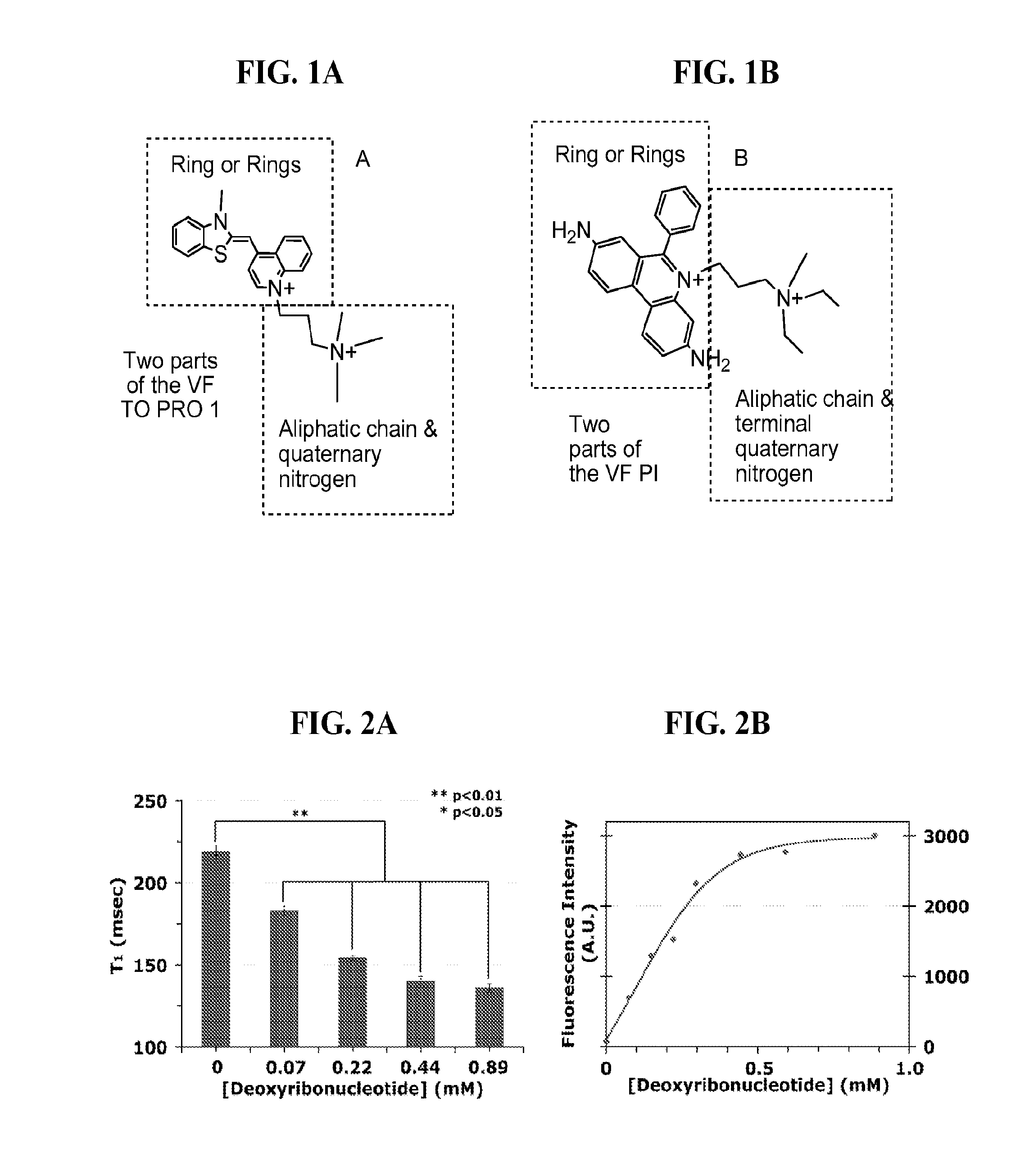
[0081]Experimental Details of the Relaxation Assay: Gadolinium thiazole orange (GadoTO) at 1 mM was employed with increasing concentrations of plasmid DNA (DNA 0.025 to 0.3 ug / uL). Relaxation times were measured on a Bruker minispec at 20 mHz. T1 values were fit to a non-linear sigmoidal dose-response regression, GraphPad® Prism) to determine the half-maximal concentration (EC50=0.089 mM, 95% confidence interval 0.078-0.101).

[0082]Experimental Details of the Fluorescence Assay: GadoTO at 2.35 uM was employed with increasing concentrations of DNA. Absorption spectra were measured on a Varian Cary 50 Bio UV-Visible spectrophotometer. The absorbance at 511 nm was measured. Fluorescence emission spectra were recorded on a Varian Cary Eclipse fluorescence spectrophometer. Fluorescence intensities were measured between 510 and 700 nm and corrected, when necessary, for matched absorbances at 511 nm. Fluorescence intensities were pl...

example b

Imaging Necrotic Cells In Vitro and In Vivo

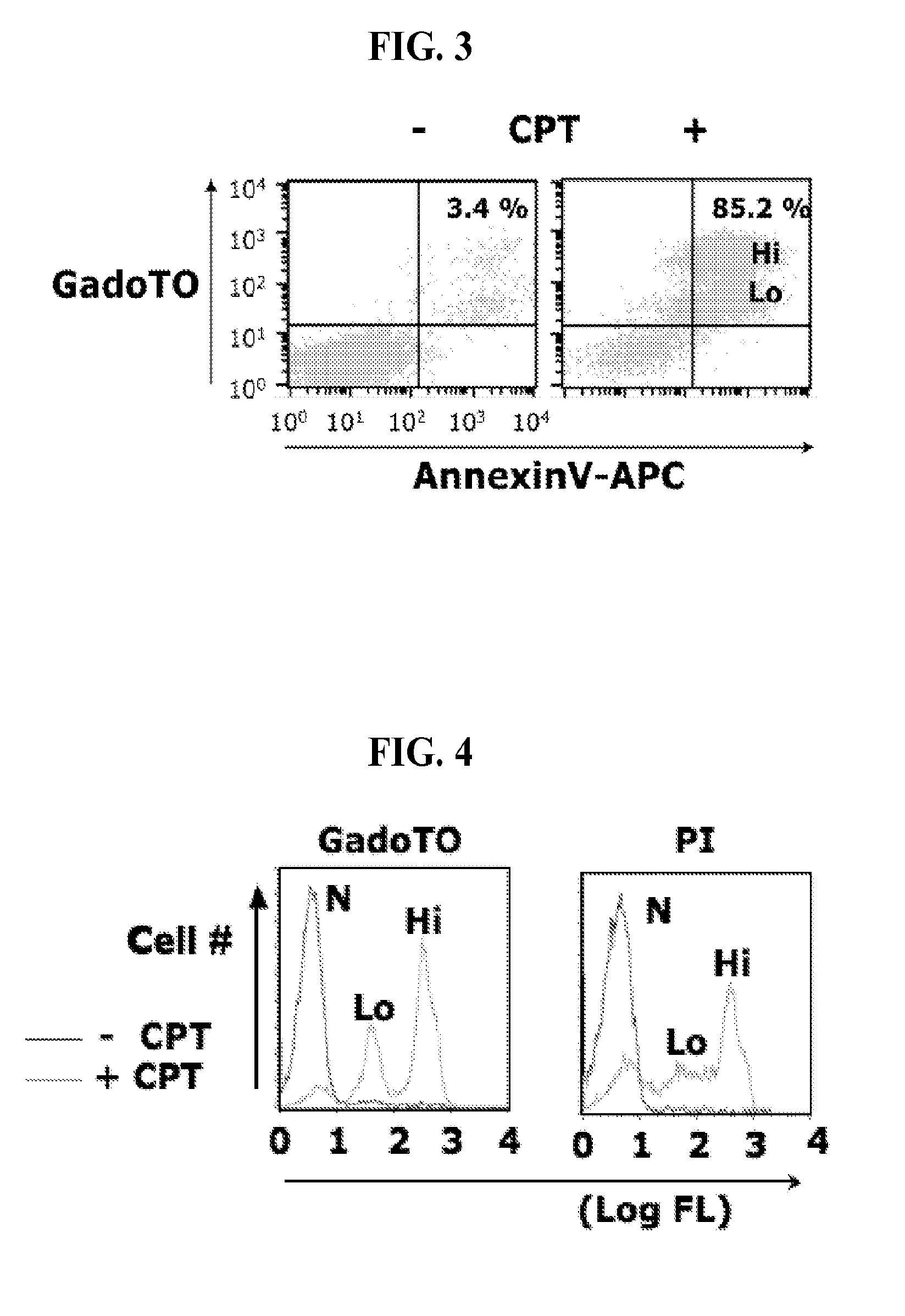
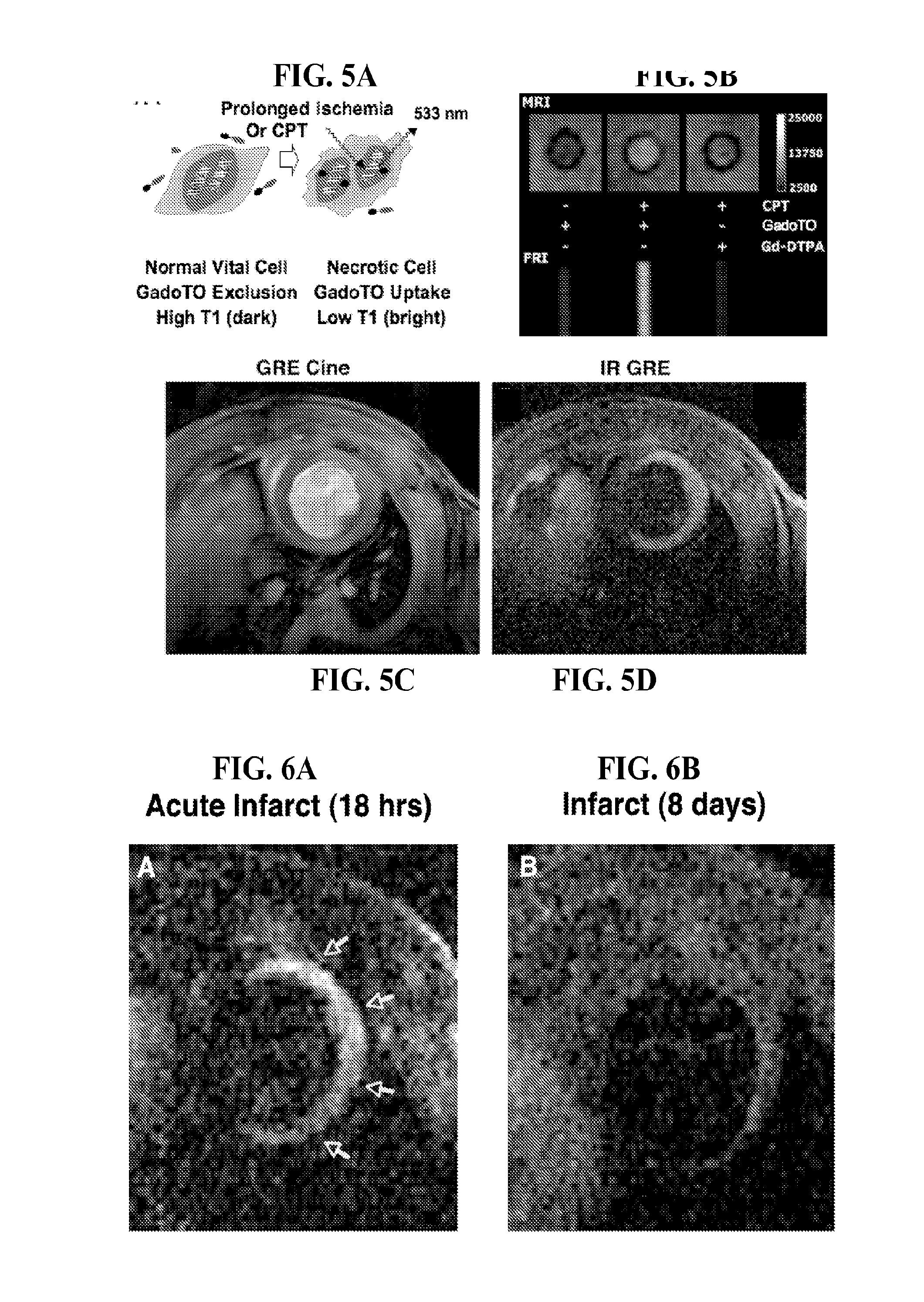
[0087]The ability to image necrotic cells in vitro and in vivo with GadoTO by MRI is shown in FIG. 5A-5D. The interaction of GadoTO with necrotic cells is shown schematically in FIG. 5A. GadoTO is excluded by vital cells. Prolonged ischemia or a 48 hour exposure to CPT induces necrosis, which causes GadoTO to enter cells and intercalate with double stranded nuclear DNA. This results in (i) nuclear cell fluorescence (emission @ 533 nm), (ii) a drop in cellular T1 and, (iii) a brightening of cells on T1 weighted MR images. For in vitro studies, we exposed Jurkat cells to CPT and GadoTO, and then obtained the T1 weighted MRI image shown in FIG. 5B. CPT treated cells took up GadoTO, and were brighter than non-CPT treated cells by MRI and fluorescence reflectance imaging (FRI). Effects on cellular T1 were confirmed by relaxometry studies using cell suspensions. CPT treatment did not induce the uptake of the non-DNA binding Gd-DTPA chelate by MRI...


PUM

PropertyMeasurementUnit
molecular weightaaaaaaaaaa
concentrationsaaaaaaaaaa
temperatureaaaaaaaaaa

Abstract

The present invention provides compositions and methods based on vital fluorochrome conjugates that are useful for imaging dying and dead cells.

Description

CROSS REFERENCE TO RELATED APPLICATION[0001]This application claims priority to U.S. Provisional Patent Application No. 61 / 184,523, filed on Jun. 5, 2009, the disclosure of which is incorporated herein by reference in its entirety.STATEMENT REGARDING FEDERALLY SPONSORED RESEARCH OR DEVELOPMENT[0002]The present invention was made with government support Under Grant Nos. R01-EB004472 and R01-EB00066 awarded by the National Institutes of Health. The United States Government has certain rights in the invention.FIELD OF THE INVENTION[0003]The present disclosure describes the design and synthesis of vital fluorochrome conjugates for the imaging of dead or dying cells.BACKGROUND OF THE INVENTION[0004]Cells often die in well-defined stages, transitioning from healthy (vital) cells to apoptotic cells and then to necrotic cells. An important class of compounds for imaging necrosis (cell death) is vital fluorochromes (VF's). VF's are widely used to visualize necrosis (or cell viability by thei...

Claims


Application Information

Patent Timeline
Patent Type & Authority Applications(United States)
IPC IPC(8): A61K51/04A61K49/10C07F5/00C07D417/06C07F1/08C07D417/14
CPCA61K49/0002A61K49/085A61K49/10A61K51/0455A61K49/106A61K49/1833A61K49/103
Inventor JOSEPHSON, LEEGARANGER, ELISABETHHILDERBRAND, SCOTTSOSNOVIK, DAVIDYUAN, HUSHAN
Owner THE GENERAL HOSPITAL CORP
Why Eureka
Try Eureka