

Serum markers associated with early and other stages of breast cancer
a breast cancer and marker technology, applied in the field of disease-specific marker identification, can solve the problems of difficult detection of low-abondance proteins, inability to separate protein complexes, and inability to achieve polyacrylamide gel electrophoresis
- Summary
- Abstract
- Description
- Claims
- Application Information
AI Technical Summary
Benefits of technology
Problems solved by technology
Method used
Image

Examples
example 1
Separation of Protein Complexes Using Two-Dimensional Membrane Electrophoresis or 2-D HPLE
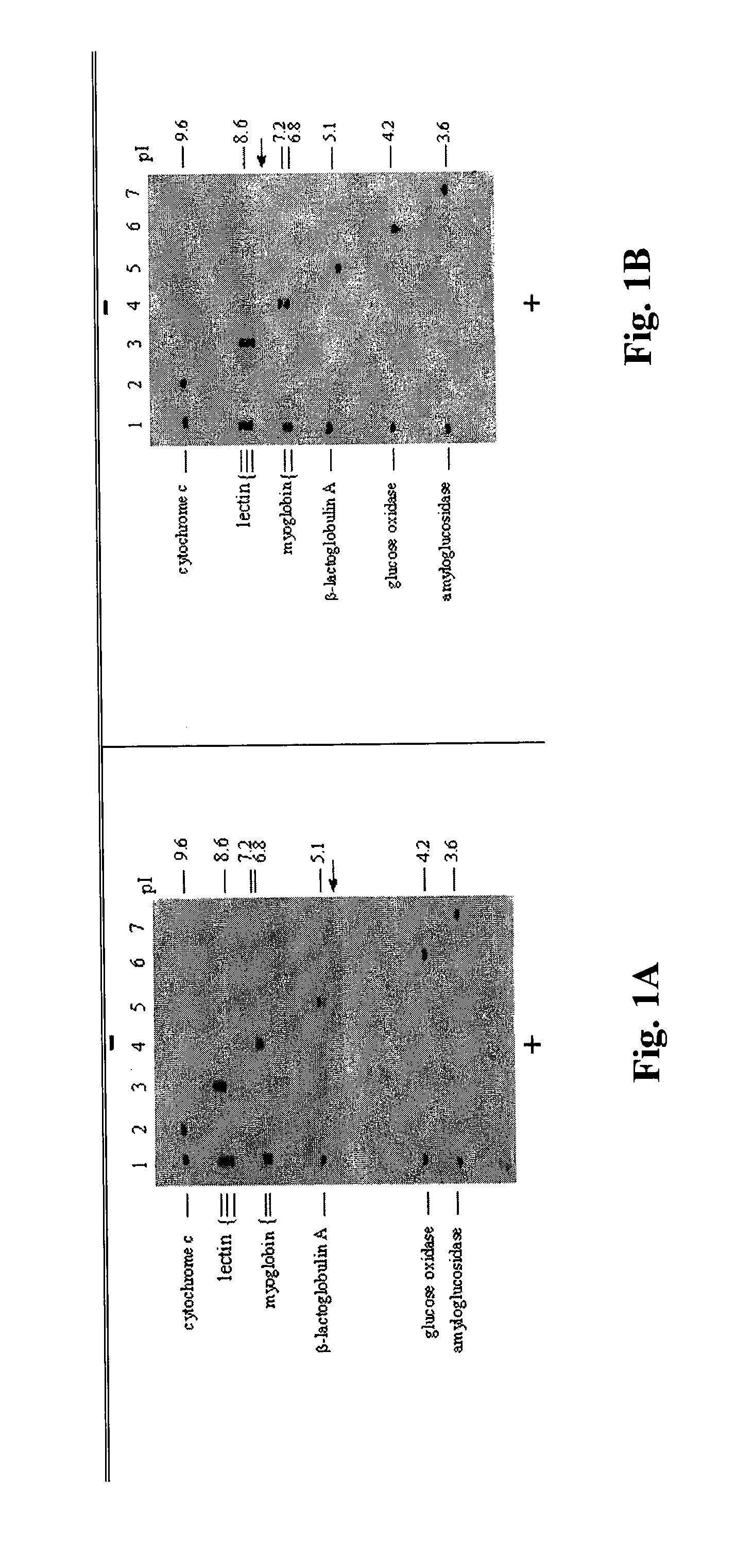
[0082]An innovative procedure for separation and detection of protein complexes containing the newly generated breast cancer markers was developed. To retain protein complexes containing low-abundance breast cancer markers, the electrophoresis process was carried out under non-denaturing conditions. Unlike the commonly used two-dimensional polyacrylamide gel electrophoresis (2-D PAGE), proteins and their complexes are separated directly on polyvinylidene fluoride (PVDF) membrane rather than in a gel. The term “2-D HPLE” (2-D High Performance Liquid Electrophoresis) will be used subsequently to described the 2-D membrane electrophoresis process. Thus, the 2-D HPLE procedure not only separates serum albumin complexes but also eliminates the blotting step required for Western blotting analysis when the conventional 2-D PAGE is used.
[0083]The 2-D HPLE is carried out using a horizontal electrophores...
example 2
Separation of Serum Albumin Complexes from Normal (Healthy) Individuals
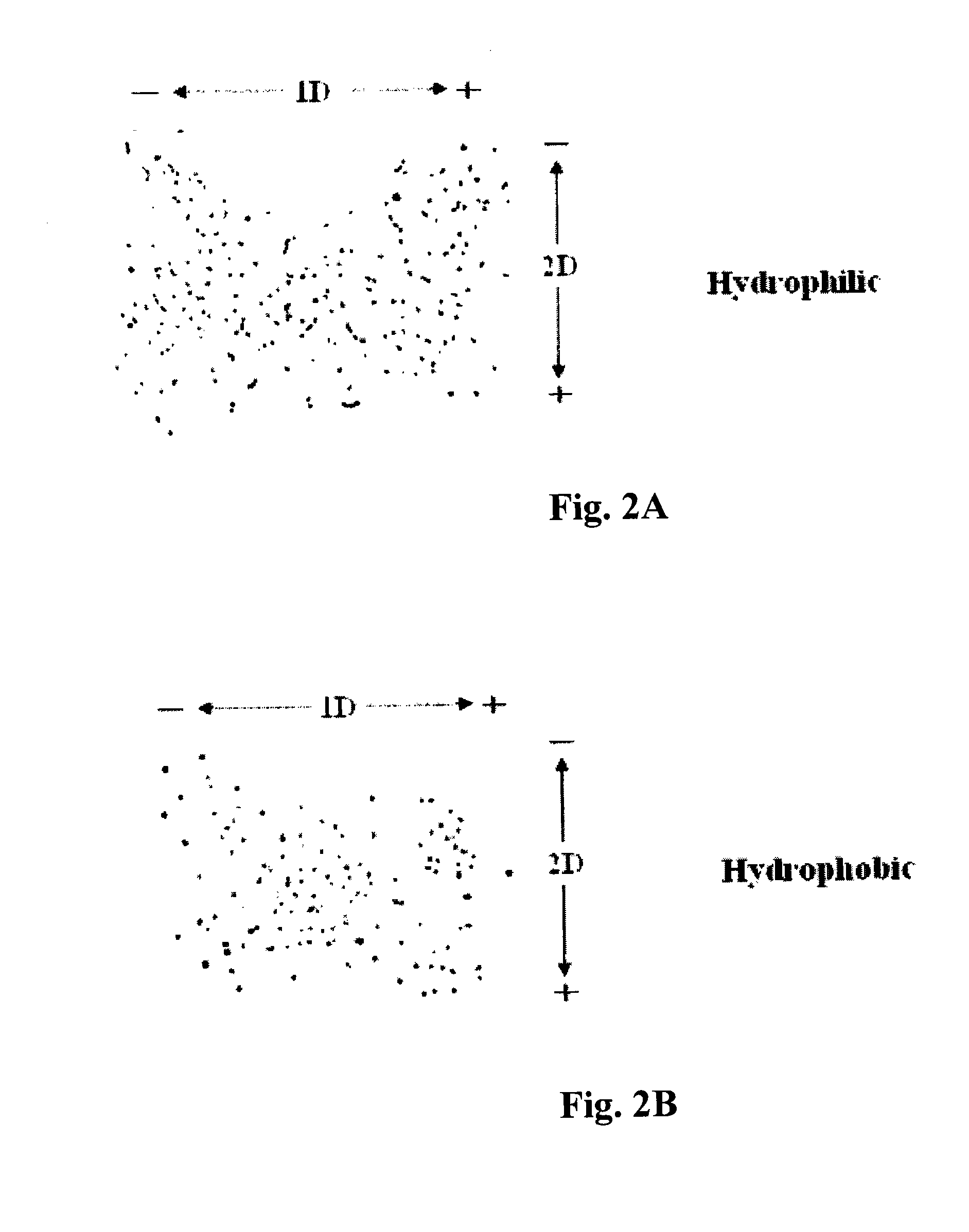
[0092]A prerequisite in identifying serum markers associated with the initiation and progression of a disease is to be able to separate the albumin complexes and to establish a 2-D albumin complex profile of normal (healthy) individuals. The profile can then serve as a reference for the detection of disease protein-containing albumin complexes.
[0093]To eliminate the crowding of albumin complex spots, serum samples were separated into hydrophilic and hydrophobic fractions by using the detergent Triton X-114 before electrophoresis (Bordier (1981) J. Biol. Chem. 256: 1604-07). Six μg of total protein from each fraction were spotted at the middle of a PVDF blot membrane (9 cm×13 cm). Upon completion of the first dimension separation at pH 5.0 which took 6 minutes to complete, the membrane was washed several minutes in deionized H2O to remove the first dimension solvent. After equilibration with the second dimension s...
example 3
Albumin Complexes Associated with Earliest Stage (Stage 0) Breast Cancer
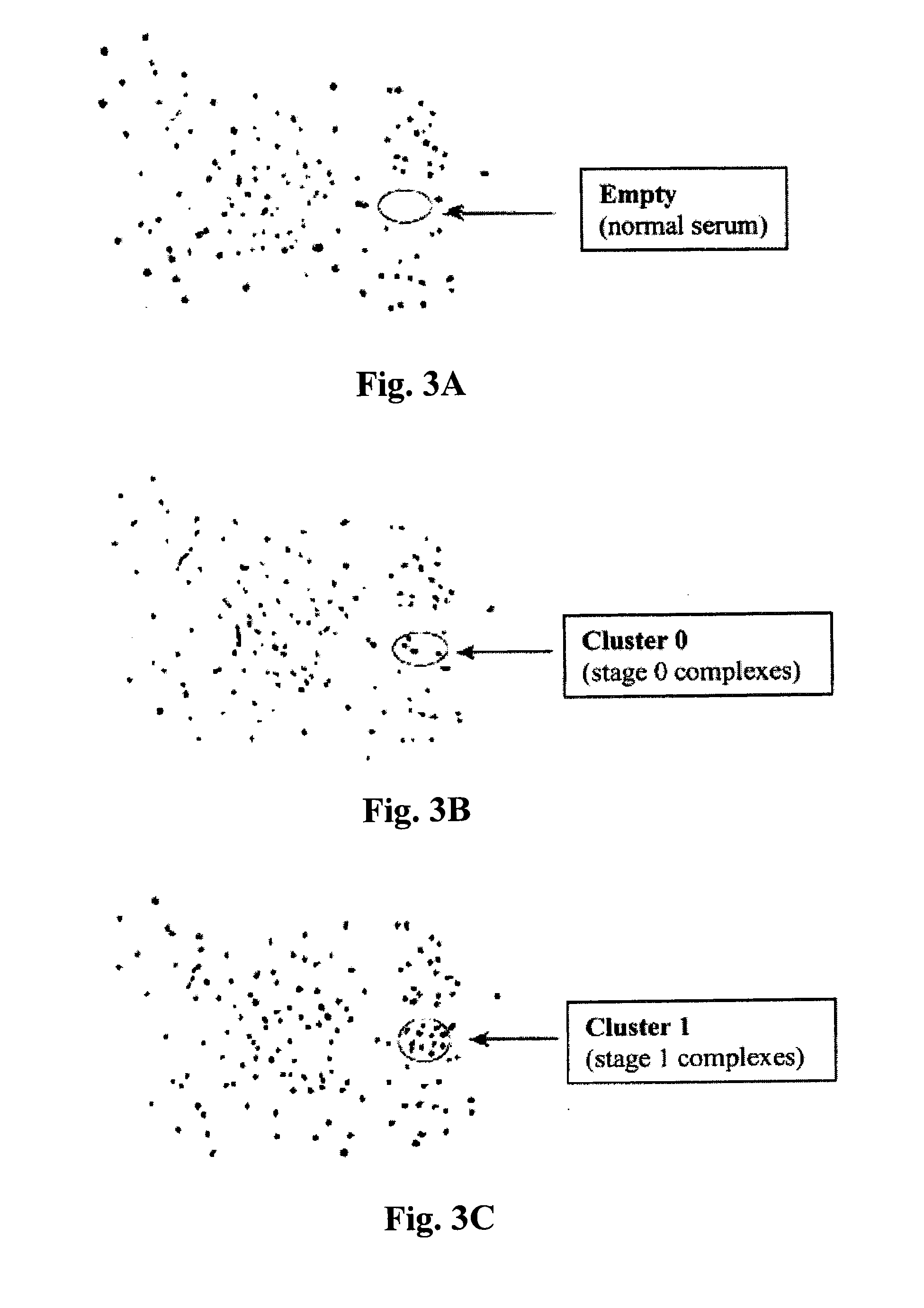
[0094]Serum samples from patients with stage 0, I, II, III, or IV breast cancer as well as normal controls were purchased from the Lombardi Comprehensive Cancer Center at Georgetown University. They house an extensive serum and tumor repository of breast cancer patients.
[0095]As indicated earlier, 2-D HPLE separates serum albumin complexes and can detect any newly formed cancer-protein containing albumin complexes among several hundred pre-existing complexes due to change in their surface charges which cause them to migrate to different locations on the PVDF membrane. This altered electrophoretic mobility allows for the detection of cancer peptide motif-containing albumin complexes associated with very early stage of breast cancer (Stage 0) before the disease is detected by mammography. Likewise, albumin complexes containing cancer peptide motifs for other more advanced stages (Stages I to IV) were also detected...
PUM
 Abstract
Description
Claims
Application Information