Methods for detection of genetic disorders

a genetic disorder and detection method technology, applied in the field of genetic disorder detection, can solve the problems of serious birth defects, fertilized eggs are formed, and errors in the formation or combination of chromosomes, and achieve the effect of reducing the disruption of the “buffy-coat"

Inactive Publication Date: 2008-10-28
RAVGEN INC
View PDF152 Cites 323 Cited by
  • Summary
  • Abstract
  • Description
  • Claims
  • Application Information

AI Technical Summary

Benefits of technology

[0189]In fact, it is an advantage of the invention that primers that copy an entire gene sequence need not be utilized. Rather, the copied locus of interest is preferably only a small part of the total gene or a small part of a non-coding region of DNA. There is no advantage to sequencing the entire gene as this can increase cost and delay results. Sequencing only the desired bases or loci of interest maximizes the overall efficiency of the method because it allows for the sequence of the maximum number of loci of interest to be determined in the fastest amount of time and with minimal cost.
[0190]Because a large number of sequences can be analyzed together, the method of the invention is especially amenable to the large-scale screening of a number of loci of interest.
[0191]Any number of loci of interest can be analyzed and processed, especially at the same time, using the method of the invention. The sample(s) can be analyzed to determine the sequence at one locus of interest or at multiple loci of interest at the same time. The loci of interest can be present on a single chromosome or on multiple chromosomes.
[0192]Alternatively, 2, 3, 4, 5, 6, 7, 8, 9, 10-20, 20-25, 25-30, 30-35, 35-40, 40-45, 45-50, 50-100, 100-250, 250-500, 500-1,000, 1,000-2,000, 2,000-3,000, 3,000-5,000, 5,000-10,000, 10,000-50,000 or more than 50,000 loci of interest can be analyzed at the same time when a global genetic screening is desired. Such a global genetic screening might be desired when using the method of the invention to provide a genetic fingerprint to identify an individual or for SNP genotyping.
[0193]The locus of interest to be copied can be within a coding sequence or outside of a coding sequence. Preferably, one or more loci of interest that are to be copied are within a gene. In a preferred embodiment, the template DNA that is copied is a locus or loci of interest that is within a genomic coding sequence, either intron or exon. In a highly preferred embodiment, exon DNA sequences are copied. The loci of interest can be sites where mutations are known to cause disease or predispose to a disease state. The loci of interest can be sites of single nucleotide polymorphisms. Alternatively, the loci of interest that are to be copied can be outside of the coding sequence, for example, in a transcriptional regulatory region, and especially a promoter, enhancer, or repressor sequence.Method for Determining the Sequence of a Locus of Interest
[0194]Any method that provides information on the sequence of a nucleic acid can be used including but not limited to allele specific PCR, PCR, gel electrophoresis, ELISA, mass spectrometry, MALDI-TOF mass spectrometry hybridization, primer extension, fluorescence detection, fluorescence resonance energy transfer (FRET), fluorescence polarization, DNA sequencing, Sanger dideoxy sequencing, DNA sequencing gels, capillary electrophoresis on an automated DNA sequencing machine, microchannel electrophoresis, microarray, southern blot, slot blot, dot blot, single primer linear nucleic acid amplification, as described in U.S. Pat. No. 6,251,639, SNP-IT, GeneChips, HuSNP, BeadArray, TaqMan assay, Invader assay, MassExtend, or MassCleave™ (hMC) method.

Problems solved by technology

Occasionally, an error occurs in either the formation or combination of chromosomes, and the fertilized egg is formed with too many or too few chromosomes, or with chromosomes that are mixed in some way.
Because each chromosome contains many genes, chromosomal abnormalities are likely to cause serious birth defects, affecting many body systems and often including developmental disability (e.g., mental retardation).
However, during germ-cell formation in the translocation-bearing individuals, the proper distribution of chromosomes in the egg or sperm occasionally fails, resulting in miscarriage, malformation, or mental retardation of the offspring.
Errors that generate too many or too few chromosomes can also lead to disease phenotypes.
Without knowledge gained by prenatal diagnosis, there could be an untoward outcome for the fetus or the mother or both.
However, the techniques that are non-invasive are less specific, and the techniques with high specificity and high sensitivity are highly invasive.
An ultrasound examination can be useful to determine the size and position of the fetus, the amount of amniotic fluid, and the appearance of fetal anatomy; however, there are limitations to this procedure.
This is a highly invasive procedure in which a needle is passed through the mother's lower abdomen into the amniotic cavity inside the uterus.
While amniocentesis can be used to provide direct genetic information, risks are associated with the procedure including fetal loss and maternal Rh sensitization.
CVS has the disadvantage of being an invasive procedure, and it has a low but significant rate of morbidity for the fetus; this loss rate is about 0.5 to 1% higher than for women undergoing amniocentesis.
Furthermore, there is also the possibility that maternal blood cells in the developing placenta will be sampled instead of fetal cells and confound chromosome analysis.
While the MSAFP test is non-invasive, the MSAFP is not 100% specific.
Therefore, results from an MSAFP test are never considered definitive and conclusive.
While the triple screen test is non-invasive, abnormal test results are not indicative of a birth defect.
This high incidence of false positives causes tremendous stress and unnecessary anxiety to the expectant mother.
While improvements have been made for the isolation and enrichment of fetal cells, it is still difficult to get many fetal blood cells.
There may not be enough to reliably determine anomalies of the fetal karyotype or assay for other abnormalities.
Furthermore, most techniques are time consuming, require high-inputs of labor, and are difficult to implement for a high throughput fashion.
However, the diagnostic and clinical applications of circulating fetal DNA is limited to genes that are present in the fetus but not in the mother (Pertl and Bianchi, Obstetrics and Gynecology 98: 483-490 (2001)).

Method used


Image

  • Methods for detection of genetic disorders
  • Methods for detection of genetic disorders
  • Methods for detection of genetic disorders

Examples


example 1

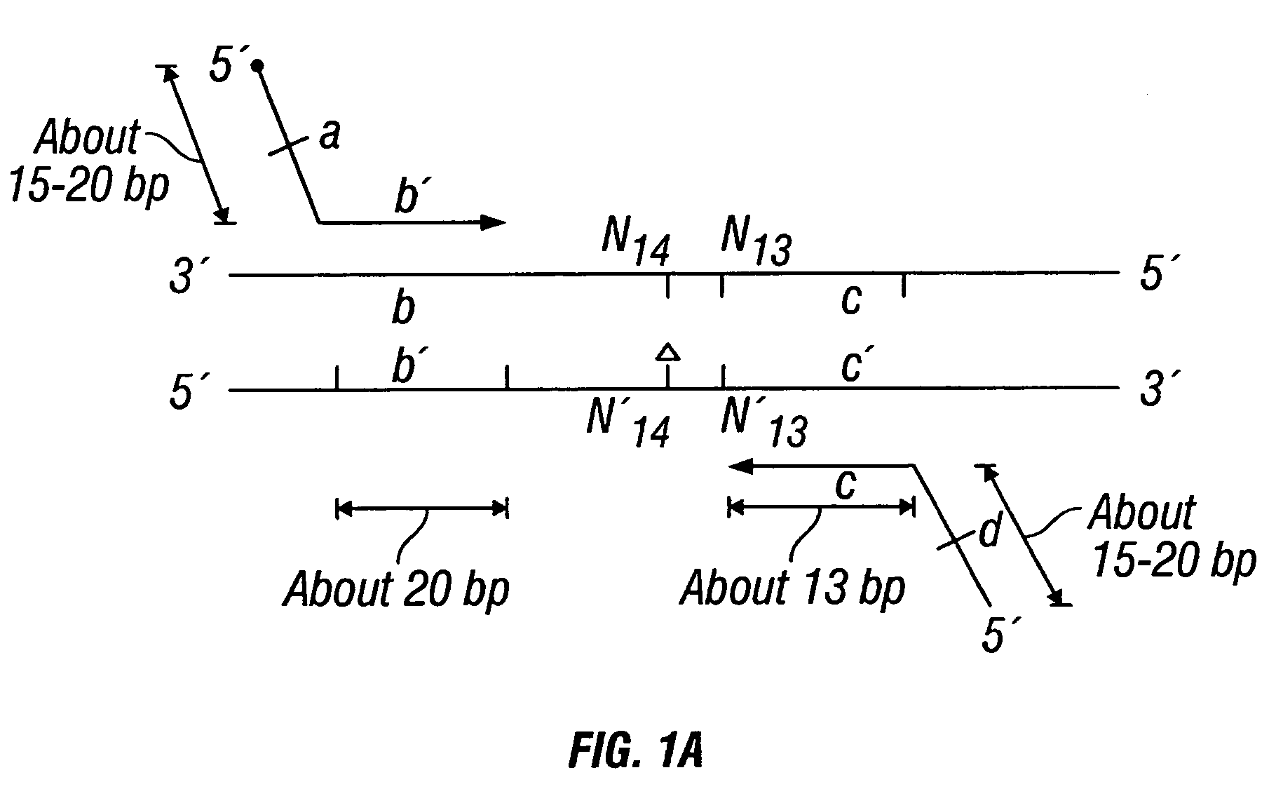
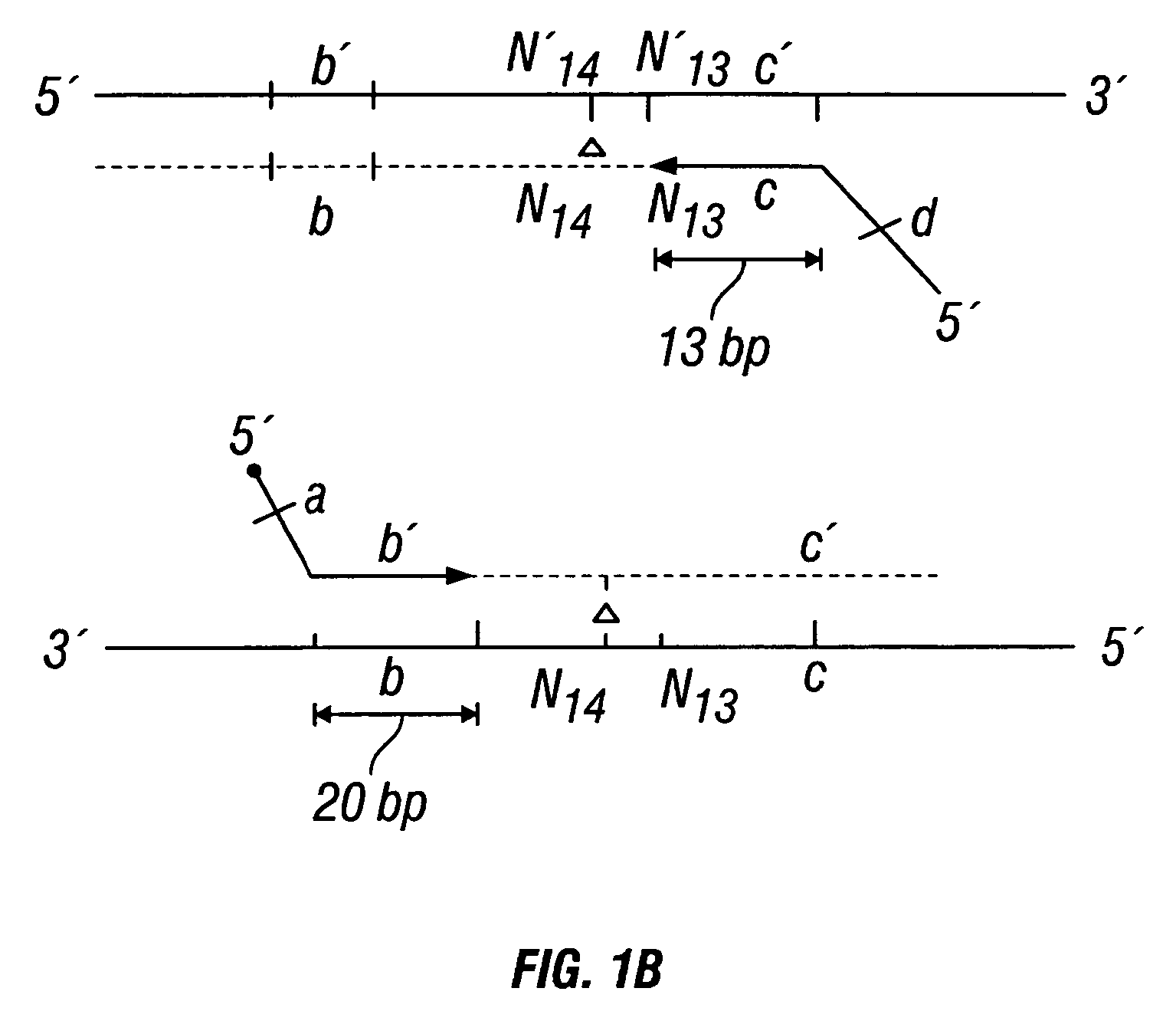
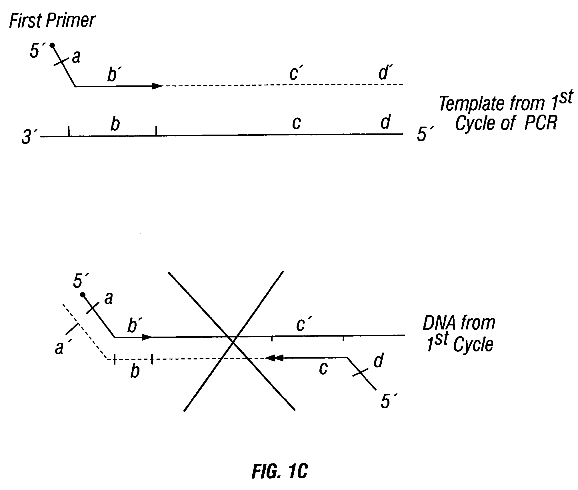
[0426]DNA sequences were amplified by PCR, wherein the annealing step in cycle 1 was performed at a specified temperature, and then increased in cycle 2, and further increased in cycle 3 for the purpose of reducing non-specific amplification. The TM1 of cycle 1 of PCR was determined by calculating the melting temperature of the 3′ region, which anneals to the template DNA, of the second primer. For example, in FIG. 1B, the TM1 can be about the melting temperature of region “c.” The annealing temperature was raised in cycle 2, to TM2, which was about the melting temperature of the 3′ region, which anneals to the template DNA, of the first primer. For example, in FIG. 1C, the annealing temperature (TM2) corresponds to the melting temperature of region “b.” In cycle 3, the annealing temperature was raised to TM3, which was about the melting temperature of the entire sequence of the second primer. For example, in FIG. 1D, the annealing temperature (TM3) corresponds to the melting temper...

example 2

[0444]SNPs on chromosomes 1 (TSC0095512), 13 (TSC0264580), and 21 (HC21S00027) were analyzed. SNP TSC0095512 was analyzed using two different sets of primers, and SNP HC21S00027 was analyzed using two types of reactions for the incorporation of nucleotides.

Preparation of Template DNA

[0445]The template DNA was prepared from a 5 ml sample of blood obtained by venipuncture from a human volunteer with informed consent. Template DNA was isolated using the QIAmp DNA Blood Midi Kit supplied by QIAGEN (Catalog number 51183). The template DNA was isolated as per instructions included in the kit. Following isolation, template DNA from thirty-six human volunteers were pooled together and cut with the restriction enzyme EcoRI. The restriction enzyme digestion was performed as per manufacturer's instructions.

Primer Design

[0446]SNP HC21S00027 was amplified by PCR using the following primer set:

[0447]

First primer:(SEQ ID NO: 17)5′ ATAACCGTATGCGAATTCTATAATTTTCCTGATAAAGG 3′Second primer:(SEQ ID NO: ...

example 3

[0487]Four loci of interest from chromosome 1 and two loci of interest from chromosome 21 were amplified in separate PCR reactions, pooled together, and analyzed. The primers were designed so that each amplified locus of interest was a different size, which allowed detection of the loci of interest.

Preparation of Template DNA

[0488]The template DNA was prepared from a 5 ml sample of blood obtained by venipuncture from a human volunteer with informed consent. Template DNA was isolated using the QIAmp DNA Blood Midi Kit supplied by QIAGEN (Catalog number 51183). The template DNA was isolated as per instructions included in the kit. Template DNA was isolated from thirty-six human volunteers, and then pooled into a single sample for further analysis.

Primer Design

[0489]SNP TSC 0087315 was amplified using the following primers:

[0490]

First primer:(SEQ ID NO: 15)5′TTACAATGCATGAATTCATCTTGGTCTCTCAAAGTGC 3′Second primer:(SEQ ID NO: 16)5′TGGACCATAAACGGCCAAAAACTGTAAG3′.

[0491]SNP TSC0214366 was am...


PUM

PropertyMeasurementUnit
compositionaaaaaaaaaa

Abstract

The invention provides a method useful for detection of genetic disorders. The method comprises determining the sequence of alleles of a locus of interest, and quantitating a ratio for the alleles at the locus of interest, wherein the ratio indicates the presence or absence of a chromosomal abnormality. The present invention also provides a non-invasive method for the detection of chromosomal abnormalities in a fetus. The invention is especially useful as a non-invasive method for determining the sequence of fetal DNA. The invention further provides methods of isolation of free DNA from a sample.

Description

CROSS-REFERENCE TO RELATED APPLICATIONS[0001]This application is a continuation of International Application No. PCT / US03 / 27308, filed Aug. 29, 2003, which is a continuation-in-part of International Application No. PCT / US03 / 06198, filed Feb. 28, 2003, which claims the priority benefit of U.S. Provisional Application Ser. No. 60 / 378,354, filed May 8, 2002, the contents of each of which are hereby incorporated by reference herein in their entirety. This application is also a continuation-in-part of U.S. patent application Ser. No. 10 / 661,165, filed Sep. 11, 2003 now U.S. Pat. No. 7,332,277, which is a continuation-in-part of International Application No. PCT / US03 / 06198, filed Feb.28, 2003, which claims the priority benefit of U.S. Provisional Application Ser. No. 60 / 378,354, filed May 8, 2002. The contents of each of the following applications are also hereby incorporated by reference in their entirety herein: U.S. patent application Ser. No. 10 / 093,618, filed Mar. 11, 2002; and U.S. ...

Claims


Application Information

Patent Timeline
Patent Type & Authority Patents(United States)
IPC IPC(8): C12Q1/68C07H19/00C07H21/00
CPCC12Q1/6827C12Q2533/101C12Q2525/313C12Q2525/131
Inventor DHALLAN, RAVINDER S.
Owner RAVGEN INC
Why Eureka
Try Eureka