Depth disambiguation of interventional instruments from a single x-ray projection image and its calibration

Inactive Publication Date: 2012-07-26
KONINKLIJKE PHILIPS ELECTRONICS NV
View PDF1 Cites 0 Cited by
  • Summary
  • Abstract
  • Description
  • Claims
  • Application Information

AI Technical Summary

Benefits of technology

[0010]Therefore, a more robust use of the width estimation is made, by using it to discriminate between possible locations using a segmentation of the anatomy, rather than estimating the absolute depth of the instrument. Furthermore, a calibration process makes it robust to possible errors in the

Problems solved by technology

A very challenging electrophysiology procedure is radio-frequency a

Method used


Image

  • Depth disambiguation of interventional instruments from a single x-ray projection image and its calibration
  • Depth disambiguation of interventional instruments from a single x-ray projection image and its calibration
  • Depth disambiguation of interventional instruments from a single x-ray projection image and its calibration

Examples


Example

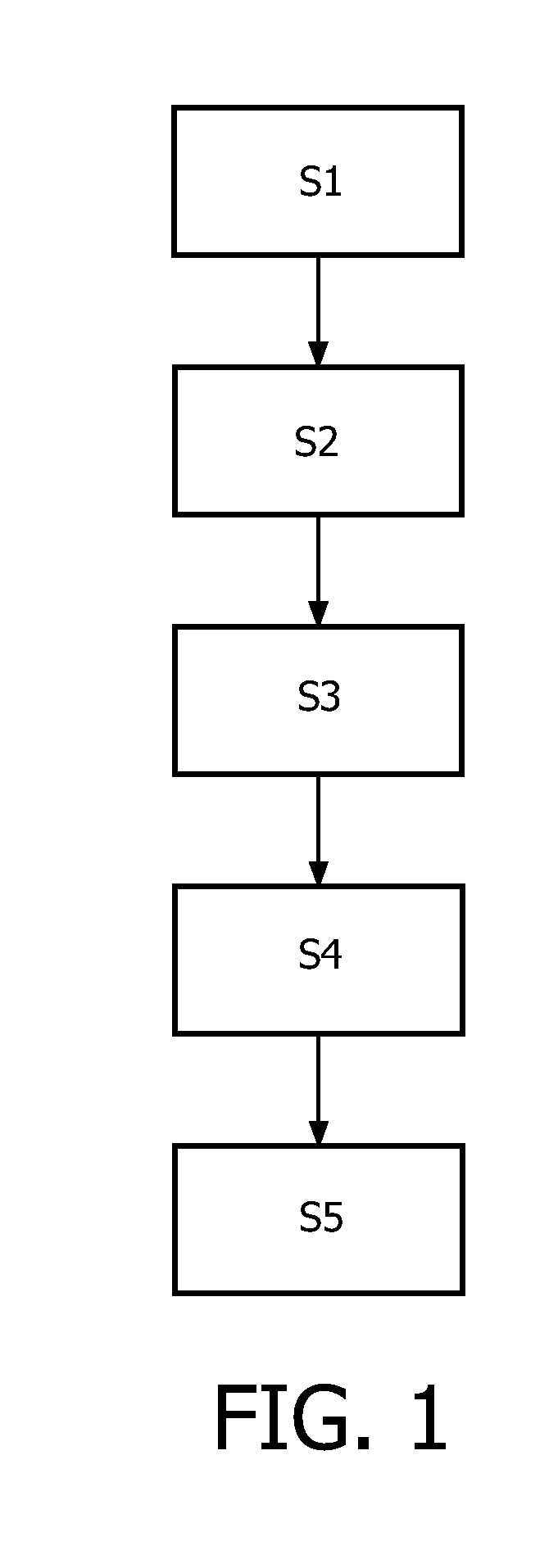
[0030]FIG. 1 is a flow chart, showing the steps of a method for determining a depth of an instrument in an object according to the invention. It will be understood that the steps described with respect to the method are major steps, wherein these major steps might be differentiated or divided into several sub steps. Furthermore, there might be also sub steps between these major steps. Therefore, a sub step is only mentioned if that step is important for the understanding of the principles of the method according to the invention.

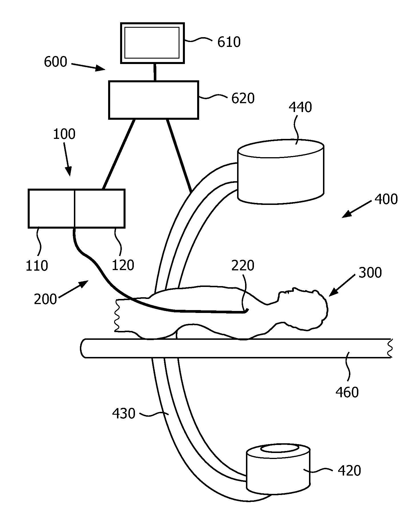
[0031]In step S1 of the method according to the invention one x-ray projection image of the instrument in the object is generated. To reduce the radiation to which for example a patient is exposed, only a single projection image is generated.

[0032]In step S2, the size of a portion of the instrument in the object is estimated. Usually, this portion will be the tip portion of an instrument, i.e. the portion of a catheter at which for example ablation electrode...


PUM


Abstract

X-ray images are projective, meaning that the 3D geometry is flattened along projection lines going from the source to the detector. In particular procedures, such as mapping or ablation, the interventional instrument lies on the wall of the organ. Using a 3D segmentation of this organ registered to the x-ray, the instrument necessarily lies on the intersection of this surface with its projection line. The line and the surface typically intersect with a segmentation surface at a discrete number of points (typically 2 for shapes such as the anterior of the LA). One then has just to disambiguate between these different possible locations to determine the exact location of the instrument. In this invention, we propose to use the apparent width of the instrument measured in x-ray images to accomplish this task.

Description

FIELD OF THE INVENTION[0001]The present invention relates to x-ray guided procedures. Especially, the invention relates to a method for determining a depth of an instrument in an object. Furthermore, the invention relates to a computer program for determining a depth of an instrument in an object as well as to an x-ray device equipped with such a computer program.BACKGROUND OF THE INVENTION[0002]Electrophysiology is a specific domain of interventional cardiology where physicians use intracardiac catheters to locate and cure electrical dysfunctions of the heart rhythm, under x-ray fluoroscopy guidance. A very challenging electrophysiology procedure is radio-frequency ablation for the treatment of an atrial fibrillation, also called AF.[0003]Electrophysiologics need a special training to perfectly know the anatomy and the excess pathways to all the sites of interest and some practice to select the correct devices and manipulate them to target. The patient's anatomy can be recorded wit...

Claims


Application Information

Patent Timeline
IPC IPC(8): A61B6/00G06K9/62
CPCA61B6/12A61B6/466G06T2207/30004G06T2207/10116G06T7/602G06T7/62
Inventor CATHIER, PASCALGOGIN, NICOLAS PIERRE BRUNOFLORENT, RAOUL
Owner KONINKLIJKE PHILIPS ELECTRONICS NV
Why Eureka
Try Eureka