
System and method for estimating vascular flow using ct imaging

a technology of vascular flow and ct imaging, applied in the field of diagnostic imaging, can solve the problems of low diagnostic value, low accuracy, and low accuracy of cta, and achieve the effect of reducing the negative predictive value of cta, and high invasive procedures

Active Publication Date: 2012-09-27
GENERAL ELECTRIC CO
View PDF15 Cites 173 Cited by
  • Summary
  • Abstract
  • Description
  • Claims
  • Application Information

AI Technical Summary

Problems solved by technology

However, false positives may still occur at undesired frequency, reducing the positive predictive value of CCTA (ratio of true positive classifications to the total number of positive classifications).
However, these procedures are highly invasive and frequently turn out to be unnecessary (diagnostic) (e.g., in approximately ⅓ of the procedures in patients with multi-vessel disease).

Method used


Image

  • System and method for estimating vascular flow using ct imaging
  • System and method for estimating vascular flow using ct imaging
  • System and method for estimating vascular flow using ct imaging

Examples


Embodiment Construction

[0021]The operating environment of embodiments of the invention is described with respect to a multi-slice computed tomography (CT) system. Moreover, embodiments of the invention will be described with respect to the detection and conversion of x-rays. However, one skilled in the art will further appreciate that the invention is equally applicable for the detection and conversion of other high frequency electromagnetic energy. Embodiments of the invention will be described with respect to a “third generation” CT scanner, but is equally applicable with other CT system configurations.

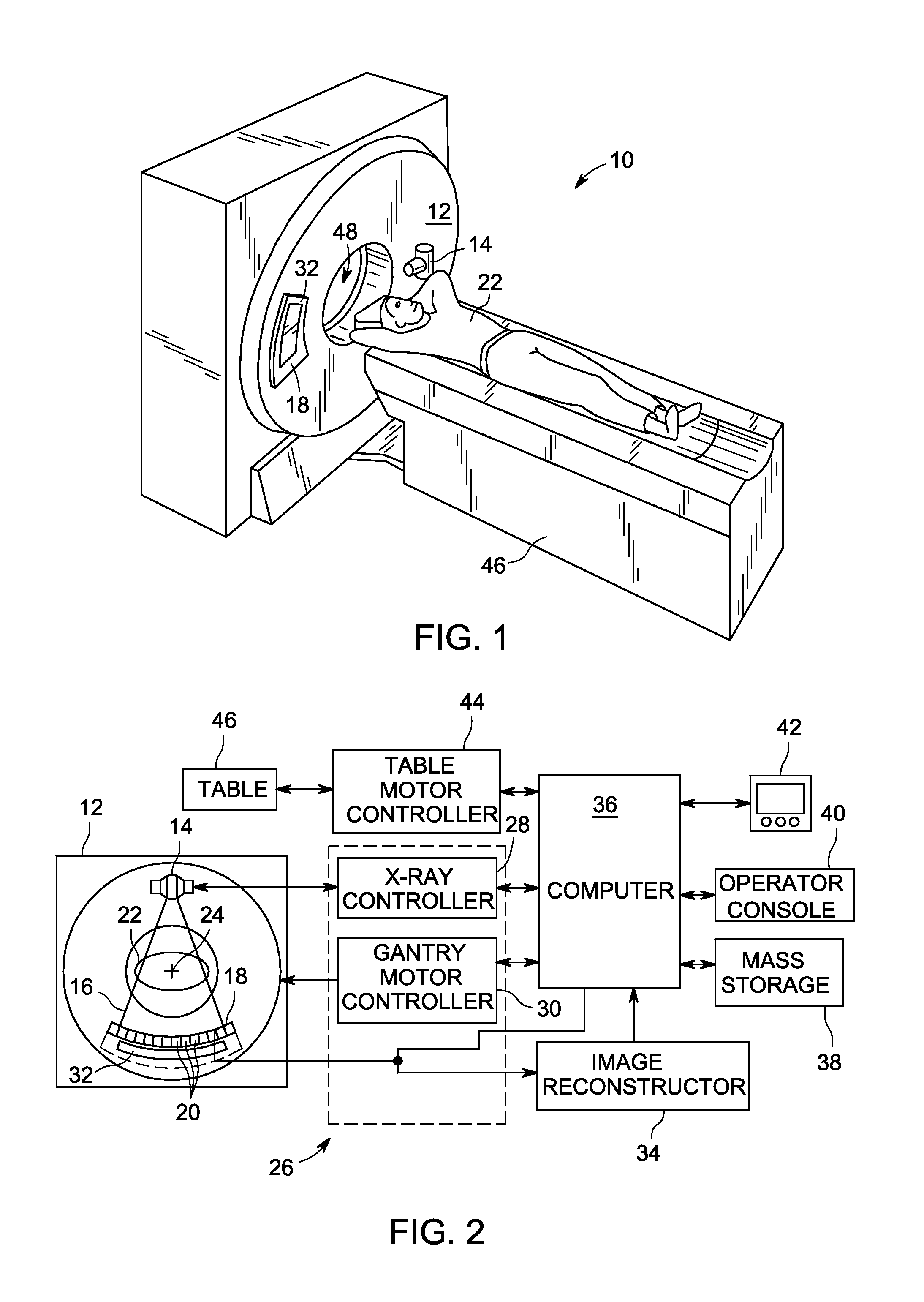
[0022]Referring to FIG. 1, a computed tomography (CT) imaging system 10 is shown as including a gantry 12 representative of a “third generation” CT scanner. Gantry 12 has an x-ray source 14 that projects a beam of x-rays toward a detector assembly and / or collimator 18 on the opposite side of the gantry 12. Referring now to FIG. 2, detector assembly 18 is formed by a plurality of detectors 20 and data acqu...


PUM


Abstract

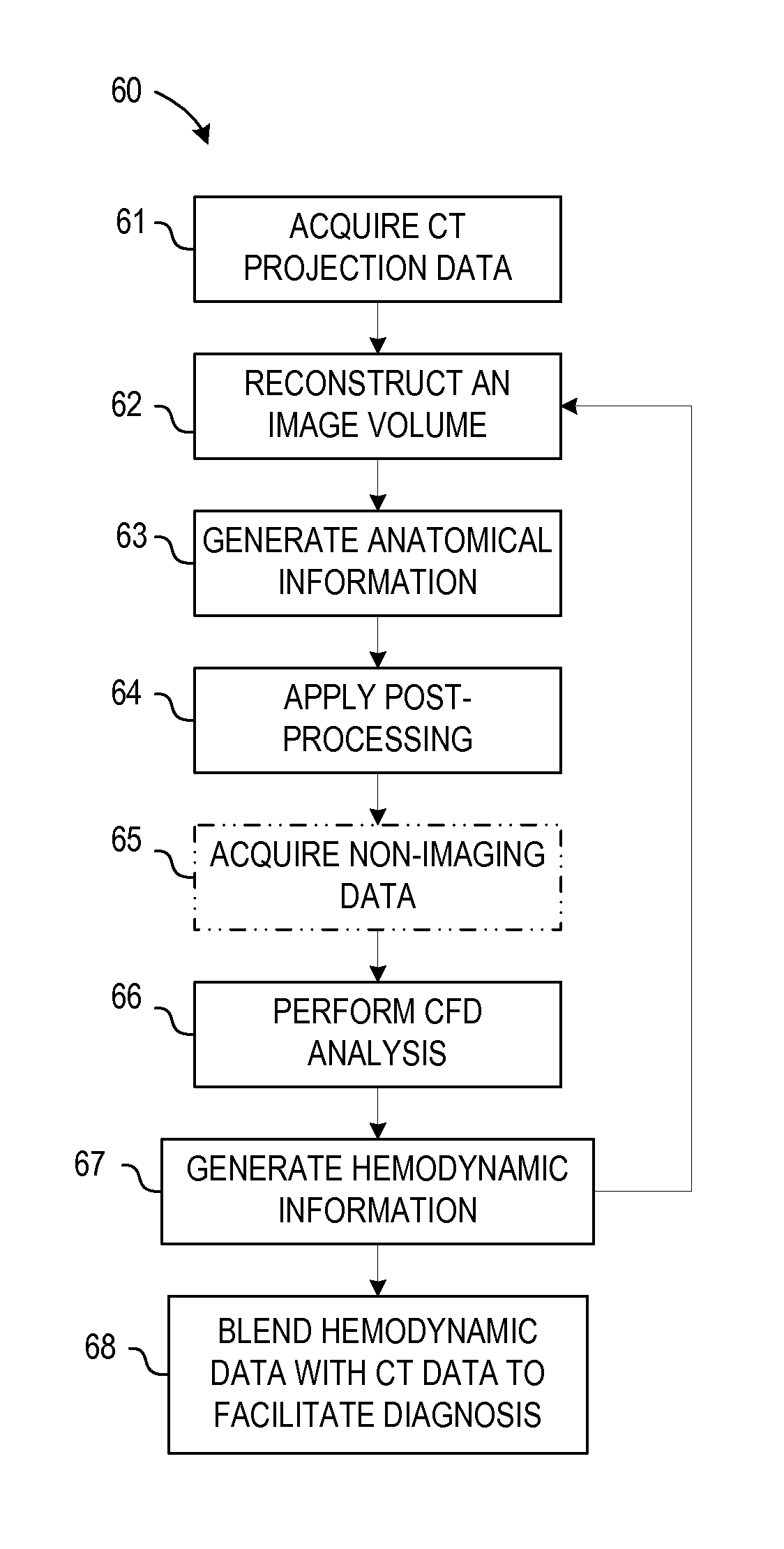
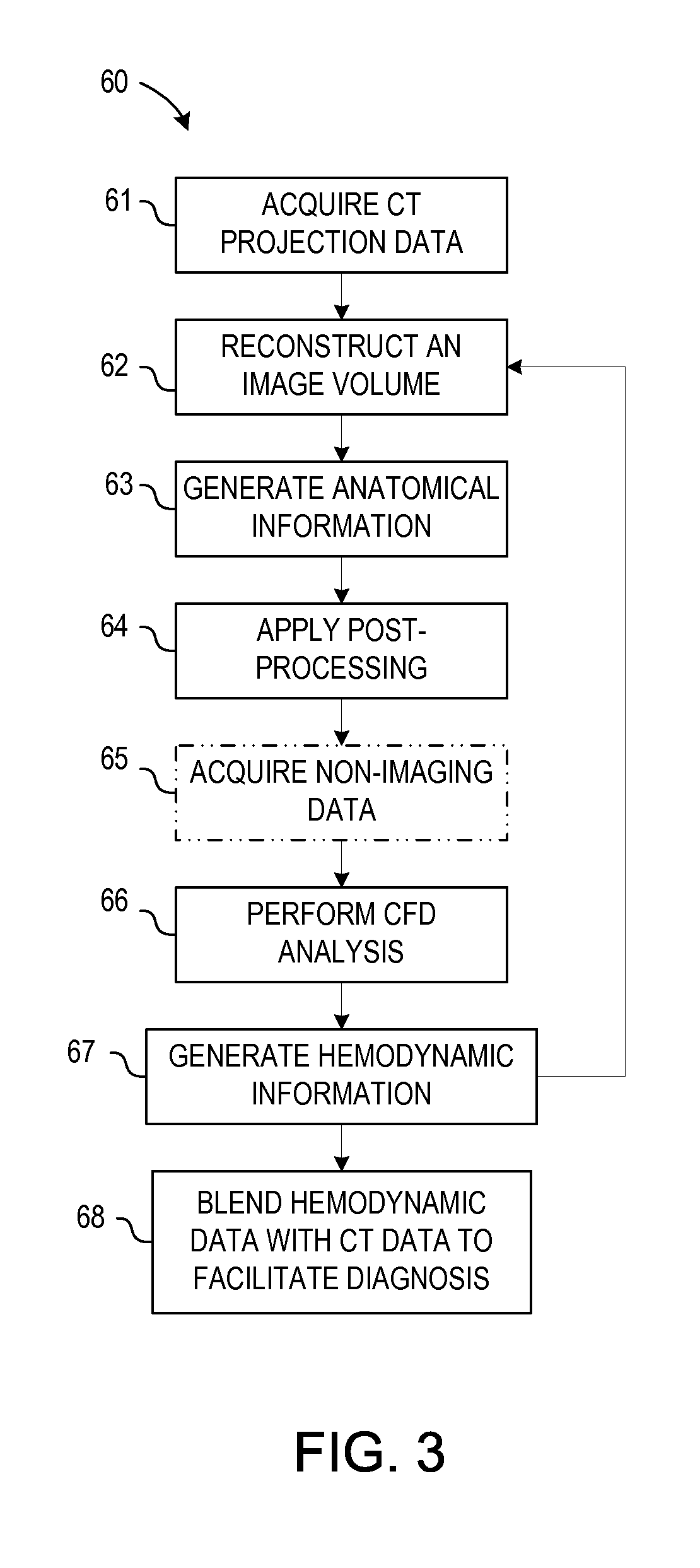
A system and method for estimating vascular flow using CT imaging include a computer readable storage medium having stored thereon a computer program comprising instructions, which, when executed by a computer, cause the computer to acquire a first set of data comprising anatomical information of an imaging subject, the anatomical information comprises information of at least one vessel. The instructions further cause the computer to process the anatomical information to generate an image volume comprising the at least one vessel, generate hemodynamic information based on the image volume, and acquire a second set of data of the imaging subject. The computer is also caused to generate an image comprising the hemodynamic information in combination with a visualization based on the second set of data.

Description

CROSS-REFERENCE TO RELATED APPLICATION[0001]The present application is a non-provisional of, and claims priority to, U.S. Provisional Patent Application Ser. No. 61 / 454,646, filed Mar. 21, 2011, the disclosure of which is incorporated herein by reference.BACKGROUND OF THE INVENTION[0002]Embodiments of the invention relate generally to diagnostic imaging and, more particularly, to a system and method of estimating hemodynamic information.[0003]Non-invasive imaging technologies allow images of the internal structures or features of a patient to be obtained without performing an invasive procedure on the patient. In particular, such non-invasive imaging technologies rely on various physical principles, such as the differential transmission of x-rays through the target volume, measurement of resonant magnetic signals, or the reflection of acoustic waves, to acquire data and to construct images or otherwise represent the observed internal features of the patient.[0004]For example, Corona...

Claims


Application Information

Patent Timeline
IPC IPC(8): G06K9/00A61B6/03G16Z99/00
CPCG06T11/008G06T2211/404A61B6/032A61B6/481A61B6/482A61B6/503G06T2207/30104A61B6/507A61B6/5217A61B6/5294G06T7/0016G06T2207/10081A61B6/504G16Z99/00G16H50/30
Inventor SENZIG, ROBERT F.AVANCHA, RAVIKANTHDORRI, BIJANDUTTA, SANDEEPGRAY, STEVEN J.HSIEH, JIANGJACKSON, JOHN IRVINJOTHIPRASAD, GIRIDHARLICATO, PAUL EDGAROKERLUND, DARIN ROBERTRIFU, TOSHIHIROSIROHEY, SAAD AHMEDTAHA, BASELEDIC, PETER MICHAELKNOPLIOCH, JEROMEBHOTIKA, RAHUL
Owner GENERAL ELECTRIC CO
Learn More