
Method and apparatus for compressing imaging data of whole blood sample analyses

a technology for imaging data and analysis, applied in image analysis, image enhancement, instruments, etc., can solve the problems of laborious blood smears, cost prohibitive, and general non-profit use of blood smears

Inactive Publication Date: 2012-10-18
ABBOTT POINT CARE
View PDF13 Cites 37 Cited by
  • Summary
  • Abstract
  • Description
  • Claims
  • Application Information

AI Technical Summary

Benefits of technology

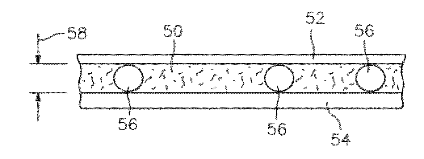
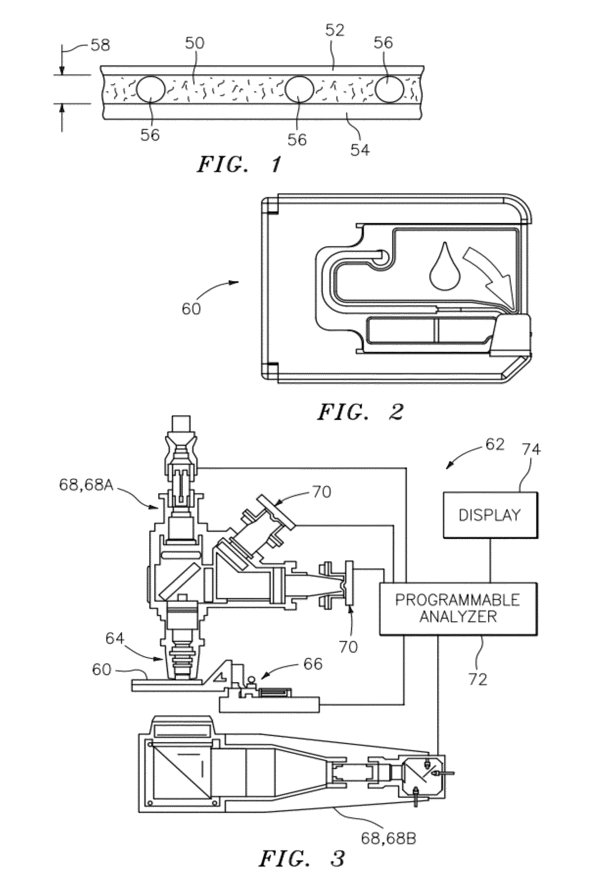
[0011]According to another aspect of the present invention, an apparatus for analyzing a whole blood sample quiescently residing within a chamber is provided. The apparatus includes an objective lens, a sample illuminator, an image dissector, and a programmable analyzer. The sample illuminator is operable to provide a fluorescent excitation light and one or more transmission lights. The image dissector is adapted to receive one or both of light fluorescing from the sample and light transmitted through the sample, and to produce signals representative of such light. The programmable analyzer ...

Problems solved by technology

Blood smears are problematic for several reasons; e.g., the cells must be killed and fixed, which process precludes many types of supravital stains and analyses whose results depend upon living cells, and blood smears are labor intensive, cost prohibitive, and time consuming For at least these reasons, blood smears are generally not favored for commercial applications.
Attempts to automate analyses of whole blood samples have met with some success, but typically have several drawbacks.
These instruments typically require fluid handling equipment and require the sample be diluted.
Analyses of biological fluid specimens using color images can be hindered by the substantial size of the image files.
Large image files can consume storage space, impede transfer to a remote location, and / or slow processing.

Method used


Image

  • Method and apparatus for compressing imaging data of whole blood sample analyses
  • Method and apparatus for compressing imaging data of whole blood sample analyses
  • Method and apparatus for compressing imaging data of whole blood sample analyses

Examples


Embodiment Construction

[0049]The present method and apparatus is directed toward creating an image of a biological fluid sample, and processing that image to create one or more image analysis data files. The processing utilizes one or more compression algorithms operable to compress an image analysis data file to a smaller size while maintaining acceptably low levels of information loss. The image analysis data is typically generated by an analysis of an image of a biological fluid sample quiescently residing within an analysis chamber. The analysis includes identifying and locating particular constituents within the sample.

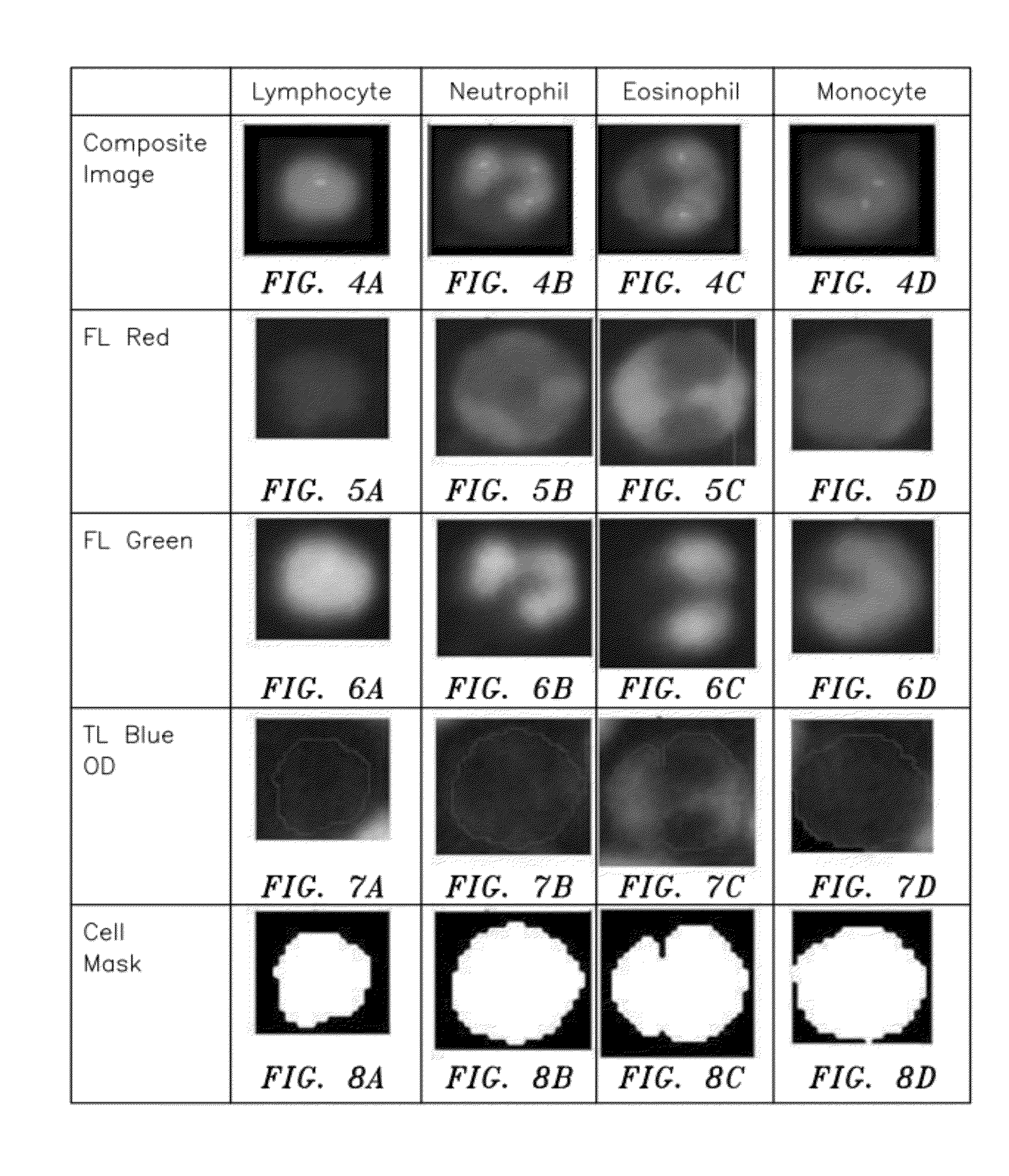
[0050]An exemplary analysis involves performing a leukocyte differential count (“LDC”) on a whole blood sample. As indicated above, an LDC is an analysis wherein the different types of WBCs are identified and enumerated. The results can be expressed in terms of the relative percentages of identified WBC types; e.g., monocytes, eosinophils, neutrophils, and lymphocytes within a blood sa...


PUM


Abstract

A method and apparatus for analyzing white blood cells (WBCs) within a whole blood sample quiescently residing within a chamber is provided. The chamber is defined by at least one transparent panel, and the whole blood sample includes at least one colorant operable to differentially identify at least one WBC type from another WBC type within the sample. The method includes the steps of: a) creating at least one image of the sample quiescently residing within the chamber; b) identifying portions of the sample image, with each portion representing a single WBC; c) compressing the sample image portions using a first compression algorithm; and d) one of compressing a remainder of the sample image not included in the portions using a second compression algorithm, or discarding the remainder.

Description

BACKGROUND OF THE INVENTION[0001]1. Technical Field[0002]The present invention relates to methods and apparatus for performing analyses on whole blood samples from microscopy images in general, and to methods and apparatus for compressing imaging data of the same in particular.[0003]2. Background Information[0004]Medical diagnostics often include analyses of a whole blood sample from a patient. One of the more popular diagnostics is a complete blood count (referred to as a “CBC”), which is a suite of tests that may include, in addition to the enumeration of the cellular components, red blood cell metrics, reticulocyte counts, and a leukocyte differential count (“LDC”; sometimes referred to as a “white blood cell differential”), which is the identification and enumeration of the types of white blood cells (WBCs) present in the blood sample.[0005]Historically, the differential aspects of the CBC have been performed using separate methods from those used for enumeration. For example, t...

Claims


Application Information

Patent Timeline
IPC IPC(8): G06K9/00
CPCG06T7/0012G06T2207/10024G06T2207/10056G06T2207/10064H04N19/167G06T2207/30242H04N19/102H04N19/17G06T2207/30024G06V20/698
Inventor XIE, MINWANG, ZHIZHOU
Owner ABBOTT POINT CARE
Learn More