Method For Measuring Trabecular Bone Parameters From MRI Images

a trabecular bone and parameter measurement technology, applied in the field of trabecular bone parameter measurement from mri images, can solve the problems of insufficient bmd diagnosis, insufficient mdct resolution, and high risk of fracture,

Inactive Publication Date: 2012-11-01
KOREA BASIC SCI INST
View PDF4 Cites 13 Cited by
  • Summary
  • Abstract
  • Description
  • Claims
  • Application Information

AI Technical Summary

Benefits of technology

[0008]In another aspect, the present disclosure provides a method for diagnosing osteoporosis, comprising: scanning an experimental group with a 3D MRI scanner; segmenting the MRI images to extract bone area and perform skeletonization of the bone area; detecting end-point, joint and branch voxels in the skelet...

Problems solved by technology

Osteoporosis is a bone disease that leads to a higher risk of fracture, especially in postmenopausal women.
Therefore, it does not sufficient to diagnose osteoporosis using only BMD.
The achievable resolution of MDCT is insufficient for the quantitative structural analysis of trabecular bone architecture in vivo secondary to limitations on radiation dose.
However, the MR images can only be obtained during the limited time that a patient can tolerate remainin...

Method used


Image

  • Method For Measuring Trabecular Bone Parameters From MRI Images
  • Method For Measuring Trabecular Bone Parameters From MRI Images
  • Method For Measuring Trabecular Bone Parameters From MRI Images

Examples


example 1

Acquisition of Micro MR Images

[0042]Ten bone specimens were extracted from the distal femoral condyle during knee joint replacement procedures at Bundang Seoul National University Hospital.

[0043]The specimens were cut using a saw (1×1×1 cm3) and were then fixed in formal in and stored. In preparation for the scan, the bone specimens were defatted, degassed, and immersed in 0.5% (volume percent) gadopentetate-doped water. 3D iso-cubic trabecular bone images of the bone specimens were obtained using a 4.7T Bruker BioSpec MRI scanner with a 40-cm bone size. A 2.5-cm birdcage coil with quadrature detection was used. Factors which impede high resolution 3D imaging of trabecular bone include long scan times and the blurring along the trabecular bone marrow interface caused by the susceptibility difference. Therefore, a 3D fast large-angle spin-echo (FLASE) sequence with a 140° pulse (TR=100 ms and TE=10 ms) was used to overcome these two problems.

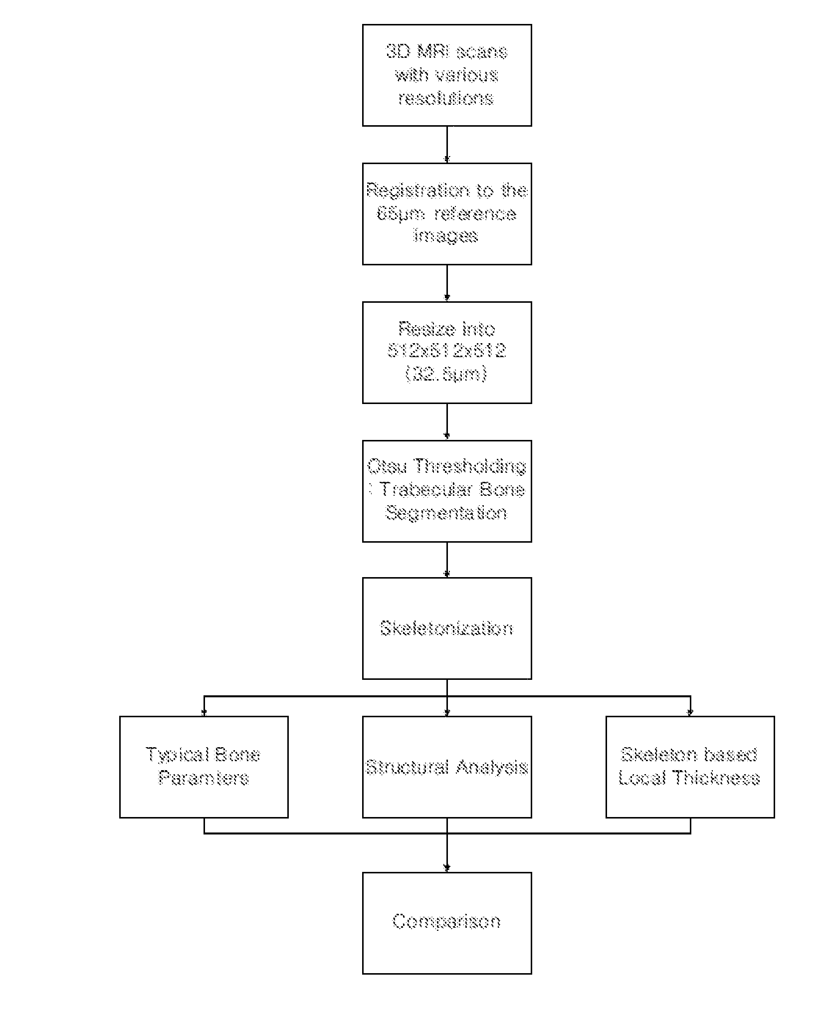
[0044]FIG. 1 shows the overall scheme of t...

example 2

Imaging Preprocessing and Trabecular Bone Segmentation

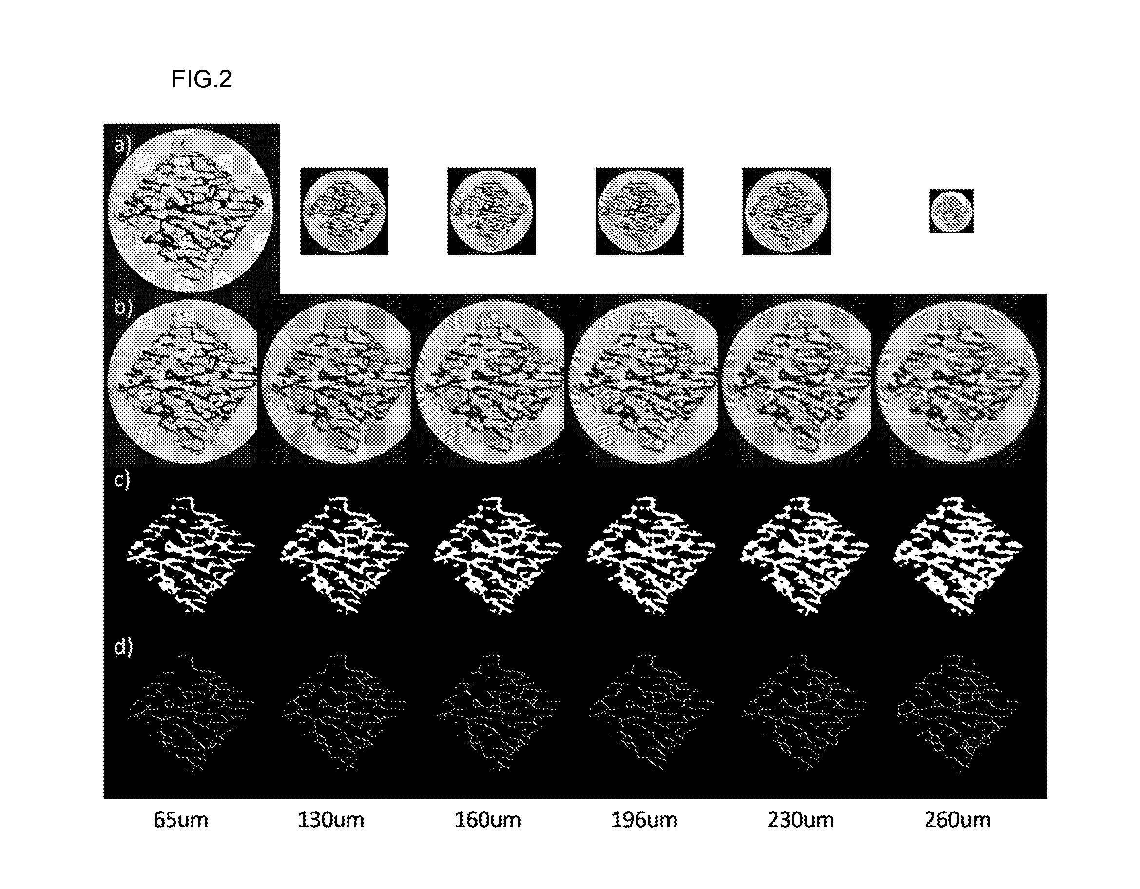
[0045]The bone specimens slightly rotated during the acquisition of the images with a 65 μm voxel size secondary to the long acquisition times and strong gradient pulses. The images acquired with larger voxel sizes did not rotate significantly. Therefore, all of the lower resolution images were registered with a high resolution reference image (65 μm cubed iso-cubic voxel size) by rigid registration based on a mean squared difference metric and cubic spline interpolation. After the registration, they were resized as 512×512×512 (iso-cubic voxel resolution: 32.5 μm) using a cubic spline interpolation algorithm. In-house software was developed for these preprocessing steps using the Insight Segmentation and Registration Toolkit (ITK). Trabecular bone extraction from images with various resolutions was performed using the Otsu thresholding method. FIGS. 2a, 2b and 2c show the original images, the re-sampled images, and the segmented...

example 3

Trabecular Bone Parameters Evaluation

[0046]To evaluate the trabecular bone parameters, skeletonization using a 3D medial surface / axis thinning algorithm was applied to detect the center line of segmented trabecular bone. Each voxel on the skeleton can be considered a structural element for bone analysis. To evaluate the trabecular bone structure, the joints and branches of the skeleton were identified. Structural bone parameters, including joint number (Joint#) and branch number (Branch#) were normalized by those of the reference images due to the large variation of joint and branch numbers between the different specimens. Therefore, normalized joint number (nJoint#), normalized branch number (nBranch#), and average branch length (avgBranchLen) were evaluated. For each voxel along the skeleton, local trabecular bone thickness was measured using a model-independent method. In addition, conventional bone parameters including trabecular thickness (TB.Th), bone density (BV / TV), trabecul...


PUM


Abstract

Disclosed is a method for measuring trabecular bone parameters from MRI images, including: scanning an experimental group with a 3D MRI scanner; segmenting the MRI images to extract bone area and perform skeletonization of the bone area; detecting end-point, joint and branch voxels in the skeleton to analyze bone structure; and measuring trabecular bone parameters based on the result of the structural analysis. The method enables diagnosing osteoporosis.

Description

BACKGROUND[0001]1. Field[0002]The present disclosure relates to a method for measuring trabecular bone parameters from MRI images.[0003]2. Description of the Related Art[0004]Osteoporosis is a bone disease that leads to a higher risk of fracture, especially in postmenopausal women. In fact, osteoporosis of the hip and osteopenia are experienced by 20% and 34-50%, respectively, of women over 50 years of age and contribute to a 40% lifetime risk of fracture of the hip, radius or spine. Clinically, dual-energy X-ray (DEXA) which measures bone mineral density (BMD) is the current and standard method used to diagnose osteoporosis. According to recent studies, BMD determines approximately 60% of trabecular bone strength. Therefore, it does not sufficient to diagnose osteoporosis using only BMD. In 1994, the World Health Organization (WHO) redefined osteoporosis as a “disease characterized by low bone mass and micro-architectural deterioration causing increased bone fragility.” The measure...

Claims


Application Information

Patent Timeline
IPC IPC(8): A61B5/055G06K9/00
CPCA61B5/055G06T7/0012G06T2207/10088A61B5/4509G06T2207/30008A61B5/4504G06T2207/20044
Inventor CHO, GYUNGGOOKIM, NAMKUGLEE, JUNE-GOOSONG, YOUNGKYUKIM, HENGJUN JCHEONG, CHAEJOON
Owner KOREA BASIC SCI INST
Why Eureka
Try Eureka