Omnidirectional super-resolution microscopy

a super-resolution, microscopy technology, applied in the field of microscopy, can solve the problems of not being very practical for routine research or intracellular structure imaging, and not being able to use non-fluorescence imaging,

Inactive Publication Date: 2013-04-18
PHOTONANOSCOPY
View PDF3 Cites 63 Cited by
  • Summary
  • Abstract
  • Description
  • Claims
  • Application Information

AI Technical Summary

Problems solved by technology

This represents a fundamental barrier to progress, for example, in cancer research where imaging is used to study changes in cytoskeletal, membrane and chromosome structure, and to visualize changes in DNA, such as patterns of methylation.
While some methods have reported resolutions down to 8 nm, their practicality is severely hampered by the need for special fluorophores and / or extreme illumination light intensities, while the other methods may generally requires thin specimens.
To date, none of these methods have been found very practical for routine research or to image intracellular structures, nor can they be used with non-fluorescence imaging.
Standing waves have also been used with total internal reflection microscopy to improve lateral resolution, but this approach is often limited to one very thin section of the specimen.
However, it does require a substantial modification of the microscope.
It is also not easy to maintain stability along both illumination paths to within a fraction of the excitation wavelength.
In practice, this setup suffers from many of the difficulties that plague 4π microscopy.
The primary limitation of standing wave microscopy sterns from the fact that the interference pattern is produced by two counter-propagating planar or nearly planar wave-fronts.
Other problems arise simply from the aliasing along the Z axis which limits sample thickness, the stability requirements, the need for closely match microscope objectives, the extensive modifications to the microscope and the need for a symmetric sample preparation between two cover-slips.

Method used


Image

  • Omnidirectional super-resolution microscopy
  • Omnidirectional super-resolution microscopy
  • Omnidirectional super-resolution microscopy

Examples


Embodiment Construction

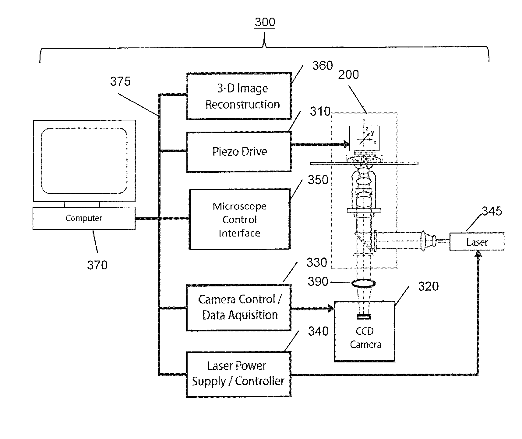
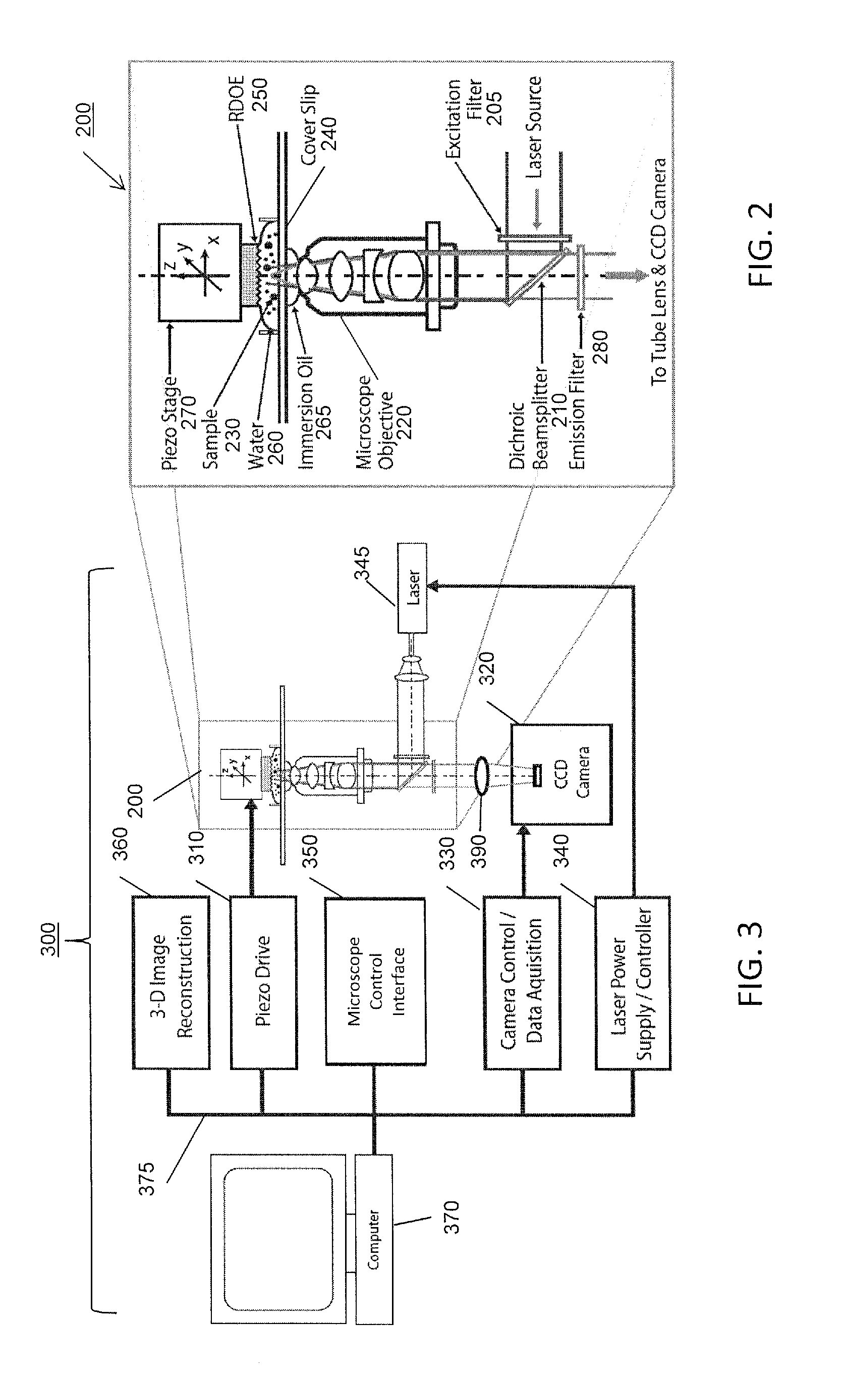
[0020]Various aspects of the present invention will be described herein with reference to drawings that are schematic illustrations of idealized configurations of the present invention. As such, variations from the shapes of the illustrations as a result, for example, manufacturing techniques and / or tolerances, are to be expected. Thus, the various aspects of the present invention presented throughout this disclosure should not be construed as limited to the particular shapes of elements (e.g., regions, layers, sections, substrates, etc.) illustrated and described herein but are to include deviations in shapes that result, for example, from manufacturing. By way of example, an element illustrated or described as a rectangle may have rounded or curved features and / or a gradient concentration at its edges rather than a discrete change from one element to another. Thus, the elements illustrated in the drawings are schematic in nature and their shapes are not intended to illustrate the ...


PUM


Abstract

A microscopy method and apparatus includes placing a specimen to be observed adjacent to a reflective holographic optical element (RDOE). A beam of light that is at least partially coherent is focused on a region of the specimen. The beam forward propagates through the specimen and is at least partially reflected backward through the specimen. The backward reflected light interferes with the forward propagating light to provide a three dimensional interference pattern that is at least partially within the specimen. A specimen region illuminated by the interference pattern is imaged at an image detector. Computational reconstruction is used to generate a microscopic image in all three spatial dimensions (X,Y,Z), simultaneously with resolution greater than conventional microscopy.

Description

BACKGROUND[0001]1. Field[0002]The present disclosure relates to microscopy, and particularly relates to super-resolution imaging microscopy in three dimensions.[0003]2. Description of Related Art[0004]Many of the features of interest in the fluorescence microscopy of cells are not resolved by a conventional optical microscope. This represents a fundamental barrier to progress, for example, in cancer research where imaging is used to study changes in cytoskeletal, membrane and chromosome structure, and to visualize changes in DNA, such as patterns of methylation. Super-resolution techniques allow the capture of images with a higher resolution than the classical diffraction limit. The recent proliferation of super-resolution methods reflects the recognition of this need. A category of super resolution techniques, known as “functional,” uses clever experimental techniques and known limitations on the matter being imaged to reconstruct a super-resolution image. Current approaches to ove...

Claims


Application Information

Patent Timeline
Patent Type & Authority Applications(United States)
IPC IPC(8): G02B21/06H04N7/18
CPCG02B21/14G02B21/0088G02B21/082G01N21/6458G02B21/33G02B21/367G02B21/26
Inventor NOWATZYK, ANDREAS G.FARKAS, DANIEL
Owner PHOTONANOSCOPY
Why Eureka
Try Eureka