Methods and apparatus for aligning sets of medical imaging data

a technology for medical imaging and sets of data, applied in image analysis, image enhancement, instruments, etc., can solve problems such as poor lv and difficult alignmen

Active Publication Date: 2013-04-18
SIEMENS MEDICAL SOLUTIONS USA INC
View PDF11 Cites 13 Cited by
  • Summary
  • Abstract
  • Description
  • Claims
  • Application Information

AI Technical Summary

Benefits of technology

[0015]This allows a more robust method of aligning the ana

Problems solved by technology

Alignment becomes difficult because the CTA images are ‘snapshots’ of single timepoints, whereas the PET data are averaged over a period of time, and therefore the question arises: what is a good alignment?
This approa

Method used


Image

  • Methods and apparatus for aligning sets of medical imaging data
  • Methods and apparatus for aligning sets of medical imaging data
  • Methods and apparatus for aligning sets of medical imaging data

Examples


Embodiment Construction

[0031]When the following terms are used herein, the accompanying definitions can be applied:

[0032]PET—Positron Emission Tomography

[0033]SUV—Standardized Uptake Value

[0034]ROI / VOI—Region / volume of interest.

[0035]LV—Left Ventricle

[0036]CT—Computed Tomography

[0037]CTA—CT Angiography

[0038]CFR—Coronary Flow Reserve

[0039]CAD—Coronary Artery Disease

[0040](N)MI—(Normalized) Mutual Information

[0041]MI—Molecular Imaging (e.g. PET)

[0042]CR—Correlation Ratio

[0043]AC—Attenuation Correction

[0044]ICP—Iterative Closest Point

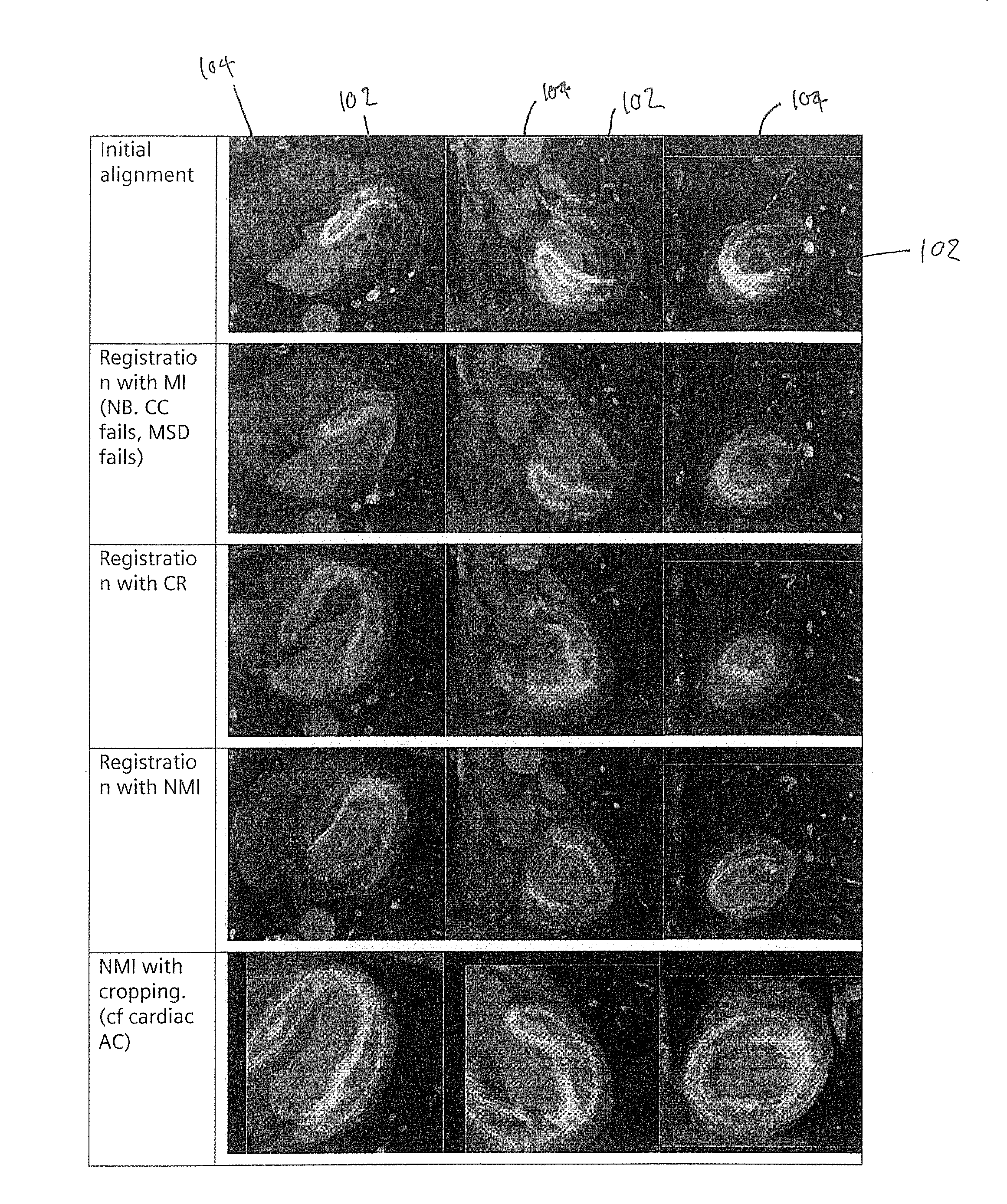
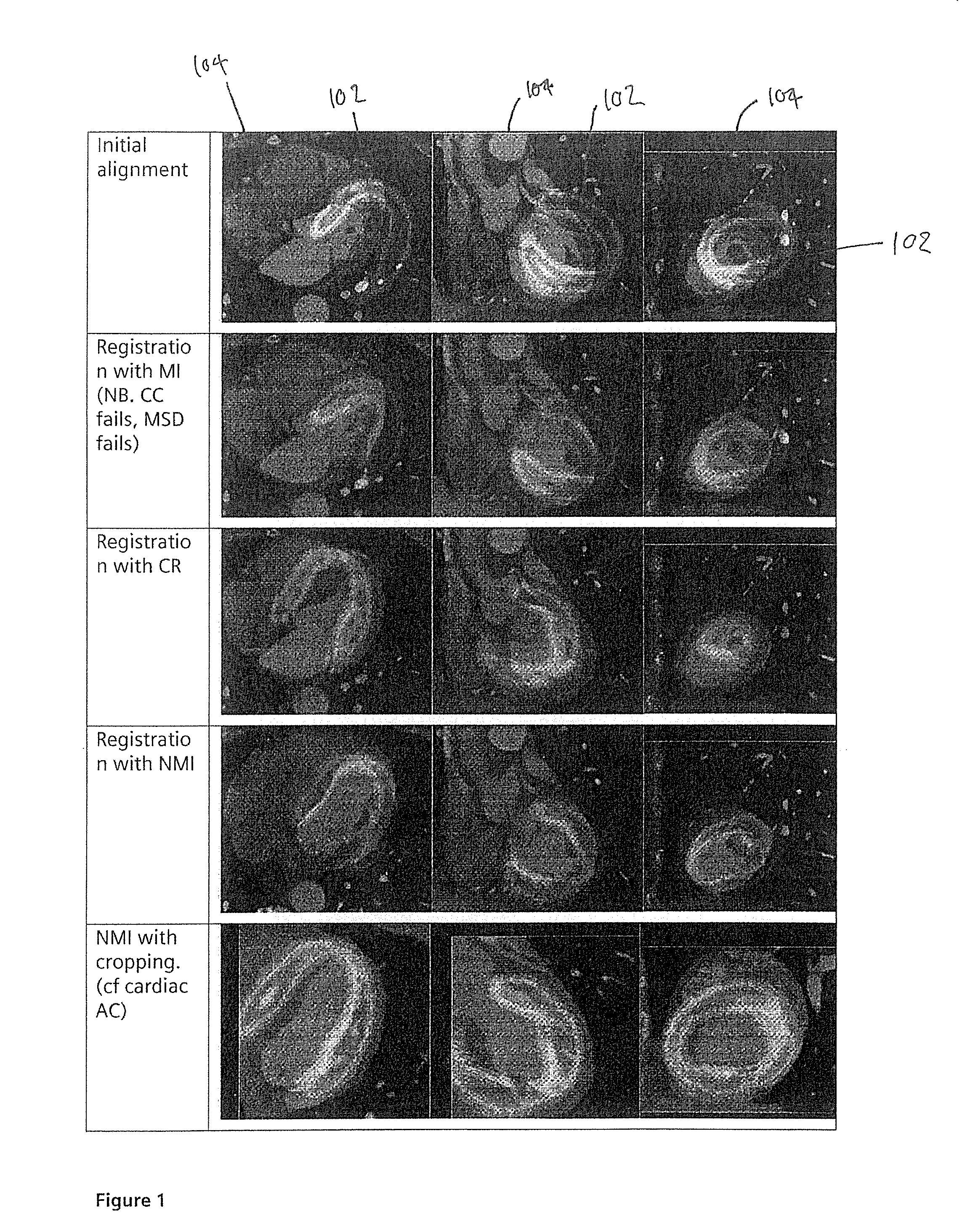
[0045]Embodiments of the invention provide methods which align the anatomical feature, such as the left ventricle, by axis and landmark points, rather than by mere comparison of data points, such as in mutual information based registration. This provides a more robust registration, as although the information in the voxels themselves in one modality may be of a different quality (for example, more blurred, if a PET image averaged over a time period and number of counts is used),...


PUM


Abstract

In a method and apparatus for aligning two sets of medical imaging data, first and second sets of image data are obtained respectively, using first and second different medical imaging modalities, of an anatomical feature of a subject. For each set, an axis of the anatomical feature and a landmark point for the anatomical feature is determined, and the first and second sets are aligned by comparing the respective axes and landmark points.

Description

BACKGROUND OF THE INVENTION[0001]1. Field of the Invention[0002]This invention is directed to methods and apparatus for aligning two sets of medical imaging data, in particular two sets, captured using different medical imaging modalities, showing an anatomical feature of a subject.[0003]2. Description of the Prior Art[0004]In the medical imaging field, several imaging schemes are known. For example PET (Positron Emission Tomography) is a method for imaging a subject in 3D using an injected radio-active substance which is processed in the body, typically resulting in an image indicating one or more biological functions. Other such functional imaging modalities are known, such as SPECT.[0005]In such functional images, many important pathologies and anatomical structures appear as very high (or low) intensities. For example, a tumor in an FDG-PET image will often appear as a bright region[0006]In cardiac medical imaging, CTA is a high resolution gated cardiac scan that enables the cli...

Claims


Application Information

Patent Timeline
IPC IPC(8): G06K9/32
CPCG06T7/0028G06T2207/10081G06T2207/30101G06T2207/10108G06T2207/10104G06T7/33G06T7/0014G06T2207/10088G06T2207/10101G06T2207/30048G06T2207/30056
Inventor BOND, SARAH
Owner SIEMENS MEDICAL SOLUTIONS USA INC
Why Eureka
Try Eureka