Method for correcting the image data that represent the blood flow

a blood flow and image data technology, applied in image data processing, surgical microscopes, sensors, etc., can solve problems such as optimal data sets to be used for reliable diagnosis, and achieve the effect of robust method for determining the shift vector

Inactive Publication Date: 2010-02-18
CARL ZEISS SURGICAL
View PDF4 Cites 20 Cited by
  • Summary
  • Abstract
  • Description
  • Claims
  • Application Information

AI Technical Summary

Benefits of technology

[0013]In one particularly advantageous embodiment, an edge image that has been updated using the edge images of the corrected individual images is generated as the reference image. Thus, only the significant components of the individual images enter into the reference image resulting in a reference image that provides a good overview over all blood vessels visible in the individual images and thus ensures a very robust method for the determination of the shift vector.

Problems solved by technology

This qualitatively allows data sets that are not optimal to be used for a reliable diagnosis.

Method used


Image

  • Method for correcting the image data that represent the blood flow
  • Method for correcting the image data that represent the blood flow
  • Method for correcting the image data that represent the blood flow

Examples


Embodiment Construction

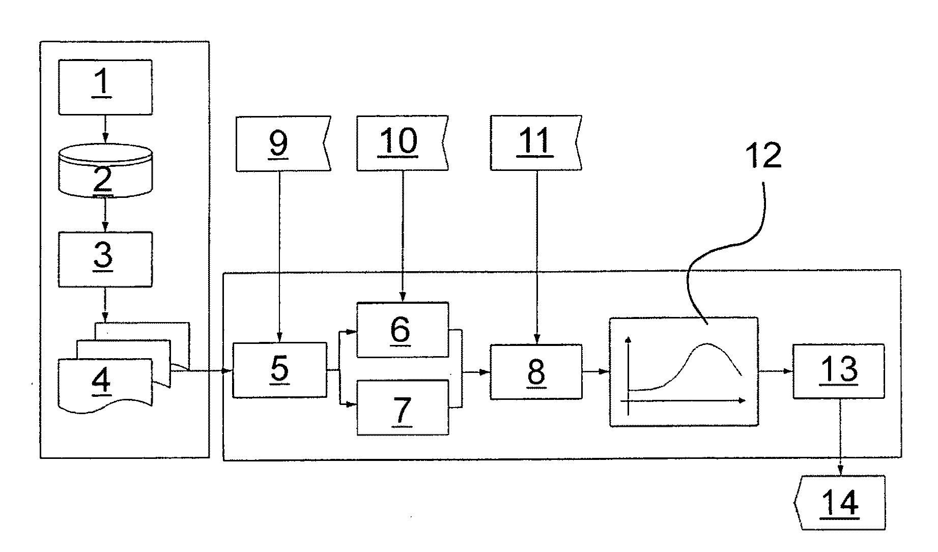
[0024]The preferred embodiments of the present invention will now be described with reference to FIGS. 1-7 of the drawings. Identical elements in the various figures are designated with the same reference numerals.

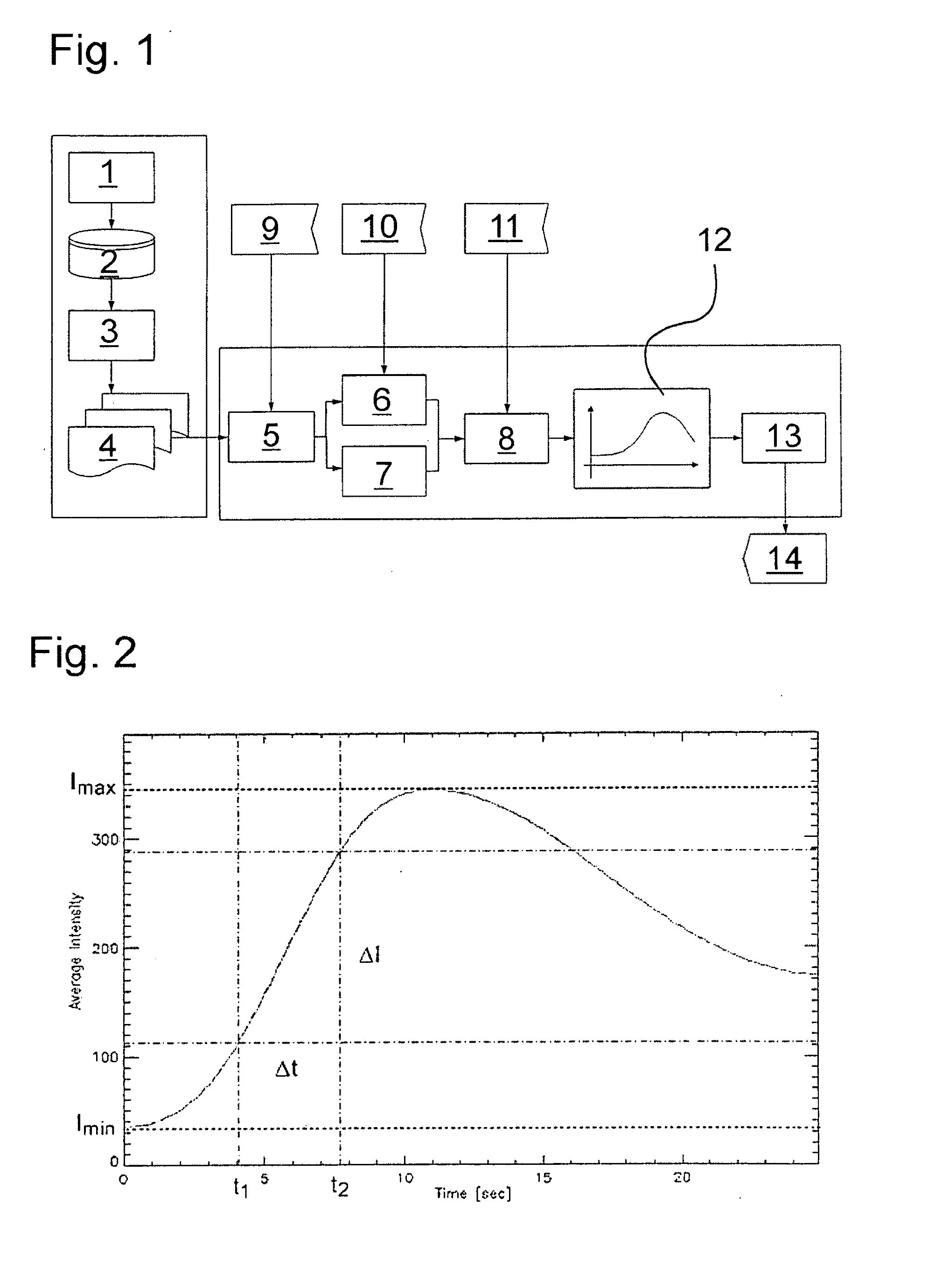
[0025]The complete system with the data flows and the individual processing steps is described in FIG. 1 and is used for representing and evaluating the blood flow. The data are recorded using a video camera 1 in the infrared range, which is arranged at the surgical microscope—not shown—or is a component thereof. The recorded infrared videos are stored in a data memory 2 and are split into individual images 4 using a video player 3. Alternatively, it is also possible to store the images of the video camera 1 directly as individual images 4. A frequency of five frames 4 per second proved to be useful. They are then corrected in a single image correction step 5. In the process, the corrections for the edge drop, the dark offset or of non-linearities of the video camera 1 are...


PUM


Abstract

A method for correcting the image data representing the blood flow for the evaluation and quantitative representation of the blood flow in a tissue or vascular region is based on the signal of a contrast agent injected into the blood. Several individual images of the signal emitted by the tissue or vascular region are recorded and stored at successive points in time. At least two individual images are correlated and a shift vector is generated based on the correlation. Thereafter, the image data of the individual images are shifted in relation to each other according to the shift vector.

Description

BACKGROUND OF THE INVENTION[0001]The invention relates to a method for correcting the image data that represent the blood flow in a patient.[0002]Several methods for observing and determining the blood flow in tissue and vascular regions are known in which a chromophore such as indocyanine green, for example, is applied. The fluorescent dye can be observed as it spreads in the tissue or along the blood vessels using a video camera. Depending on the area of application, the observation can be non-invasive or in the course of surgery, for example, via the camera of a surgical microscope.[0003]Many methods are known, where only the relative distribution of the fluorescent dye in the tissue or in the blood vessels is examined qualitatively in order to draw conclusions concerning their blood flow. For example, conclusions are made about the blood flow and diagnoses are provided by watching an IR video recorded during surgery. It is also known to record an increase in the brightness of th...

Claims


Application Information

Patent Timeline
Patent Type & Authority Applications(United States)
IPC IPC(8): A61B6/00G06K9/00
CPCA61B5/0261A61B5/0275A61B19/5223G06T2207/30104G06T7/0026G06T2207/10016G06T2207/10048G06T7/0012A61B90/20G06T7/32
Inventor SCHUHRKE, THOMASMECKES, GUENTER
Owner CARL ZEISS SURGICAL
Why Eureka
Try Eureka