Radiation imaging apparatus and control method thereof, and radiation imaging system

a control method and imaging apparatus technology, applied in the direction of material analysis using wave/particle radiation, instruments, applications, etc., can solve the problems of reducing the image quality of the x-ray image, affecting the effect of the main imaging, and affecting the transmission delay and propagation delay between the source controllers

Active Publication Date: 2013-05-16
FUJIFILM CORP
View PDF9 Cites 28 Cited by
  • Summary
  • Abstract
  • Description
  • Claims
  • Application Information

AI Technical Summary

Benefits of technology

[0011]An object of the present invention is to provide a radiation imaging apparatus that can always perform radiography with an optimal radiation dose with minimum time and effort, a control method of the radiation imaging apparatus, and a radiation imaging system having the radiation imaging apparatus.
[0023]According to the present invention, the automatic exposure control is performed in the preliminary imaging. The main imaging is carried out with the main irradiation time or the current-time product that is determined based on a result of the preliminary imaging. Thus, the present invention eliminates the need for measuring the thickness of a breast for the preliminary imaging, calculating a radiation dose, calculating an index value based on image data obtained in the preliminary imaging, and the like.
[0024]Therefore, it is possible to carry out the main imaging easily with an appropriate radiation dose.

Problems solved by technology

In the AEC, if the source controller delays performing X-ray stop operation, the image quality of the X-ray image is degraded and moreover the patient is exposed to an unnecessary dose.
However, in actual fact, the transmission delay and propagation delay between the source controller and the X-ray image detecting device tends to retard the X-ray stop operation.
However, the U.S. Pat. No. 7,639,779 takes time and effort to measure the thickness of the breast and the pressing pressure on the breast, for the purpose of determining the radiation dose for the preliminary imaging.

Method used


Image

  • Radiation imaging apparatus and control method thereof, and radiation imaging system
  • Radiation imaging apparatus and control method thereof, and radiation imaging system
  • Radiation imaging apparatus and control method thereof, and radiation imaging system

Examples


Embodiment Construction

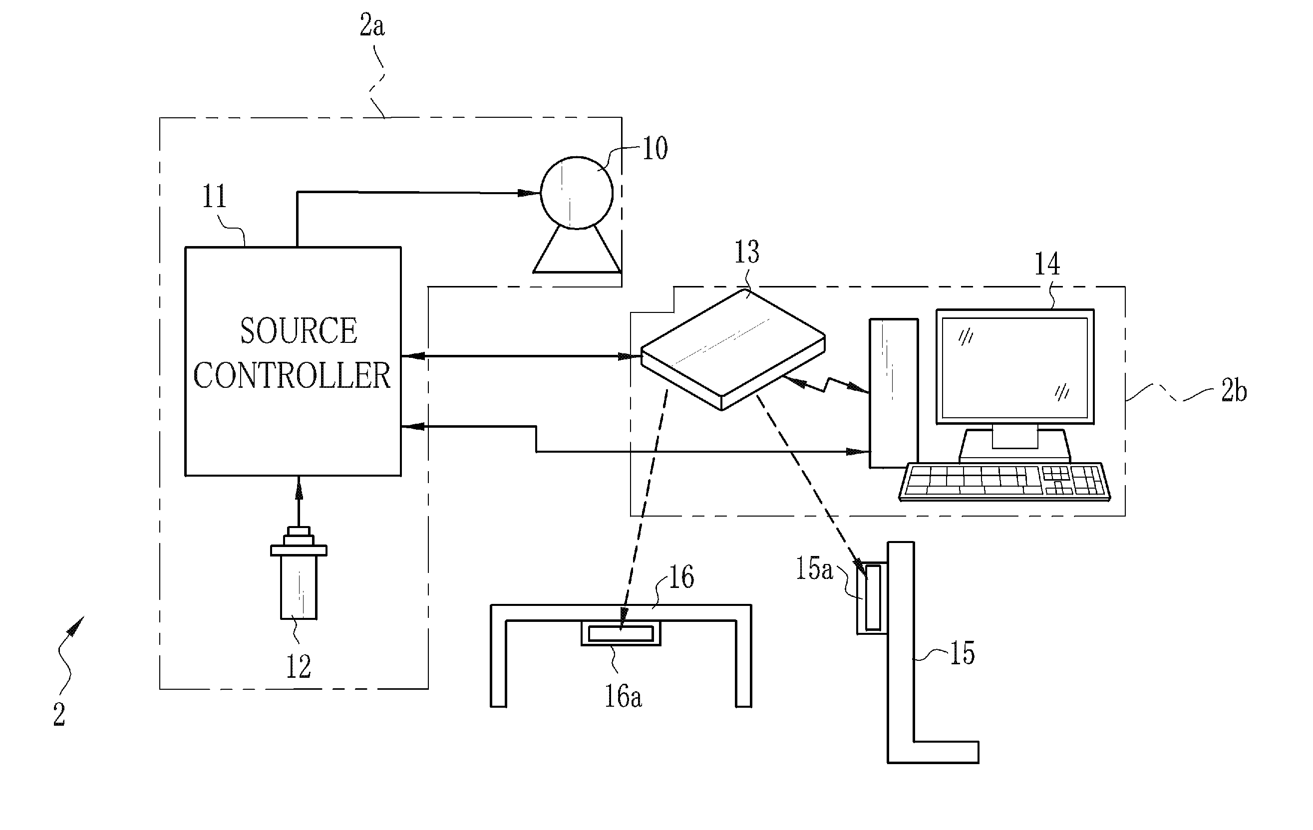
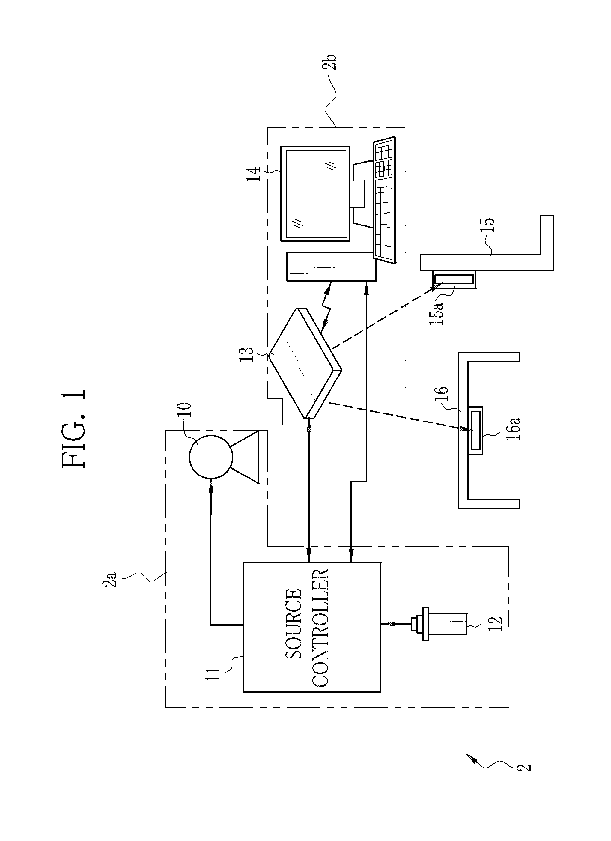
[0038]As shown in FIG. 1, an X-ray imaging system 2 is constituted of an X-ray source 10, a source controller 11, an emission switch 12, an electronic cassette 13, a console 14, and an imaging stand 15, and an imaging table 16. The X-ray source 10 contains an X-ray tube for emitting X-rays. The source controller 11 controls the operation of the X-ray source 10. The emission switch 12 commands the start of X-ray emission. The electronic cassette 13 detects the X-rays that have passed through an object e.g. a patient's body to output an X-ray image. The console 14 controls the operation of the electronic cassette 13, and performs image processing on the X-ray image. The imaging stand 15 and the imaging table 16 are used in performing radiography of the patient in a standing position and a lying position, respectively. In addition to above, the X-ray imaging system 2 has a source shift mechanism (not shown) for setting the X-ray source 10 in a desired orientation and position. Referenc...


PUM


Abstract

Besides normal pixels, a plurality detection pixels are arranged in an imaging surface of an FPD. In preliminary imaging, X-rays are emitted to an imaged body portion of a patient. The detection pixels receive the X-rays passed through the body portion, and output AEC detection signals. If an integral value of the AEC detection signals has reached a threshold value, X-ray emission is stopped and the preliminary imaging is completed. A main exposure condition determination unit determines a main irradiation time, being an irradiation time with the X-rays during the main imaging, based on an irradiation time with the X-rays during the preliminary imaging and the integral value of the AEC detection signals. The main imaging is performed using the main irradiation time. The normal pixels continue a charge accumulation operation over the preliminary imaging and the main imaging to produce an X-ray image for use in diagnosis.

Description

BACKGROUND OF THE INVENTION[0001]1. Field of the Invention[0002]The present invention relates to a radiation imaging apparatus that performs preliminary imaging and main imaging in sequence, a control method of the radiation imaging apparatus, and a radiation imaging system.[0003]2. Description Related to the Prior Art[0004]In a medical field, a radiation imaging system, for example, an X-ray imaging system using X-rays is widely known. The X-ray imaging system is constituted of an X-ray generating apparatus for generating the X-rays, and an X-ray imaging apparatus for taking an X-ray image by reception of the X-rays. The X-ray generating apparatus includes an X-ray source for emitting the X-rays to a patient's body, a source controller for controlling the operation of the X-ray source, and an emission switch for issuing an emission start command of the X-rays. The X-ray imaging apparatus includes an X-ray image detecting device for detecting the X-ray image based on the X-rays pass...

Claims


Application Information

Patent Timeline
Patent Type & Authority Applications(United States)
IPC IPC(8): A61B6/00
CPCA61B6/542A61B6/548A61B6/4283A61B6/4233
Inventor TAJIMA, TAKASHI
Owner FUJIFILM CORP
Why Eureka
Try Eureka