Apparatus and methods for characterization of lung tissue by raman spectroscopy

a lung tissue and raman spectroscopy technology, applied in the field of tissues, can solve the problems of many false positive identifications, high false positives, and often fatal lung cancer, and achieve the effect of fast and objective measures

Inactive Publication Date: 2013-09-05
BRITISH COLUMBIA CANCER AGENCY BRANCH
View PDF3 Cites 18 Cited by
  • Summary
  • Abstract
  • Description
  • Claims
  • Application Information

AI Technical Summary

Benefits of technology

[0039]Some embodiments of the invention provide methods and apparatus for acquiring and analyzing point Raman spectra to provide objective measures for evaluating tissues, for example, tissues at candidate locations in the lungs or bronchial tree. Some embodiments provide fast and objective measures of whether a lesion is preneoplastic, malignant or neither.

Problems solved by technology

Lung cancer is often fatal.
However, the average reported specificity of WLB+AFB is only 60% which leads to many false positive identifications as explained, for example, in: Lam S.
However, a main reason for the high number of false positives is the low specificity inherent with AFB.

Method used


Image

  • Apparatus and methods for characterization of lung tissue by raman spectroscopy
  • Apparatus and methods for characterization of lung tissue by raman spectroscopy
  • Apparatus and methods for characterization of lung tissue by raman spectroscopy

Examples


example application

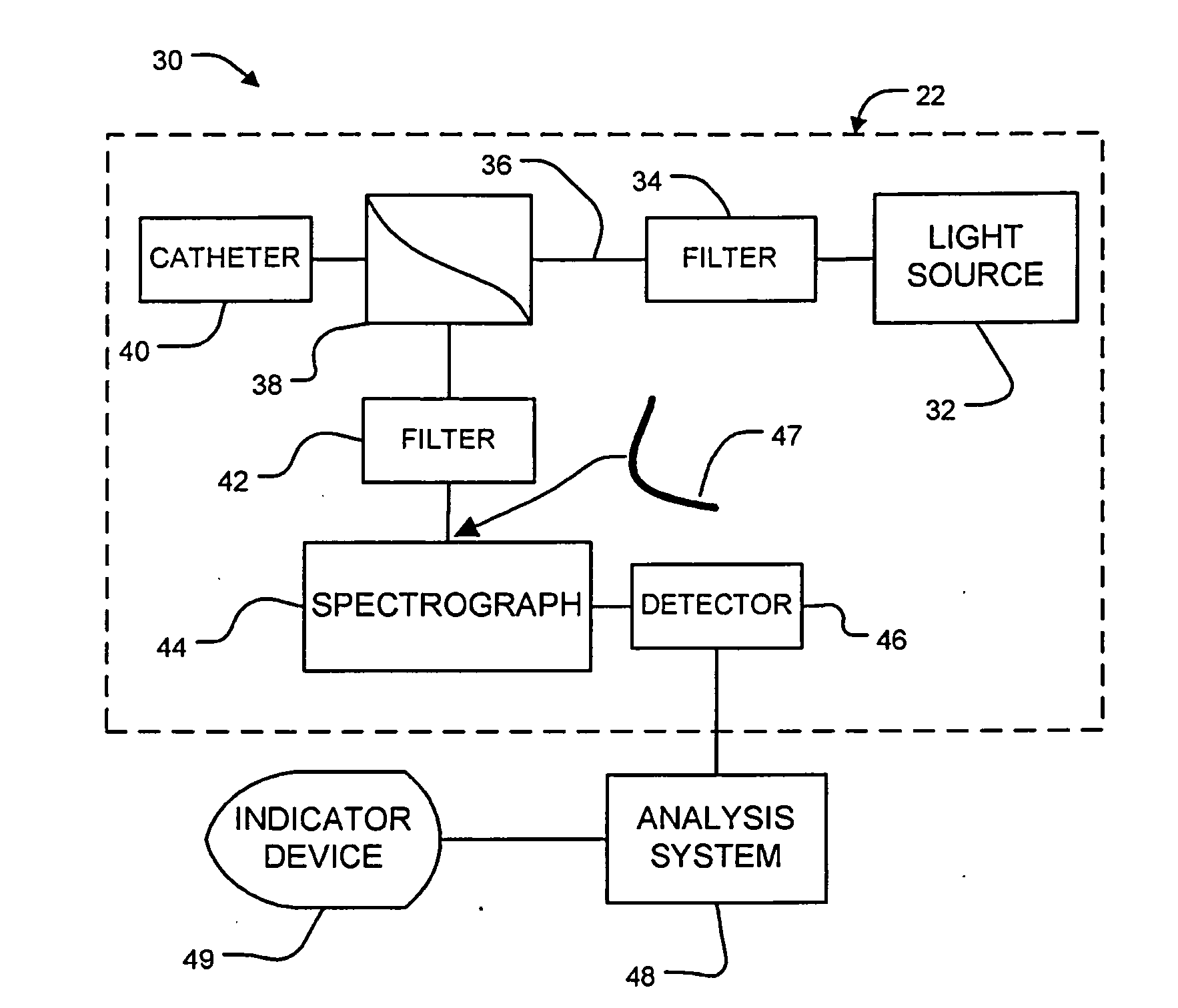
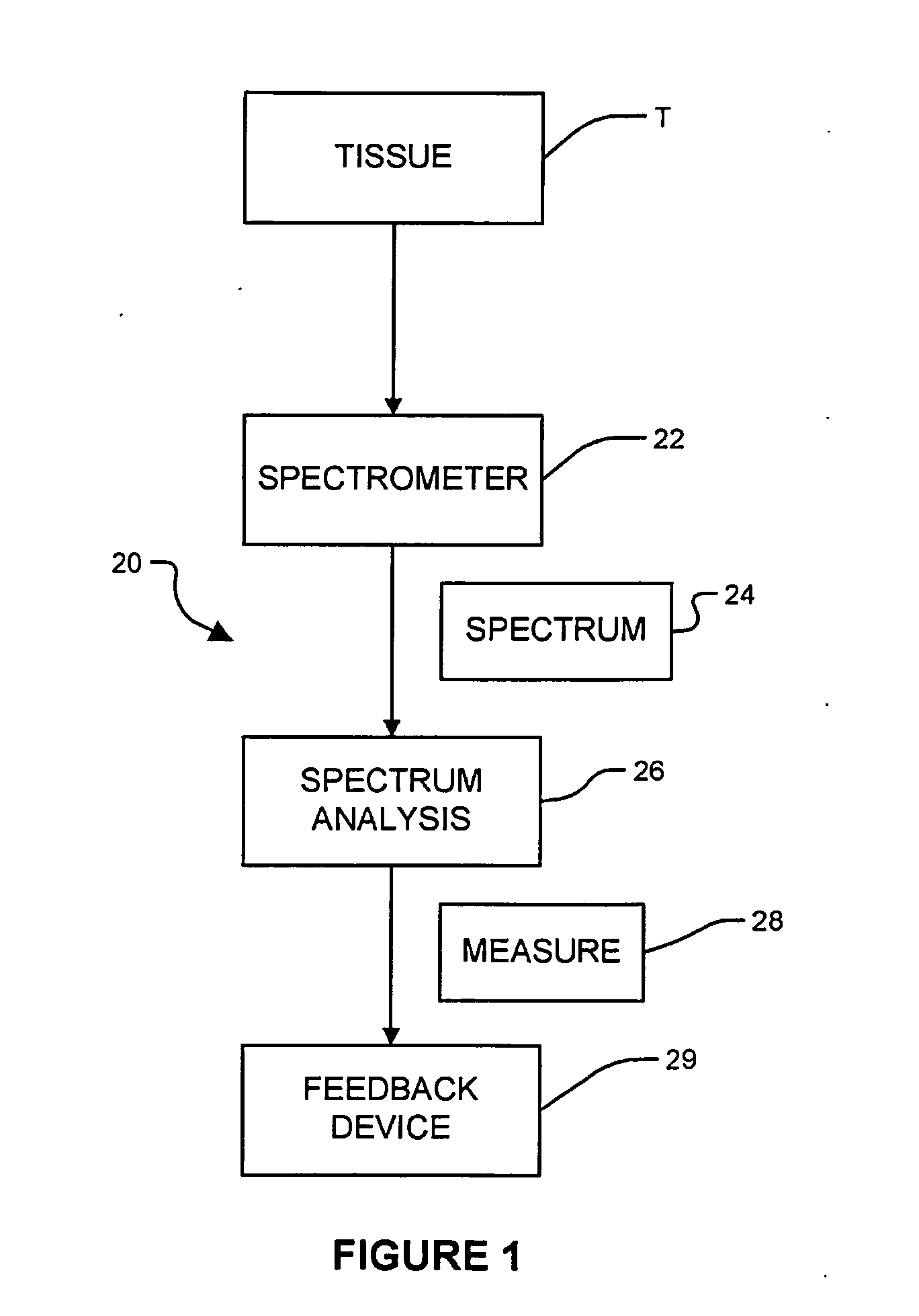
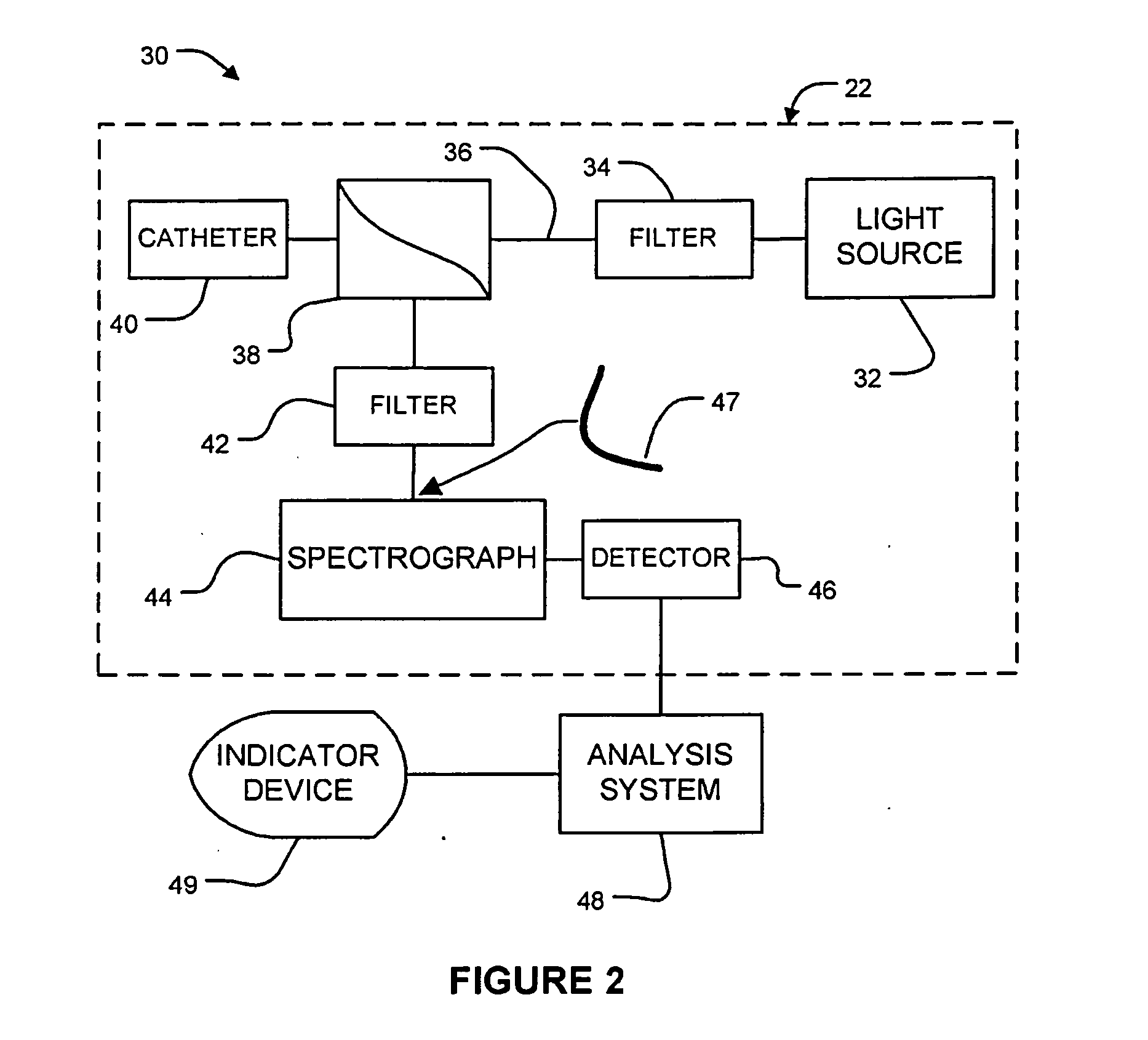
[0125]A bronchoscopist performs a bronchoscopy on a patient and uses a range of imaging modalities (for example AFB+WLB) to identify lesions that merit further investigation. The bronchoscopist is using a bronchoscope equipped with Raman spectroscopy apparatus as described herein. The bronchoscopist places the bronchoscope so that the end of the Raman catheter is adjacent to a lesion of interest and operates the Raman spectroscopy apparatus to acquire one or more Raman spectra for tissue in the lesion. The apparatus analyzes the Raman spectrum in real time and attempts to classify the tissue based on the spectrum. The apparatus generates a signal to the bronchoscopist based on the result of the analysis. As a simple example, the apparatus may display a green light if the analysis indicates a classification of ≦MILD and a red light if the analysis indicates a classification of ≧MOD. In some embodiments, the apparatus may indicate a yellow light if the classification cannot be establi...


PUM


Abstract

Near-infrared Raman spectroscopy can be applied to identify preneoplastic lesions of the bronchial tree. Real-time in vivo Raman spectra of lung tissues may be obtained with a fiber optic catheter passed down the instrument channel of a bronchoscope. Using prototype apparatus, preneoplastic lesions were detected with sensitivity and specificity of 96 and 91% respectively. The use of Raman spectroscopy apparatus and methods in conjunction with other bronchoscopy imaging modalities can substantially reduce the number of false positive results.

Description

CROSS-REFERENCE TO RELATED APPLICATION[0001]This application claims priority from U.S. patent application No. 61 / 297,486 entitled ENDOSCOPIC LASER RAMAN SPECTROSCOPY FOR IMPROVING LUNG CANCER DETECTION and filed on 22 Jan. 2010 and 61 / 390,723 entitled LASER RAMAN SPECTROSCOPY REDUCES FALSE POSITIVE BIOPSIES OF AUTOFLUORESCENCE BRONCHOSCOPY and filed on 7 Oct. 2010. For purposes of the United States, this application claims the benefit under 35 U.S.C. §119 of U.S. patent application No. 61 / 297,486 filed on 22 Jan. 2010 and No. 61 / 390,723 filed on 7 Oct. 2010, both of which are hereby incorporated by reference herein.TECHNICAL FIELD[0002]The invention relates to the characterization of tissues. The invention may be applied, for example, to provide methods and apparatus for assessing lung tissue for cancer. An example embodiment provides endoscopic apparatus which may be used by a physician to evaluate the likelihood that lesions in lung tissue are cancerous.BACKGROUND[0003]Lung cancer...

Claims


Application Information

Patent Timeline
Patent Type & Authority Applications(United States)
IPC IPC(8): A61B5/00
CPCA61B5/0071G01N21/65A61B5/0084A61B5/0075
Inventor ZENG, HAISHANSHORT, MICHAELLAM, STEPHENMCWILLIAMS, ANNETTE
Owner BRITISH COLUMBIA CANCER AGENCY BRANCH
Why Eureka
Try Eureka