
Portable x-ray computed tomography (CT) scanner

Inactive Publication Date: 2013-09-19
HALO IMAGING
View PDF3 Cites 14 Cited by
  • Summary
  • Abstract
  • Description
  • Claims
  • Application Information

AI Technical Summary

Benefits of technology

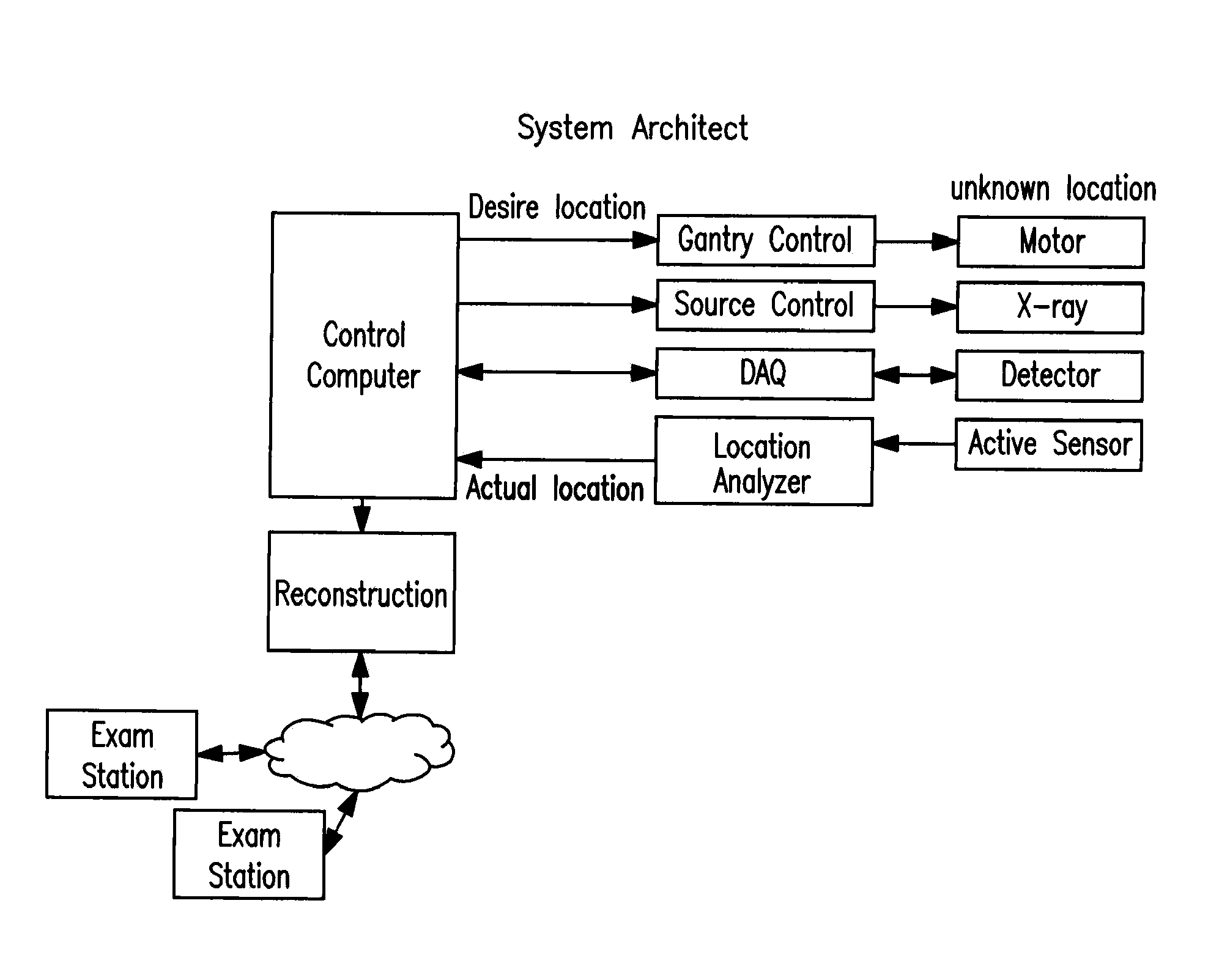
The present invention is about a mobile medical imaging system that can be easily moved around. It is a portable x-ray CT system that can provide useful images for diagnostic purposes.

Problems solved by technology

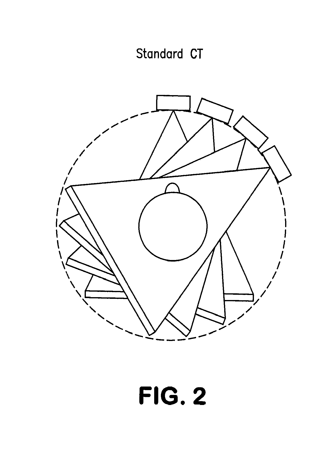
CT imaging is typically required to make the diagnosis, but the complexity and physical size of existing CT equipment often limits its availability and can cause delays in defining treatment strategies.
Smaller, so-called portable, CT scanners have been developed for use in the hospital ward or emergency room setting, but these are also massive, and are portable only in the sense that they have wheels.
They are not intended or capable of being used in mobile vehicles or moved outside the hospital environment.

Method used


Image

  • Portable x-ray computed tomography (CT) scanner
  • Portable x-ray computed tomography (CT) scanner
  • Portable x-ray computed tomography (CT) scanner

Examples


Embodiment Construction

[0027]The present invention provides a portable x-ray CT system as well as methods of use thereof. The invention addresses the need for a miniaturized mobile CT system which may be used, for example, in ground / air ambulances or field hospitals for imaging of the skull and cranial contents. The device allows for simple, quick scans which are compensated for movement artifact and transmitted electronically to local or regional medical facilities in advance of the patient's arrival. The information will significantly facilitate the development of a treatment plan for brain injury, which will save critical time once the patient reaches the hospital

[0028]Before the present device and method are described, it is to be understood that this invention is not limited to particular device and methodology described. It is also to be understood that the terminology used herein is for purposes of describing particular embodiments only, and is not intended to be limiting, since the scope of the pr...


PUM

PropertyMeasurementUnit
Weightaaaaaaaaaa
Lengthaaaaaaaaaa
Lengthaaaaaaaaaa

Abstract

The present invention provides a portable x-ray CT scanner for generating diagnostically meaningful images of a subject, such as a patient's cranium. The system includes a gantry ring which is configured to be portable or hand-held which is tether to a power supply. The device allows for simple, quick scans which are compensated for movement artifact and may then be transmitted electronically to local or regional medical facilities in advance of the patient's arrival.

Description

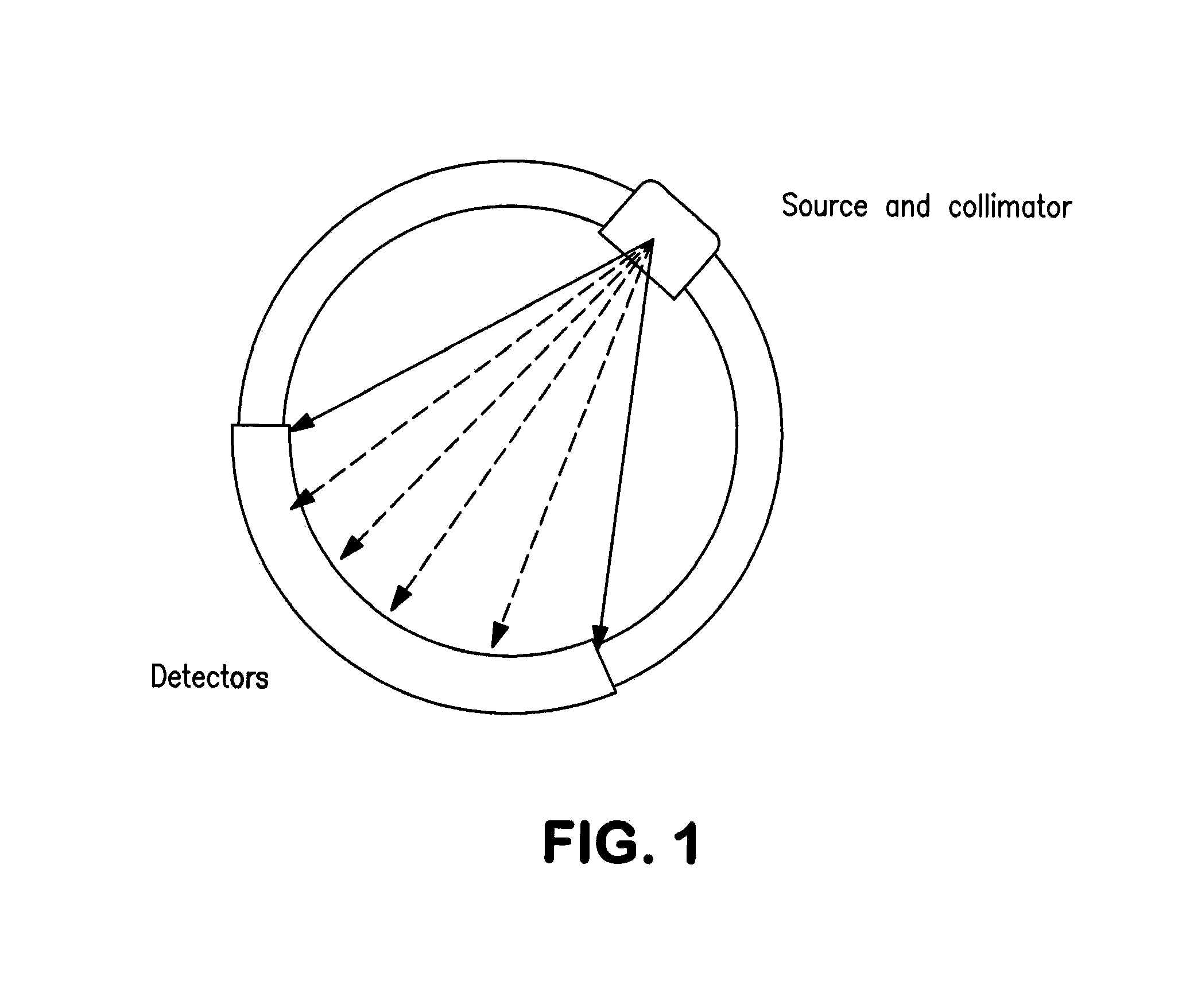
CROSS-REFERENCE TO RELATED APPLICATION(S)[0001]This application claims benefit of priority under 35 U.S.C. §119(e) of U.S. Ser. No. 61 / 604,363, filed Feb. 28, 2012, the entire contents of which are incorporated herein by reference in its entirety.BACKGROUND OF THE INVENTION[0002]1. Field of the Invention[0003]The present invention relates generally to medical imaging and more specifically to a mobile x-ray computed tomography (CT) scanning device.[0004]2. Background Information[0005]X-rays penetrating biological tissue are absorbed in proportion to the electron density of the tissue elements. Imaging media ranging from photographic film to solid-state electron-sensing materials placed on the opposite side of the tissue can sense the remaining signal and record the two-dimensional shadows of the tissue elements (FIG. 1). This is known as a plain x-ray (also called plane or planer x-ray) image and is characteristically used to diagnose bone trauma, pleural effusions and extravasated b...

Claims


Application Information

Patent Timeline
IPC IPC(8): A61B6/03
CPCA61B6/035A61B6/032Y10T29/49002Y10T29/49826A61B6/4405
Inventor SHIH, ANG
Owner HALO IMAGING
Learn More