

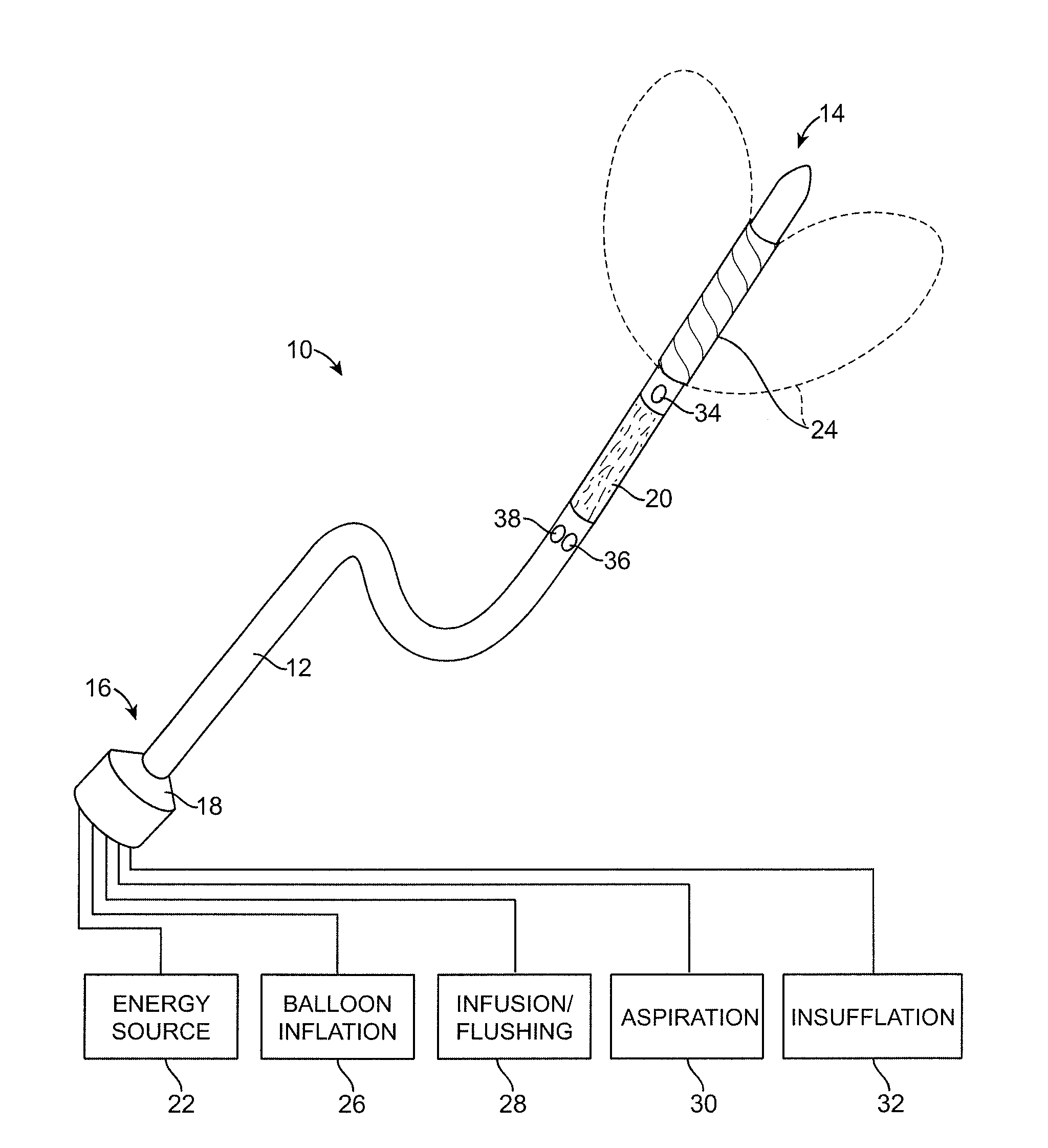
Minimally Invasive Devices for Multi-Fluid Tissue Ablation
a multi-fluid tissue and minimally invasive technology, applied in the field of medical methods and devices, can solve the problems of not disclosing patents that do not disclose the expansion of the urethra, and patents that do not disclose the use of fluid streams to resect non-coagulated tissue, etc., to reduce the need for catheterization, reduce the need for resection, and reduce the effect o
- Summary
- Abstract
- Description
- Claims
- Application Information
AI Technical Summary
Benefits of technology
Problems solved by technology
Method used
Image

Examples
example 1
Exemplary Critical Pressures of Different Kidney Tissue Compositions
[0076]Tissue critical pressures were measured in pig kidneys. Kidney tissue was chosen because its composition is similar to that of the prostate tissue. A columnar fluid stream of approximately 200 microns in diameter was used for tissue resection. The glandular tissue (the pink outer portion of the kidney) is very soft, and easily tears with finger pressure, while the inside of the kidney comprises tougher vascular tissue. The critical pressure for the glandular tissue with this fluid stream was found to be about 80 psi, and about 500 psi for the vascular tissue, as seen in Table 1 below.
TABLE 1Different critical pressures of glandularand vascular tissues in pig kidneyTissuePcrit (psi)Glandular80Vascular500
[0077]For example, experiments show that when resecting pig kidney using a nozzle of approximately 200 microns in diameter with liquid source pressure of about 500 psi, the rate of resection over a 10 cm2 area i...
example 2
Prostate Penetration Using a Diverging Fluid Stream
[0095]Measured data for resecting tissue of a canine prostate capsule are shown in FIG. 11. The penetration time through the capsule is measured as a function of tissue distance to the fluid delivery element. The angle of divergence of the fluid stream was approximately 7 degrees. The penetration time is plotted as the time to penetrate the capsule, with the capsule having a thickness of less than 1 mm.
[0096]FIG. 11 shows an increase in penetration time as the tissue distance to the fluid delivery element increases. It is noted that this effect is stronger at lower fluid source pressures. It is also noted that the penetration time of the columnar fluid stream is generally independent of tissue distance to the fluid delivery element.
example 3
Critical Pressures and Prostate Tissue Resection Using a Diverging Fluid Stream
[0097]The variation of critical pressure in diverging-fluid-stream resection as a function of different distances is shown in FIG. 12, as measured on canine prostate capsule tissue. The rate of resection was measured as the inverse of the time taken to resect (i.e., to penetrate) the entire thickness of the capsule. The rate of resection was measured as a function of fluid source pressure and tissue distance from the fluid delivery element. The rate of resection increases at higher pressures for greater distances from the fluid delivery element. This increase in rate of resection is indicative of the critical pressure. The increase in critical pressure as a function of distance shows that in a diverging fluid stream the resection effectiveness decreases with distance.
[0098]FIG. 13 illustrates the rate of resection of canine glandular tissue by a diverging fluid stream as a function of source pressure and ...
PUM
 Abstract
Description
Claims
Application Information