Ultrasound Probe for Laparoscopy

a technology of ultrasound and laparoscopy, applied in the field of ultrasound probe for laparoscopy, can solve the problems of limiting the use of intra-operative ultrasound, confusion, and difficulty, and achieve the effect of reducing the difficulty of ultrasound detection

Active Publication Date: 2013-12-19
SCHNEIDER CAITLIN MARIE +3
View PDF16 Cites 12 Cited by
  • Summary
  • Abstract
  • Description
  • Claims
  • Application Information

AI Technical Summary

Benefits of technology

[0010]In this embodiment of the mechanical interface, the grasper is oriented such that its jaws close over the probe, on the proximal side of the probe, nearest to the cable connection. The jaws of the grasper interact with mechanical stops on the grasping interlace, which, once engaged, prevent the grasper from dropping the probe.

Problems solved by technology

Unfortunately, during laparoscopic surgical procedures and in particular during robotic laparoscopic procedures, the ultrasound transducer is often controlled by an assistant.
This causes additional confusion as the ultrasound operator must position the ultrasound transducer to image tissue that is manipulated (retracted, cauterized, cut, sutured etc.) by the surgeon, and must follow the surgeon's verbal instructions or anticipate the surgeon's needs.
This is clearly difficult and limits the use of intra-operative ultrasound.

Method used


Image

  • Ultrasound Probe for Laparoscopy
  • Ultrasound Probe for Laparoscopy
  • Ultrasound Probe for Laparoscopy

Examples


Embodiment Construction

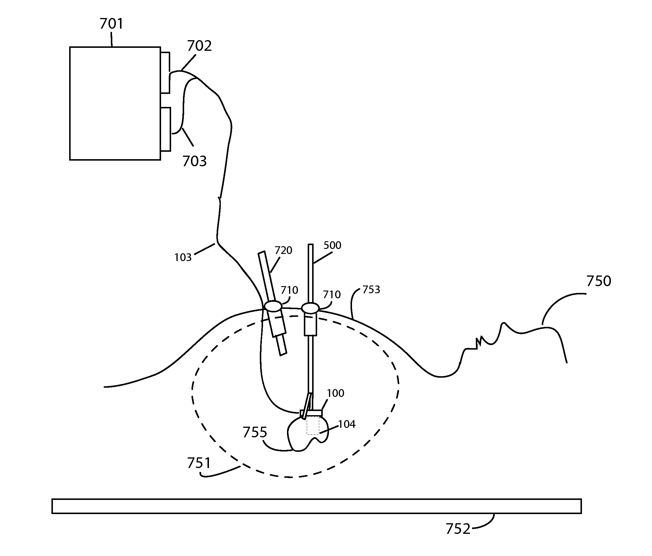
[0023]Detailed descriptions of embodiment of the invention are provided herein. It is to be understood, however, that the present invention may be embodied in various forms. Therefore, the specific details disclosed herein are not to be interpreted as limiting, but rather as a representative basis for teaching one skilled in the art how to employ the present invention in virtually any detailed system, structure, or manner. Prior disclosure of descriptions of the embodiment of the invention was made in the publication Caitlin Schneider, Julian Guerrero, Christopher Y. Nguan, Robert Rohling, Septimiu E. Salcudean: Intra-operative “Pick-Up” Ultrasound for Robot Assisted Surgery with Vessel Extraction and Registration: A Feasibility Study”, was made in the 2nd International Conference on Information Processing in Computed Assisted Interventions”, page 122-132, Jun. 22, 2011, the entirety of which is hereby incorporated by reference.

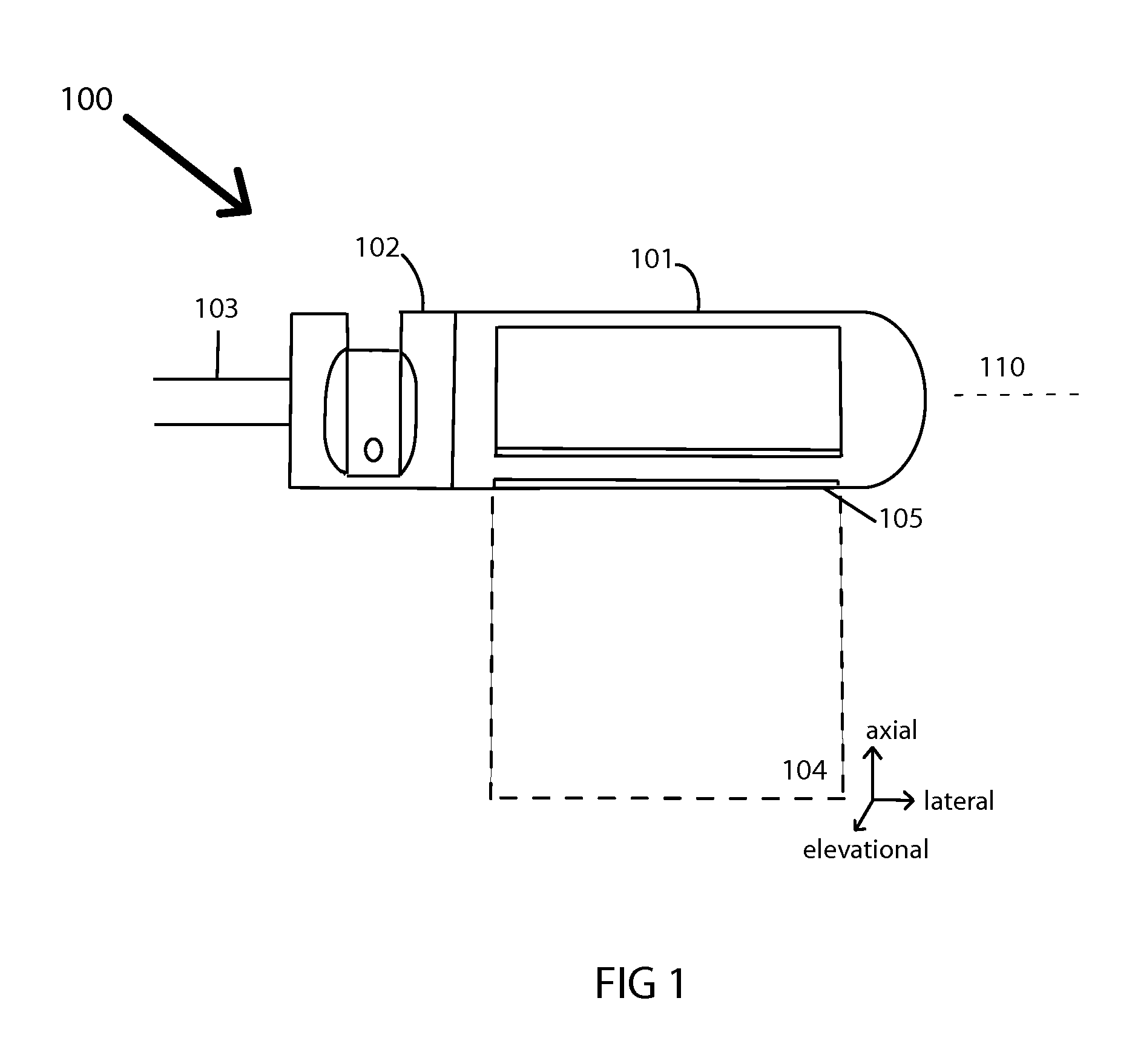
[0024]FIG. 1 shows a side view of an intra-operative ul...


PUM


Abstract

An intra-operative ultrasound probe for use with a robotic and laparoscopic surgical systems that allows for direct surgeon control over the position and orientation of the ultrasound image is presented. The transducer is designed to interface with the laparoscopic grasper so that it is easy to pick up in a locking, self-aligning and repeatable manner. The transducer is tracked in space using either forward kinematics or electromagnetic sensing, allowing multiple 2D images to be combined in order to create 3D ultrasound volumes. The 3D volumes can be further processed and displayed on the surgeon's console, or used to register and display acquired pre-operative images at the correct spatial location within the patient.

Description

FIELD OF THE INVENTION[0001]The present invention relates to the integration of ultrasound into a surgical robotic system, and is useful during minimally invasive procedures.BACKGROUND OF THE INVENTION[0002]Minimally invasive surgery (“MIS”) minimizes the overall trauma of a surgery by performing the procedure through small incisions in the abdominal wall. In comparison to traditional open surgical techniques, MIS offers the patient minimal tissue damage, reduced recovering time and quicker return to normal activities.[0003]One type of MIS is laparoscopy, in which the surgeon performs the operation through small incisions into the abdominal, thoracic or pelvic cavities. Long instruments and a camera are inserted through these incisions and grant the surgeon access to the patient's internal organs with much reduced trauma to the body. An absorbable gas, such as carbon dioxide is used to distend the patient's cavity and provide a larger working space. Many types of surgical procedures...

Claims


Application Information

Patent Timeline
Patent Type & Authority Applications(United States)
IPC IPC(8): A61B8/13
CPCA61B8/4218A61B8/4455A61B6/5247A61B8/12A61B2017/00283A61B2017/2926A61B1/3132A61B1/00193
Inventor SCHNEIDER, CAITLIN MARIESALCUDEAN, SEPTIMIU E.ROHLING, ROBERT N.NGUAN, CHRISTOPHER YEE CHAUNG
Owner SCHNEIDER CAITLIN MARIE
Why Eureka
Try Eureka