Apparatus and methods for anchoring electrode leads adjacent to nervous tissue

Active Publication Date: 2013-12-19
MAINSTAY MEDICAL
View PDF14 Cites 57 Cited by
  • Summary
  • Abstract
  • Description
  • Claims
  • Application Information

AI Technical Summary

Benefits of technology

[0015]The present invention overcomes the drawbacks of previously-known by providing apparatus for neuromuscular electrical stimulation including an elongated member having a proximal region and a distal region, one or more electrodes disposed at the distal region of the elongated member, and first and/or second fixation elements disposed at the distal region of the elongated member. The first fixation element may be shaped and sized to contact, but not penetrate, tissue (e.g., a muscle, ligament, tendon, fascia) within a patient without damaging the tissue, so as to secure the one or more electrodes in or adjacent to a desired anatomical s

Problems solved by technology

One drawback of a percutaneous approach is that relatively large and complex anchoring mechanisms cannot be deployed through the incision or using a needle, catheter, or cannula.
Additionally, in many cases, there is no convenient anatomical structure to which the medical device may be anchored.
Small changes in electrode position may in some cases adversely impact the system's ability to effectively deliver therapy.
It may not be practical or feasible to provide an anchoring mechanism inside the spinal canal to anchor a lead of the SCS.
This technique, while in common use, suffers from drawbacks including significant incidence of lead dislodgement.
Another drawback is that the superficial tissue is often an undesirable distance from the target tissue of stimulation.
Any change in patient posture which results in a change in the relative distance between th

Method used


Image

  • Apparatus and methods for anchoring electrode leads adjacent to nervous tissue
  • Apparatus and methods for anchoring electrode leads adjacent to nervous tissue
  • Apparatus and methods for anchoring electrode leads adjacent to nervous tissue

Examples


Embodiment Construction

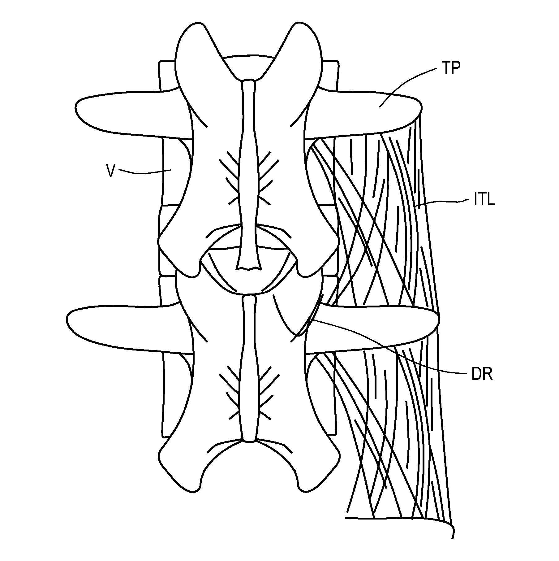
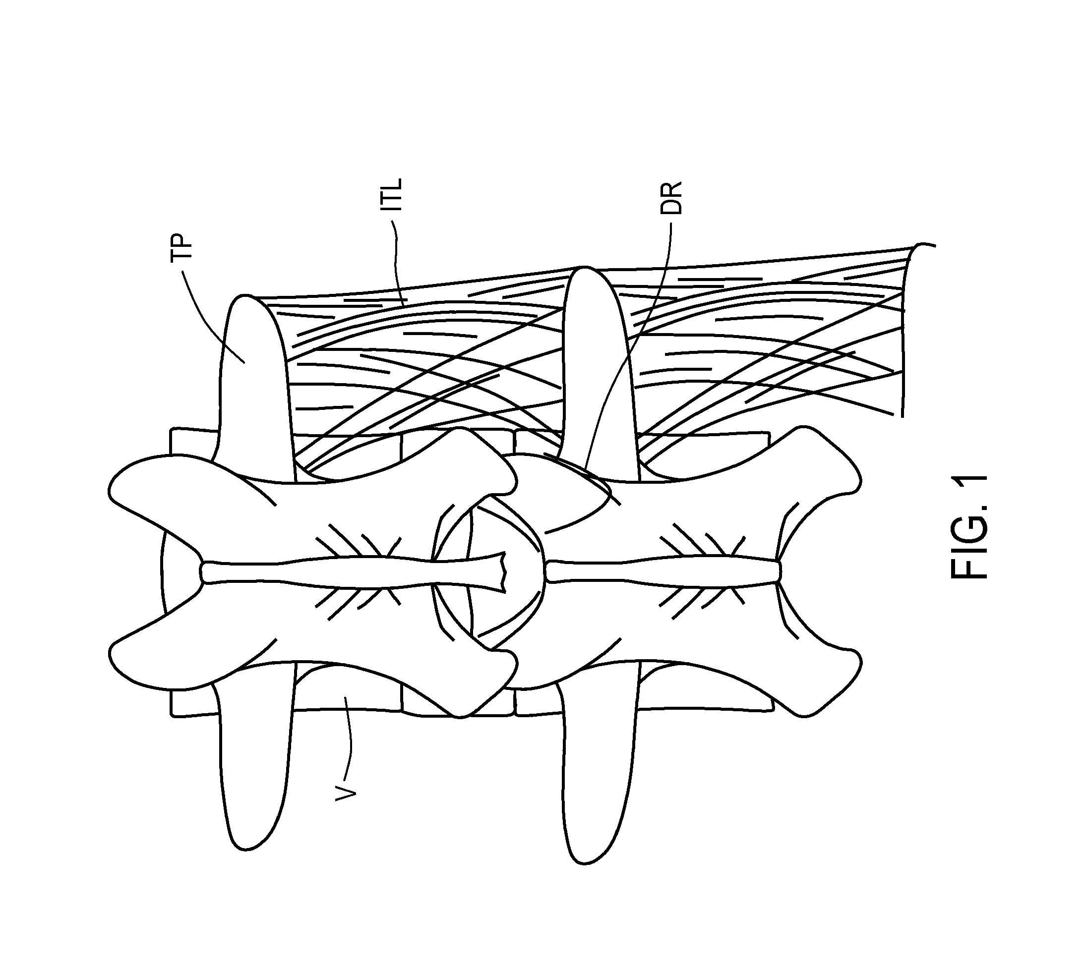
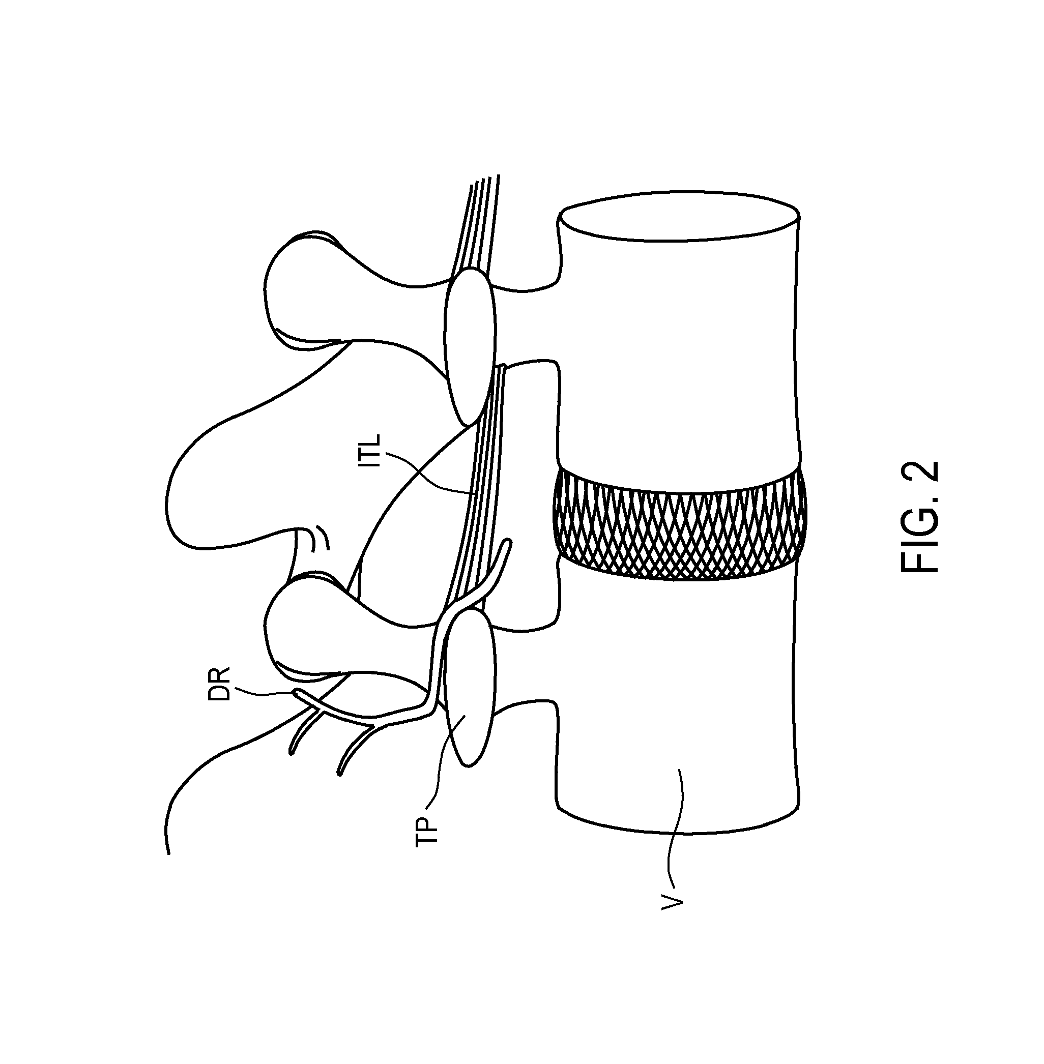
[0039]The present invention is directed to methods and apparatus for anchoring electrode leads suitable for use with an implantable neuromuscular electrical stimulation (“NMES”) device, such as described in the above-incorporated U.S. Patent Application Publication Nos. 2008 / 0228241 to Sachs and 2011 / 0224665 to Crosby. The devices described in those applications supply electrical pulses to nerves innervating the spinal muscles, such as the multifidus muscles, and stimulate the nerves controlling those muscles to effect a therapy designed to restore neural control and rehabilitation of the muscle. The implantable stimulator is disposed subcutaneously, and is coupled to one or more electrode leads having electrodes in contact with the target muscle, or nerves innervating the target muscles, or other anatomical structures associated with the muscle, such as ligaments and tendons. The NMES stimulation supplied by the stimulator applies a pulse regime that is very different than those em...


PUM


Abstract

Apparatus for neuromuscular electrical stimulation and methods for anchoring the same are provided. The apparatus may include an elongated member having one or more electrodes disposed at the distal region of the elongated member and at least one fixation element disposed at the distal region of the elongated member. The fixation element may be shaped and sized to be deployed between tissue layers, such as muscle layers, without damaging the tissue layers so as to secure the one or more electrodes in or adjacent to a desired anatomical site within a patient. An additional fixation element may be disposed at the distal region of the elongated member so that tissue, such as a muscle, may be sandwiched between the fixation elements without damaging the tissue.

Description

I. CROSS-REFERENCE TO RELATED APPLICATIONS[0001]This application claims the benefit of priority of U.S. Provisional Application Ser. No. 61 / 659,334, filed Jun. 13, 2012.II. FIELD OF THE INVENTION[0002]This application generally relates to apparatus and methods for anchoring a medical device, such as an electrical stimulation lead, catheter or other generally elongated or tubular device in the body.III. BACKGROUND OF THE INVENTION[0003]Many medical devices incorporate an elongated or tubular element that is required to be positioned at a particular anatomical site. Such devices include pacemakers, spinal cord stimulators, peripheral nerve stimulators, and drug delivery catheters.[0004]In the case of a pacemaker, for example, the leads may be threaded through a vein, and then anchored using a fixation element at the distal tip of the lead to prevent dislodgement. Such a fixation element may be a tine, fin, or screw that is secured in the trabeculae or muscle tissue of the ventricle.[0...

Claims


Application Information

Patent Timeline
IPC IPC(8): A61N1/05
CPCA61N1/0558A61N1/36057A61N1/36071
Inventor SHIROFF, JASON ALANDEMORETT, HENRY THOMASRAWAT, PRASHANT BRIJMOHANSINGHHEEMELS, JOHANNES PETRUSCROSBY, PETER ANDREW
Owner MAINSTAY MEDICAL
Why Eureka
Try Eureka