Visualization and characterization of pulmonary lobar fissures

a technology visualization and characterization, which is applied in the field of visualization and characterization of pulmonary lobar fissures, can solve the problems of difficult to accurately detect and characterize lobar fissures in diseased lungs, difficult to fully utilize the anatomic information available in image data, and risky patients, etc., and achieves easy consumption and understanding. , the effect of easy consumption

Active Publication Date: 2014-04-17
VIDA DIAGNOSTICS
View PDF1 Cites 48 Cited by
  • Summary
  • Abstract
  • Description
  • Claims
  • Application Information

AI Technical Summary

Benefits of technology

[0018]Using objective, quantitative measures of disease obtained from imaging modalities like computerized tomography (CT) in routine clinical practice requires providing accurate and immediate information to a physician in a busy practice. Anatomic structural deformities in lung fissure can be more easily understood within a visual context. Embodiments of the invention therefore provide easy and immediate access to pertinent measures of disease and enhanced multidimensional visual models, for easy consumption by the clinician such as the pulmonary physician.

Problems solved by technology

Current treatment methods for severe emphysema include lung volume reduction (LVR) surgery, which is highly invasive, and can be risky and uncomfortable for the patient.
However, accurately detecting and characterizing fissures in diseased lungs is difficult.
However, neither of these approaches makes full use of the anatomic information available in the image data.

Method used


Image

  • Visualization and characterization of pulmonary lobar fissures
  • Visualization and characterization of pulmonary lobar fissures
  • Visualization and characterization of pulmonary lobar fissures

Examples


Embodiment Construction

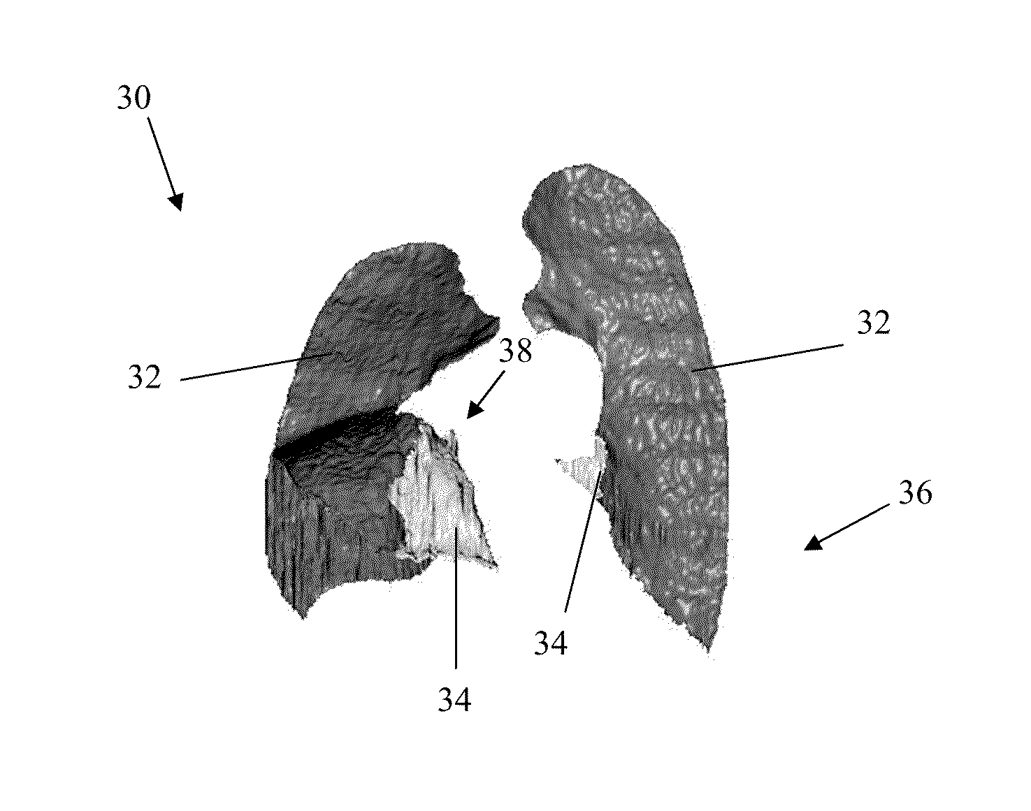
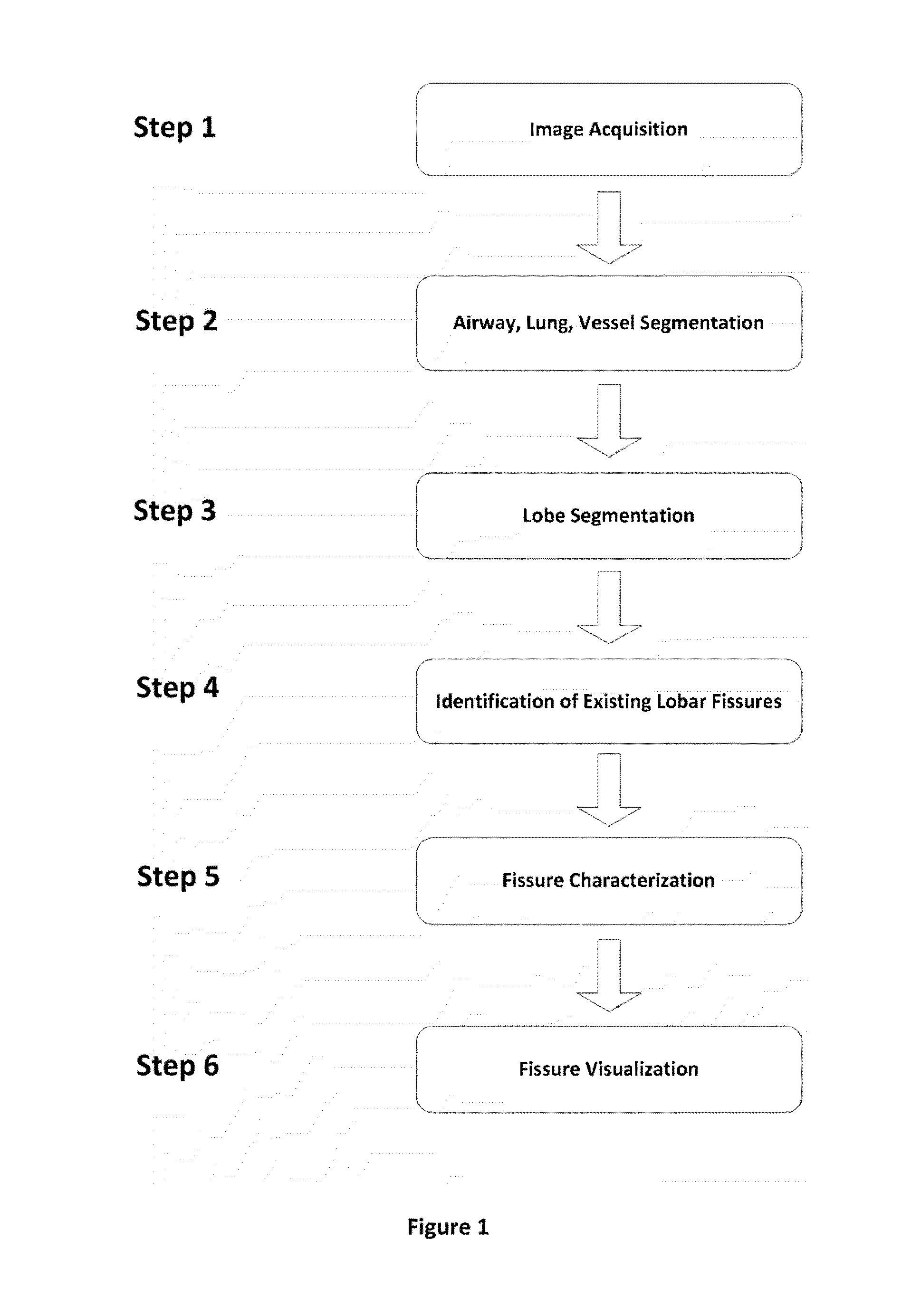
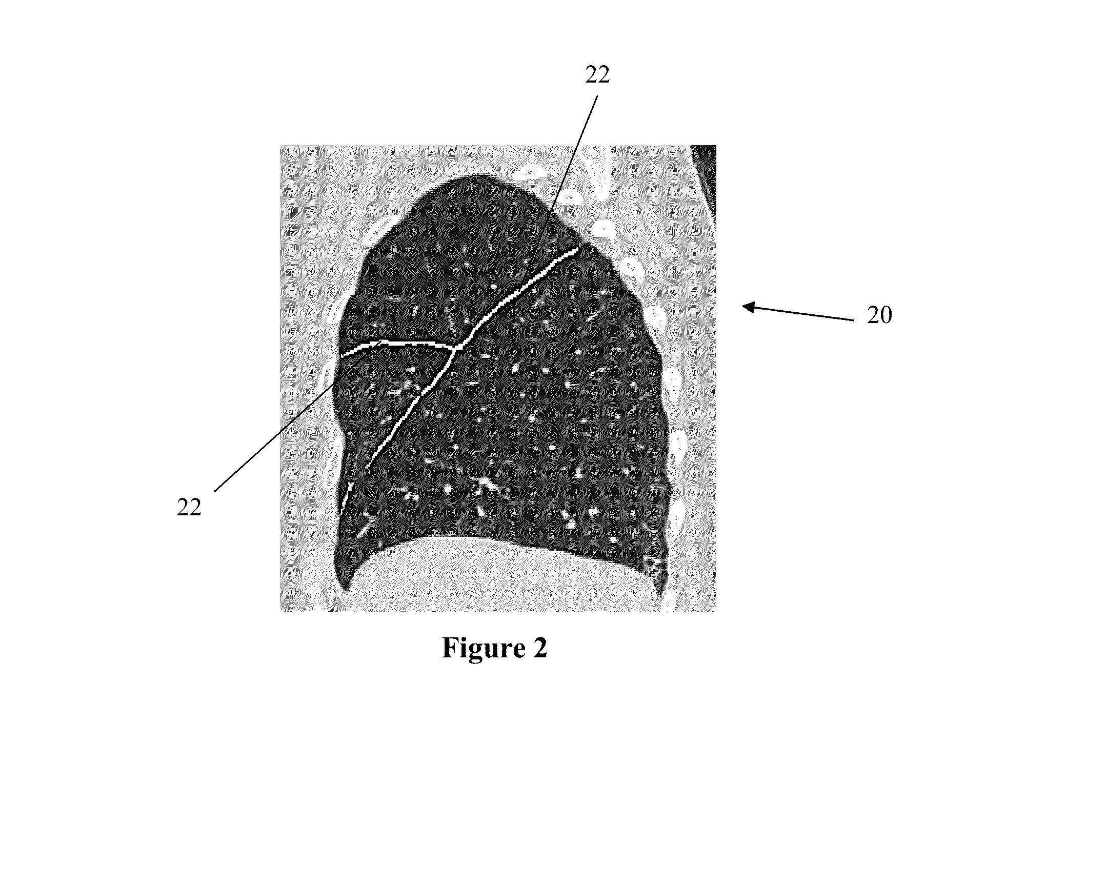
[0030]The invention describes a process to automate, display, interact with and characterize the fissures of the lung in multiple dimensions. When the human lung is imaged in vivo with an imaging acquisition device, like CT, that image can be reconstructed and evaluated to depict normal and diseased states. Because of the various subclasses of disease and the various depictions (phenotypes) of a disease entity, evaluation of lobular regions of the lung and the fissures separating them are important to accurately characterize disease and predict response to BLVR therapy.

[0031]This disclosure includes methods to provide visualization of the fissures in two and three dimensions, define the fissure boundaries, characterize their morphologic characteristics which may be used for identifying a disease phenotype, and visualize regions of intact and missing fissures, and observe the difference between normal and diseased lung in an instantaneous and automated way to enable clinical decision...


PUM


Abstract

Systems and methods for visualizing pulmonary fissures including a processor and software instructions for creating a 3 dimensional model of the fissures. Creating the 3 dimensional model includes accessing volumetric imaging data of the patient's lungs, analyzing the volumetric imaging data to segment the lungs into lobes, using the segmented lobes to identify locations at which pulmonary fissures should be present where the lobes abut each other, analyzing the volumetric images to identify locations at which pulmonary fissures actually are present as existing fissure, comparing the locations at which pulmonary fissures should be present to the locations at which pulmonary fissures are present to identify locations of missing fissure, and creating a visual display comprising a 3 dimensional model of the pulmonary fissures including existing fissure portions and missing fissure portions, with the existing fissure portion visually distinct from the missing fissure portions.

Description

PRIORITY CLAIM[0001]This application claims priority to U.S. Provisional Application No. 61 / 712,700, filed Oct. 11, 2012 and entitled Visualization and Characterization of Pulmonary Lobar Fissures, the disclosure of which is hereby incorporated by reference.FIELD OF THE INVENTION[0002]Embodiments of the invention generally relate to visualization and characterization of pulmonary lobar fissures.BACKGROUND OF THE INVENTION[0003]Severe emphysema is a debilitating disease that limits quality of life of patients and represents an end state of Chronic Obstructive Pulmonary Disease (COPD). It is believed that 3.5 million people in the US have the severe emphysematous form of COPD, and it is increasing in both prevalence and mortality. Current treatment methods for severe emphysema include lung volume reduction (LVR) surgery, which is highly invasive, and can be risky and uncomfortable for the patient. New treatment methods for treating emphysema include bronchoscopy guided LVR (BLVR) devi...

Claims


Application Information

Patent Timeline
Patent Type & Authority Applications(United States)
IPC IPC(8): A61B5/08
CPCA61B5/08G06T7/0012A61B6/032G06T19/20G06T2200/24G06T2207/10072G06T2207/30061G06T2210/41G06T2219/2012
Inventor YIN, YOUBINGNEWELL, JR., JOHN D.RAFFY, PHILIPPEWOOD, SUSAN A.
Owner VIDA DIAGNOSTICS
Why Eureka
Try Eureka