
Automated analysis of multi-lead electrocardiogram data to identify the exit sites of physiological conditions

a multi-lead electrocardiogram and analysis technology, applied in the field of automatic analysis of multi-lead electrocardiogram data, can solve the problems of heart muscle tissue damage or death (infarction), myocardium, life-threatening arrhythmias, etc., to improve the accuracy of vt exit site measurement, increase sensitivity and specificity, and enhance the effect of ecg determination

Inactive Publication Date: 2014-04-17
RGT UNIV OF MICHIGAN
View PDF6 Cites 20 Cited by
  • Summary
  • Abstract
  • Description
  • Claims
  • Application Information

AI Technical Summary

Benefits of technology

This patent describes methods for identifying the location where ventricular arrhythmias start in the heart. These methods use a special electrocardiogram (EKG) that can be done before or during a medical procedure on a patient. The EKG helps to create a 3D map of the heart, which is then used to determine the specific areas where arrhythmias can start. This helps with diagnosis and treatment of the condition. The patent also includes algorithm-based techniques that improve the accuracy of identifying the origin of arrhythmias. Overall, this patent provides a better way to locate and treat ventricular arrhythmias, which can help to improve patient outcomes.

Problems solved by technology

The resulting restriction in blood supply and ensuing oxygen shortage, if left untreated for a sufficient period of time, can cause damage or death (infarction) of heart muscle tissue (myocardium).
The presence of scar tissue can cause life threatening arrhythmias.
While VT is a regular, albeit fast regular rhythm, if left untreated, some forms of VT may get worse and lead to ventricular fibrillation, which is a fast and irregular beating that can result in death.
In ventricular fibrillation, the heart beats are so fast and irregular that the heart stops pumping blood, which can causes cardiovascular collapse and death.
While most often these PVCs are infrequent, they can occur frequently and may cause cardiomyopathy (weakening of the heart muscle).
While there are techniques to assess the origin of VT, the techniques are limited in pinpointing, with sufficient accuracy, the particular tissue or regions of the heart that cause the VT.

Method used


Image

  • Automated analysis of multi-lead electrocardiogram data to identify the exit sites of physiological conditions
  • Automated analysis of multi-lead electrocardiogram data to identify the exit sites of physiological conditions
  • Automated analysis of multi-lead electrocardiogram data to identify the exit sites of physiological conditions

Examples


example

Patient Characteristics

Table 1

[0021]Table 1 provides characteristics of the 34 patients examined in an example study for training data purposes. Mapping and radiofrequency ablation were performed in a consecutive series of 34 patients (31 males, age: 68±9 years, ejection fraction: 0.25±0.12) with post-infarction VT. Left ventricular pace-mapping was performed in these patients. Eighteen patients (53%) had a prior inferior, 9 (26%) had an anterior and 7 (21%) had an inferior and anterior wall myocardial infarction. Pace-maps were collected in these patients and used as training data. The training data were subsequently (see below) validated in another 33 consecutive post-infarction patients (32 males, age 67±10 years, ejection fraction: 0.26±0.12).

TABLE 1Characteristics of Patients used for Training and Validation of DataValidationVariableTraining datadatap-valuePatients, n3433Induced VTs, n8 ± 48 ± 50.71Age, years68 ± 9 67 ± 100.63Male / female, n31 / 332 / 10.61Left ventricular ejection ...


PUM


Abstract

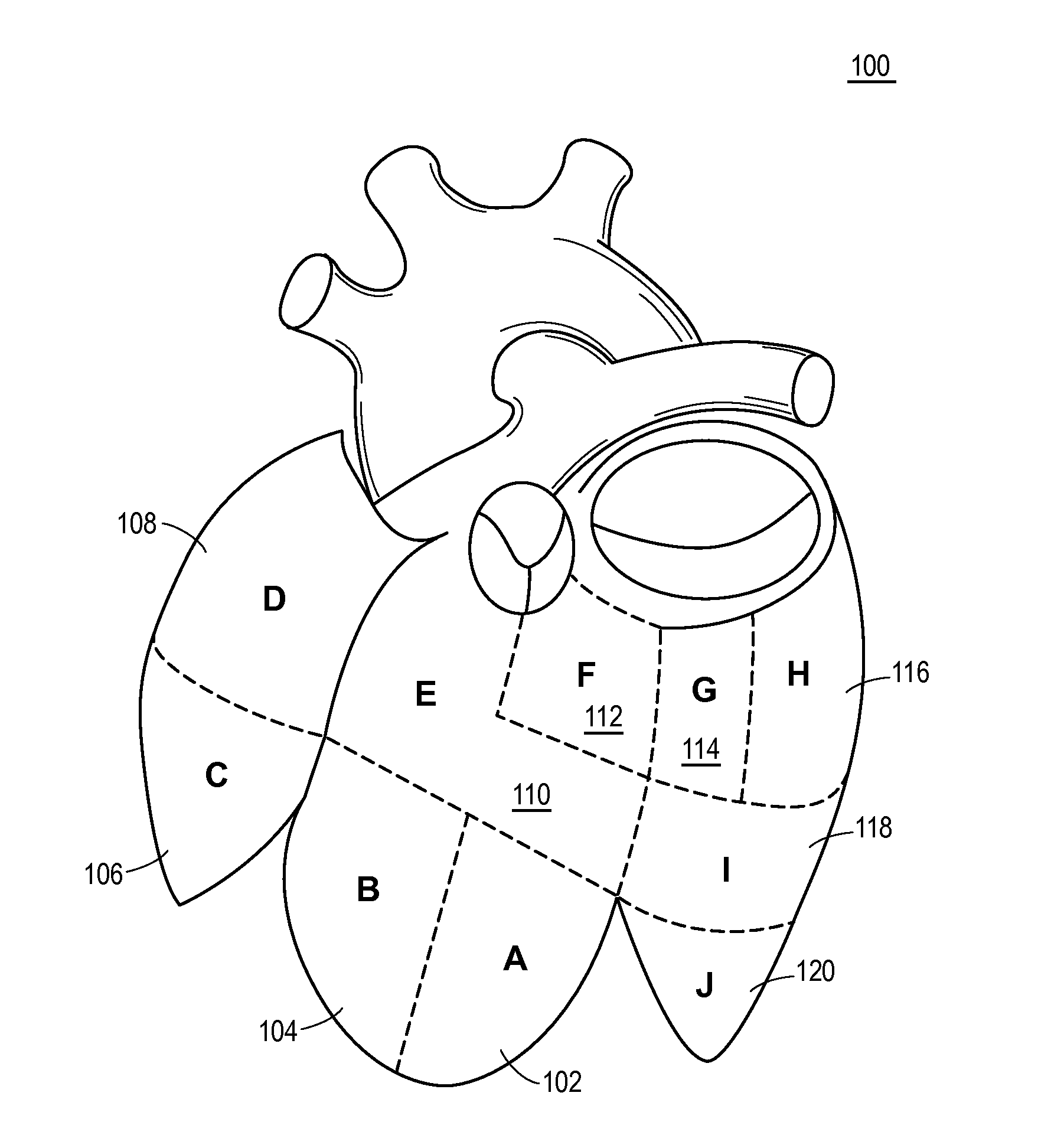
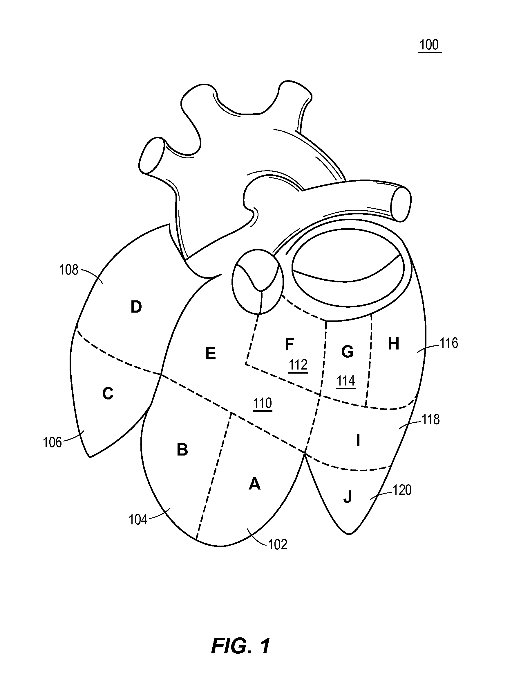
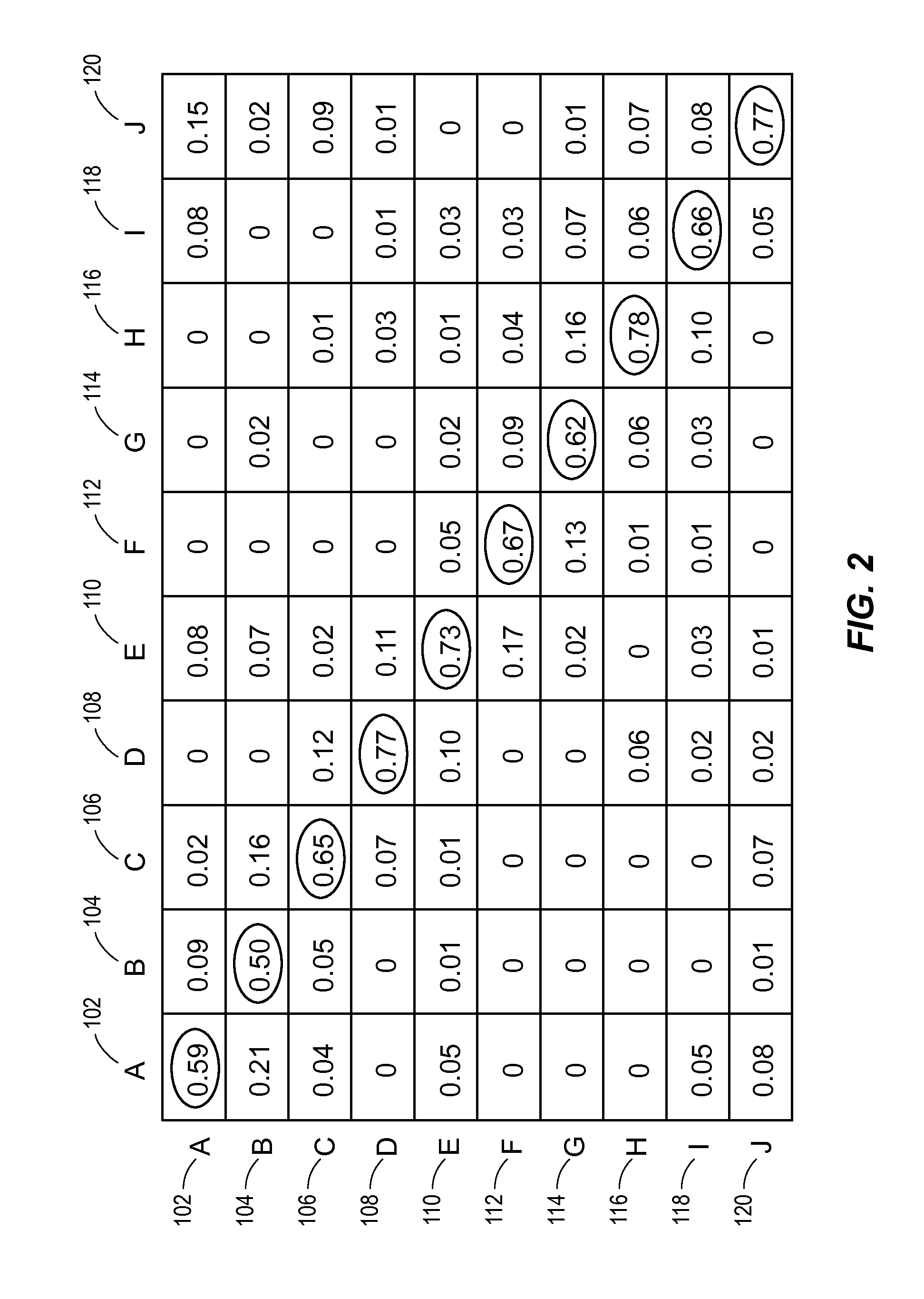
Techniques identify origins of ventricular arrhythmias (e.g., ventricular tachycardia or premature ventricular complexes) including exit sites or other sites using a single or multi-lead electrocardiogram (ECG) assembly. The ECG assembly is used to map an organ into a series of different three-dimensional (3D) regions. Pace maps or ventricular arrhythmia signals are used in form of ECG signals along with a supervised learning methods to pinpoint the potential origin of VT, i.e., exit sites, in the various regions.

Description

CROSS-REFERENCE TO RELATED APPLICATION[0001]This application claims the benefit of U.S. Application Ser. No. 61 / 710,440, filed Oct. 5, 2012, entitled “Automated Analysis of Multi-Lead Electrocardiogram Data to Identify the Exit Sites of Physiological Conditions,” which is hereby incorporated by reference in its entirety.BACKGROUND[0002]Myocardial infarction or acute myocardial infarction (AMI), commonly known as a heart attack, results from the interruption of blood supply to a part of the heart, causing heart cells to die. This is most commonly due to occlusion (blockage) of a coronary artery following the rupture or introduction of a vulnerable atherosclerotic plaque. The resulting restriction in blood supply and ensuing oxygen shortage, if left untreated for a sufficient period of time, can cause damage or death (infarction) of heart muscle tissue (myocardium). The presence of scar tissue can cause life threatening arrhythmias.[0003]One particular arrhythmia associated with infar...

Claims


Application Information

Patent Timeline
IPC IPC(8): A61B5/0464A61B5/04A61B5/363
CPCA61B5/04012A61B5/0464A61B5/7485A61N1/3621A61B5/283A61B5/363A61B5/316
Inventor BOGUN, FRANKSCOTT, CLAYTONHERO, ALFREDLIU, TZU-YU
Owner RGT UNIV OF MICHIGAN
Learn More