Imaging systems, cassettes, and methods of using the same

a slide cassette and imaging system technology, applied in the field of imaging, can solve the problems of wasting time and effort in analyzing a large number of specimens, delay in accurate diagnosis and treatment, and conventional microscopes are often not capable of capturing high resolution and high quality images

Inactive Publication Date: 2014-06-26
VENTANA MEDICAL SYST INC
View PDF11 Cites 90 Cited by
  • Summary
  • Abstract
  • Description
  • Claims
  • Application Information

AI Technical Summary

Benefits of technology

[0007]In some embodiments, a microscope slide holding cassette includes a main body, a plurality of shelves, and a plurality of support members. The main body is configured to surround and protect microscope slides and includes a first sidewall and a second sidewall. The shelves are capable of supporting microscope slides and are positioned between the first and second sidewall

Problems solved by technology

It is often time-consuming to analyze a large number of specimens due the handling time of each slide.
Long processing times may delay an accurate diagnosis and treatment.
Additionally, conventional microscopes are often not capable of capturing high resolution and high quality images suitable for archiving and later use.
Unfortunately,

Method used


Image

  • Imaging systems, cassettes, and methods of using the same
  • Imaging systems, cassettes, and methods of using the same
  • Imaging systems, cassettes, and methods of using the same

Examples


Embodiment Construction

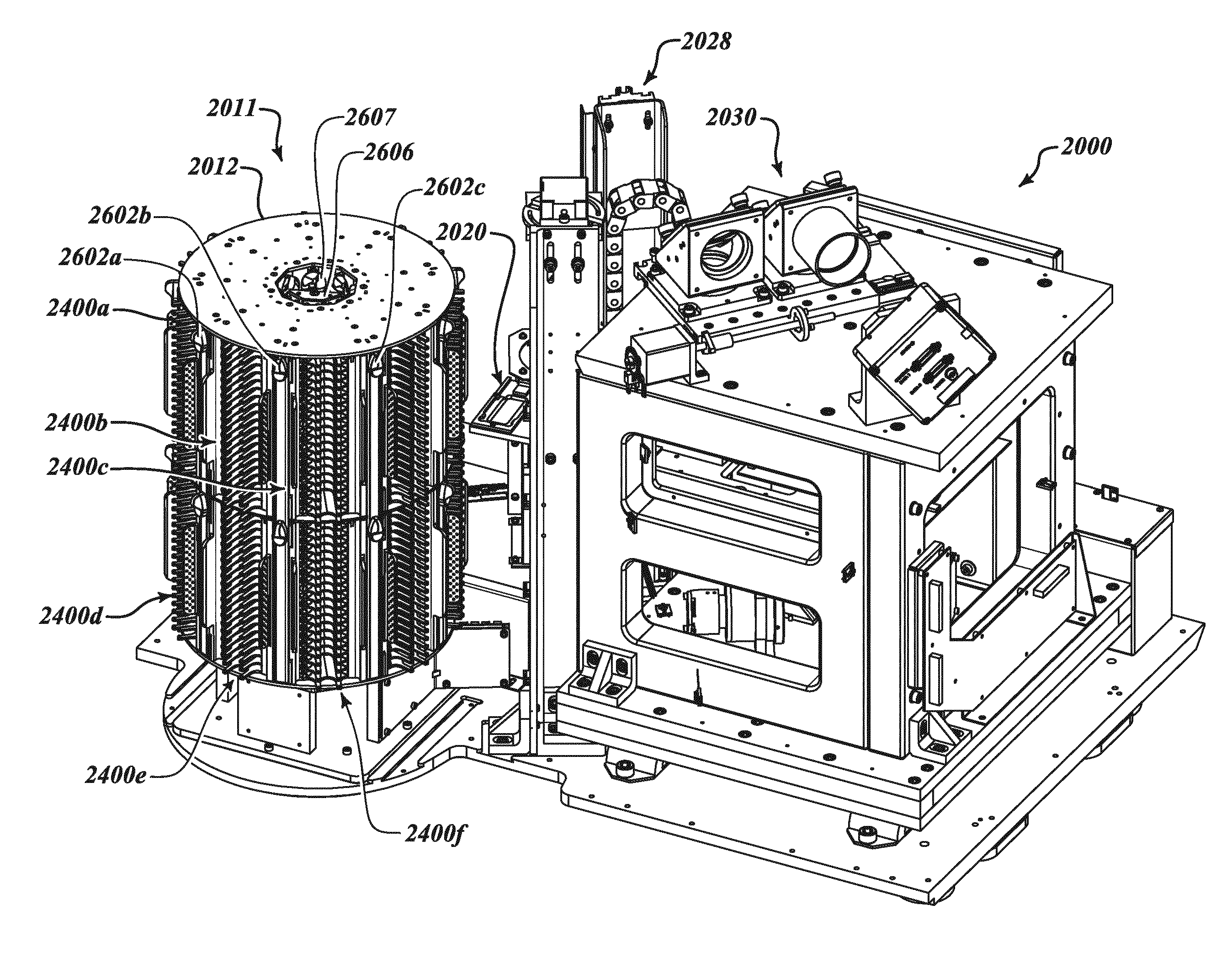
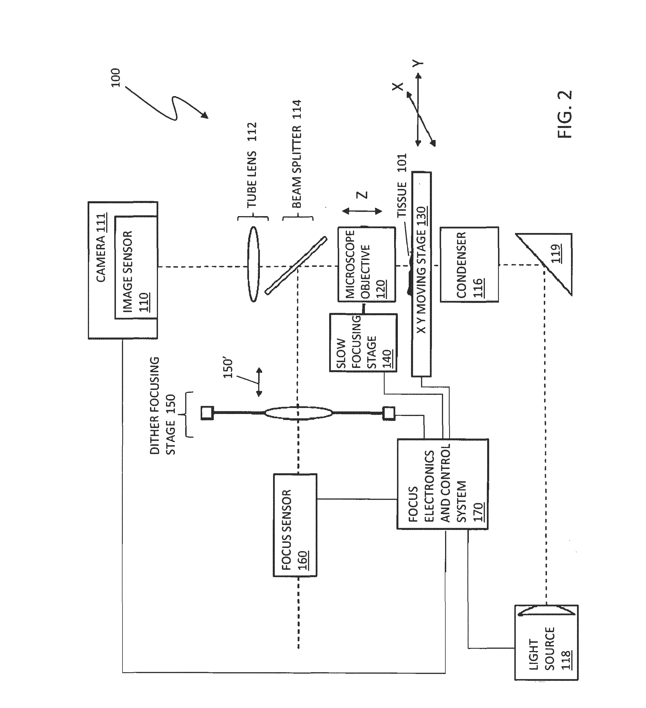
[0071]FIG. 1 is a schematic illustration of an imaging system 5 of a scanning microscope and / or other scanning device that may include various component devices used in connection with digital pathology sample scanning and imaging according to various embodiments of the system described herein. The imaging system 5 may include an imaging device with focusing system 10, a slide stage system 20, a slide caching system 30 and an illumination system 40, among other component systems 50, as further discussed in detail elsewhere herein. It is also noted that the system described herein may be used in connection with microscope slide scanning instrument architectures and techniques for image capture, stitching and magnification as described in U.S. Patent App. Pub. No. 2008 / 0240613 A1 to Dietz et al., entitled “Digital Microscope Slide Scanning System and Methods,” which is incorporated herein by reference, including features in connection with reconstituting an image with a magnification ...


PUM


Abstract

A microscope slide holding cassette includes a main body for surrounding and protecting microscopes, and shelves capable of holding the microscope slides. The shelves have support members that extend outwardly in order to support label ends of slides. Catches carried at the end of the support members are capable of limiting movement of the slides during transport of the cassette. To transport slides to different workstations, a pickup device can use a vacuum to pick up a slide without contacting areas of the slide that may have glue. The pickup device can load and unload the cassette. The pickup device can have a low-profile configuration in order to access relatively small spaces for processing flexibility.

Description

BACKGROUND[0001]1. Technical Field[0002]This application relates to the field of imaging and, more particularly, to imaging systems, slide cassettes, and methods of processing biological samples on slides.[0003]2. Description of the Related Art[0004]Molecular imaging identification of changes in the cellular structures indicative of disease is often important to better understanding in medicinal science. Microscopy applications are applicable to microbiology (e.g., gram staining, etc.), plant tissue culture, animal cell culture (e.g., phase contrast microscopy, etc.), molecular biology, immunology (e.g., ELISA, etc.), cell biology (e.g., immunofluorescence, chromosome analysis, etc.), confocal microscopy, time-lapse and live cell imaging, series imaging, and three-dimensional imaging.[0005]In manual methods of microscopy, specimen-bearing slides are manually loaded into a microscope and viewed through the ocular lens of the microscope. In medical applications, a pathologist can insp...

Claims


Application Information

Patent Timeline
IPC IPC(8): G01N35/02G02B21/34
CPCG02B21/34G01N35/025B01L9/52G02B21/24B01L2200/025B01L2200/026B01L2200/143B01L2200/18B01L2300/021B01L2300/0822B01L2300/0841G02B21/26
Inventor HEBERT, RAPHAELLONEY, GREGORY C.MORAVICK, KEITHMORICONI, DAVIDTODD, CHRISTOPHER EUGENE
Owner VENTANA MEDICAL SYST INC
Why Eureka
Try Eureka