Method and systems for cell-level fish dot counting

a cell-level fish and dot technology, applied in the field of cell-level fish dot counting, can solve the problems of time-consuming and tiring manual counting of fish dots, and the difficulty of automatic fish dot detection

Active Publication Date: 2014-07-24
LEICA MICROSYSTEMS CMS GMBH
View PDF5 Cites 25 Cited by
  • Summary
  • Abstract
  • Description
  • Claims
  • Application Information

AI Technical Summary

Problems solved by technology

However, in addition to being subjective, manual counting of FISH dots is time-consuming and tiring.
In spite of the use of these algorithms, automatic FISH dot detection is still challenging, especially in the presence of background artifacts (FIG. 1A), dense clusters of cell nuclei and FISH dots (FIG. 1B) and / or low FISH dots contrast (see FIG. 1C).

Method used


Image

  • Method and systems for cell-level fish dot counting
  • Method and systems for cell-level fish dot counting
  • Method and systems for cell-level fish dot counting

Examples


Embodiment Construction

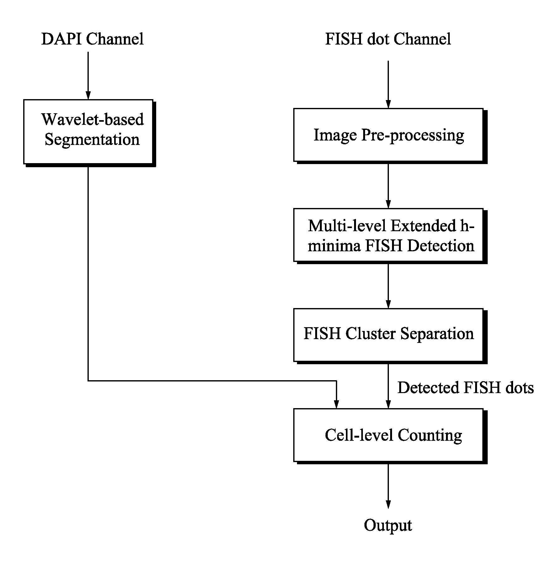
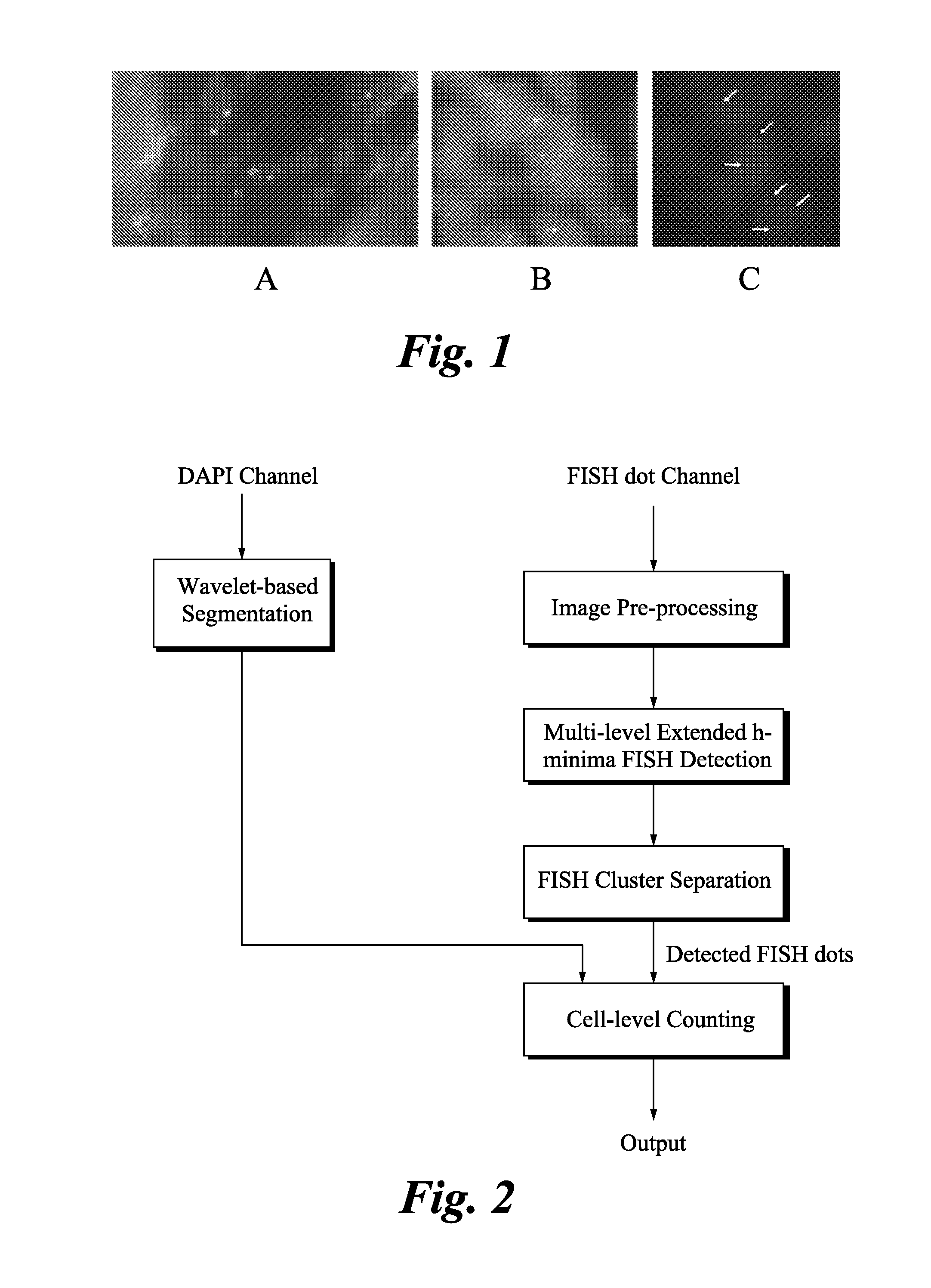
[0015]Disclosed are methods for cell-level counting of FISH dots in 2-D fluorescence images of a biological sample. As used herein, the term “biological sample” refers to a sample obtained from a biological subject, including sample of biological tissue or fluid origin obtained in vivo or in vitro. Such samples can be, but are not limited to, body fluid (e.g., blood, blood plasma, serum, or urine), organs, tissues, fractions, cells isolated from mammals including, humans and cell organelles. Biological samples also may include sections of the biological sample including tissues (e.g., sectional portions of an organ or tissue). Biological samples may also include extracts from a biological sample. Biological samples may comprise proteins, carbohydrates or nucleic acids.

[0016]A biological sample may be of prokaryotic origin, archaeal origin, or eukaryotic origin (e.g., insects, protozoa, birds, fish, and reptiles). In some embodiments, the biological sample is mammalian (e.g., rat, mo...


PUM


Abstract

The invention relates to a computer implemented method and systems for cell level fish dot counting. FISH (fluorescence in situ hybridization) dot counting is the process of enumerating chromosomal abnormalities in the cells which can be used in areas of diagnosis and cancer research. The method comprises in part overlaying images of a biological sample comprising a nuclear counterstain mask and a FISH binary mask. The FISH binary mask is extracted using a multi-level extended h-maxima or h-minima.

Description

CROSS-REFERENCE TO RELATED APPLICATIONS[0001]This application claims priority to U.S. provisional patent application No. 61 / 756,025 filed Jan. 24, 2013; the disclosure of which is incorporated herein by reference in its entirety.BACKGROUND[0002]Fluorescence in situ hybridization (FISH) dot counting is the process of enumerating chromosomal abnormalities in interphase cell nuclei. This process is widely used in many areas of biomedical research, especially in cancer diagnosis and treatment decisions. For example, breast cancer is the most common cancer for women worldwide. Human Epidermal Growth Factor Receptor 2 (HER2) gene copy number is important for breast cancer patients when determining eligibility for adjuvant targeted therapy and / or chemotherapy medications. 1 FISH testing can be used to see if the cells have extra copies of the HER2 gene: a cell having more copies of the HER2 gene is known to have more HER2 receptors, which receive signals that stimulate the growth of breast...

Claims


Application Information

Patent Timeline
Patent Type & Authority Applications(United States)
IPC IPC(8): G06K9/00
CPCG06K9/00127G06V20/69
Inventor PADFIELD, DIRK RYANSEPPO, ANITTI ELJASAL-KOFAHI, YOUSEF AHMED
Owner LEICA MICROSYSTEMS CMS GMBH
Why Eureka
Try Eureka