Methods For Predicting Mammalian Embryo Viability

a mammalian embryo and viability technology, applied in the field of methods for predicting mammalian embryo viability, can solve the problems of costly approach and inevitable risks, and achieve the effects of accurate measurement of sub-pixel movements, high developmental potential, and high developmental potential

Inactive Publication Date: 2014-07-24
CAMBRIDGE ENTERPRISE LTD
View PDF0 Cites 9 Cited by
  • Summary
  • Abstract
  • Description
  • Claims
  • Application Information

AI Technical Summary

Benefits of technology

[0101]Another advantage of the present invention is that it provides a way of assessing the developmental potential of an embryo very early on, from measurements taken over a short period of time from embryos in the very earliest stage of development before the first embryonic cell division. In some cases, the relevant measurements are all taken within 6 hours, more preferably within 5, 4, 3, 2 or 1 hour of fertilization. In some cases the measurements are all taken before pronuclei formation in the embryo. More preferably the measurements are all taken before regression of the FC. For example the time lapse images of the embryo may all be captured within these periods of time. In some cases the method is completed, including the prediction of developmental potential, within 6 days, more preferably within 4, 3, 2 or 1 day of fertilization, or within 12, 6, 4, 3, 2 or 1 hours of fertilization.
[0102]In some cases the quantitative image analysis uses particle image velocimetry (PIV). PIV is a standard analysis technique used extensively in fluid dynamics21-24 that involves cross-correlating image sub-regions between sequential pairs of images. Using PIV analysis each image can be divided into square interrogation regions and the cross-correlation coefficient distribution can be calculated within a larger region in the next image in the sequence. The peak of the cross-correlation coefficient distribution gives the most likely location in the second image of the material in the interrogation region in the first image. This can then be used to calculate the displacement vector over the time between images (FIG. 1A). As the displacement of the pattern of contrast of an area of pixels is measured over many pixels it is possible to accurately measure sub-pixel movements with this technique22, 24, 48
[0103]In some cases in accordance with the invention, one or more embryos in a population of embryos can be graded according to their developmental potential. The developmental potential of each embryo can be graded relative to the developmental potential of the other embryos in the population. This may provide a useful way of determining which embryos from the population of embryos are most suitable for a particular subsequent intended use, for example transfer to a maternal recipient with the aim of a resulting pregnancy. Embryos can thereby be selected according to their developmental potential.
[0104]The method may involve calculating the population mean interval or average interval between speed peaks or FC pulsations, and / or the population mean average basal cytoplasmic speed. The method may involve calculating the population standard deviations for these parameters. Obvious outliers in the population may be excluded from the calculations. For example, embryos that have a calculated interval or average interval between speed peaks or FC pulsations or a calculated average basal cytoplasmic speed that is more than 1 standard deviation, or more than 1.5 standard deviations, or more than 2 standard deviations, or more than 3 standard deviations away from the mean, or that are in the top and / or bottom 10% of calculated values may be excluded. The population mean and / or standard deviations can be used to quantify the relative developmental potential of one or more of the embryos in the population. For example having a calculated interval or average interval between speed peaks that is longer than the population mean interval or average interval between speed peaks, and / or having an average basal cytoplasmic speed that is faster than the population mean average basal cytoplasmic speed may be indicative of an embryo having high developmental potential.
[0105]Conversely having a calculated interval or average interval between speed peaks that is shorter than the population mean interval or average interval between speed peaks, and / or having an average basal cytoplasmic speed that is slower than the population mean average basal cytoplasmic speed, may be indicative of an embryo having high developmental potential. Similarly having a calculated interval or average interval between speed peaks that is at least half, or at least a third or at least a quarter or at least one, two or three standard deviations longer or shorter than the population mean interval or average interval between speed peaks, and / or having an average basal cytoplasmic speed that is at least half, or at least a third, or at least a quarter, or at least one, two or three standard deviation faster or slower than the population mean average basal cytoplasmic speed of the population may be indicative of an embryo having high or low developmental potential respectively.
[0106]Combining a rapid imaging of the freshly fertilized mouse egg with advanced image analysis based on particle image velocimetry reveals that fertilization induces rhythmical cytoplasmic movements that coincide with pulsations of the protrusion forming above the sperm head. We found that these movements are caused by contractions of the actomyosin cytoskeleton triggered by Ca2+ oscillations induced by fertilization. Most importantly, the relationship between the movements and the events of egg activation makes it possible to use the movements alone to predict whether a zygote will develop successfully. This method offers thus far the earliest and fastest, non-invasive way to predict the vitality of eggs fertilized in vitro and therefore can potentially improve greatly the prospects for IVF treatment.

Problems solved by technology

Not only is this approach costly but there are inevitable risks from such long term in vitro culture to the blastocyst stage.

Method used


Image

  • Methods For Predicting Mammalian Embryo Viability
  • Methods For Predicting Mammalian Embryo Viability
  • Methods For Predicting Mammalian Embryo Viability

Examples


example 1

Fertilization of the Mouse Egg Results in Bursts of Rhythmic Cytoplasmic Motion

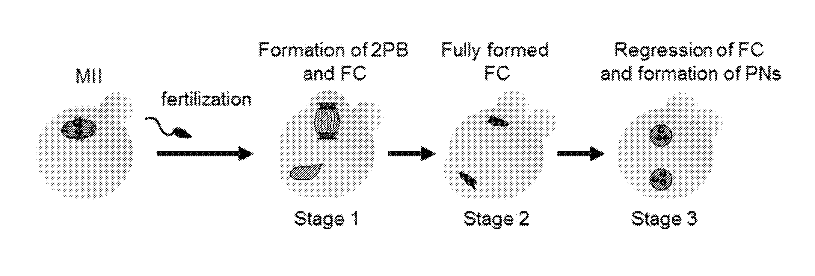
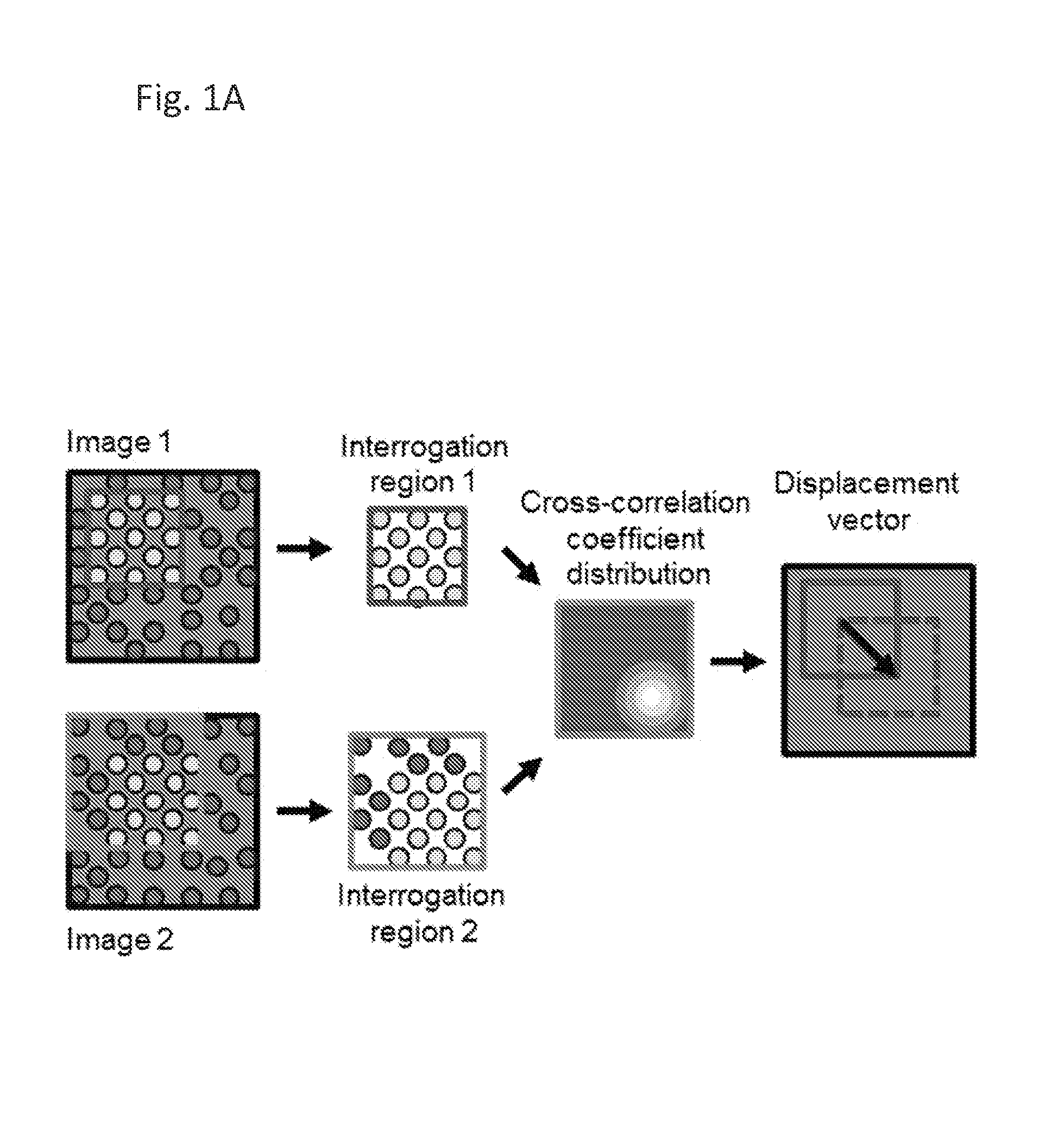
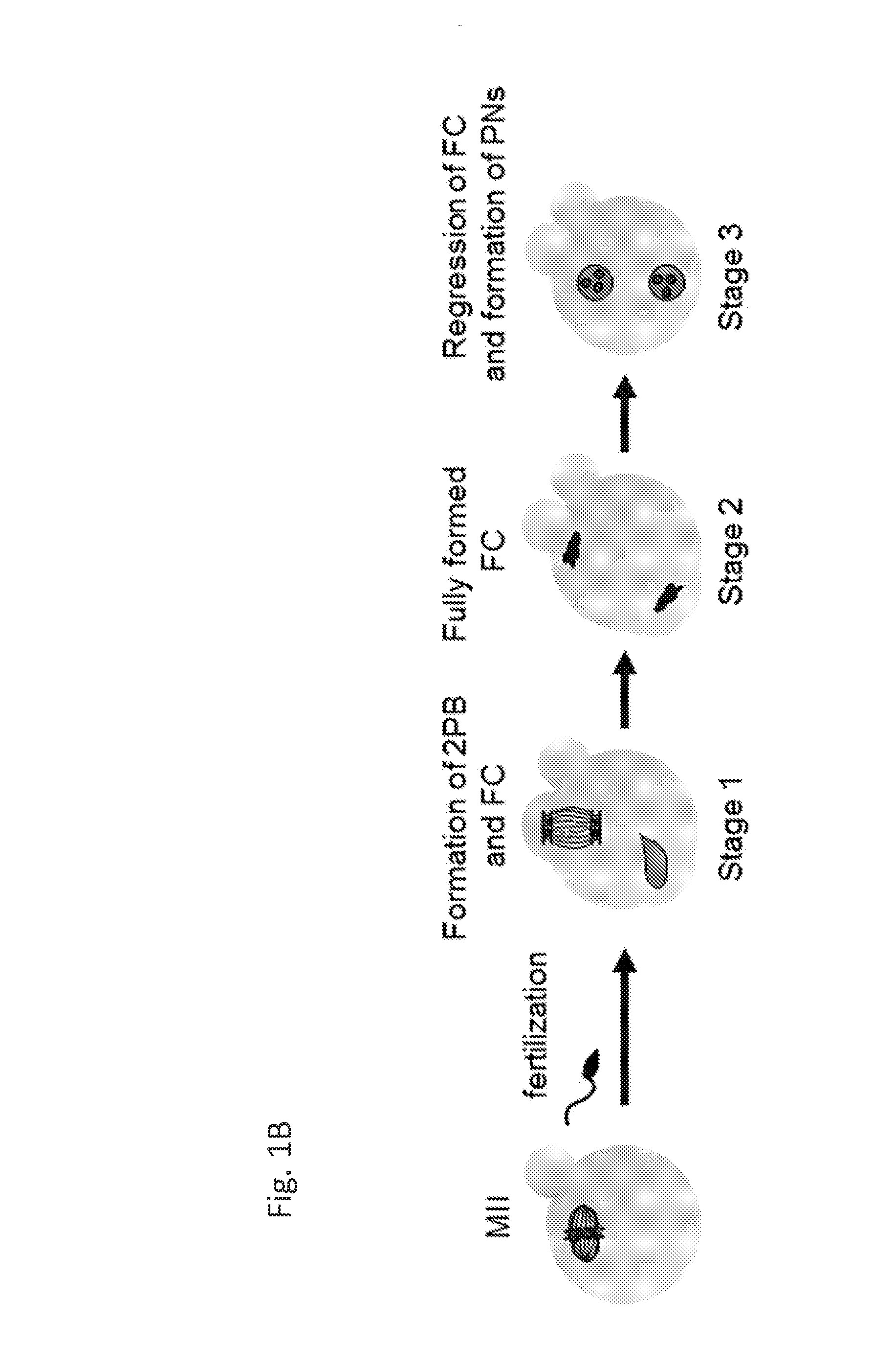
[0135]To characterise cytoplasmic movements upon fertilization of the mouse egg and to evaluate whether, and if so how, they relate to other events associated with fertilization we first filmed eggs from the time immediately after fertilization until pronuclei formation. We then used advanced image analysis based on the PIV method21-24 to analyse and quantify these movements (FIG. 1A). This revealed that fertilization of the mouse egg was followed by dramatic periodic increases and decreases in velocity of the cytoplasmic movements (that we termed speed-peaks; FIG. 1B-D, n=55 unfertilized oocytes and n=125 eggs fertilized in vivo). This sequence of fast repetitive movements throughout the egg cytoplasm lasted for approximately 4 hours, until pronuclei formation. The speed-peaks were initially of relatively low amplitude (between anaphase of meiosis II and early FC outgrowth; Stage 1); but subsequently dev...

example 2

High Amplitude Cytoplasmic Speed-Peaks Correlate with Pulsations of the FC and Depend on the Actomyosin Cytoskeleton

[0137]Since the direction of the abrupt movements at Stage 2 suggested that the FC could be involved in their generation, we next examined the morphology of the FC throughout this period (69 speed-peaks, 14 zygotes). This showed that at each speed-peak, the apex of the FC sank inwards and the region where the FC merged into the convex outline of the zygote widened. As a consequence, the diameter of the zygote along the axis of the FC (hereinafter called the ‘FC-diameter’) decreased by 1.28+ / −0.66 μm (1.55+ / −0.79% of its length) (FIG. 2A, C). The shape of the FC was restored during the inter-peak interval (FIG. 2C). Interestingly, the extent of reduction of the FC diameter showed a linear association with speed-peak amplitude (20 zygotes, 71 speed-peaks analyzed) (FIG. 2B).

[0138]Since the actomyosin cytoskeleton is enriched in the FC region17, 25, we considered that thi...

example 3

Cytoplasmic Movements Correlate with and Depend on Ca2+ Transients

[0141]As fertilization is known to initiate oscillations of free Ca2+ 10, 29, we next wished to address whether the actomyosin-mediated speed-peaks might depend upon these Ca2+ pulses. To this end, we loaded fertilized eggs with the Ca2+ sensitive fluorescent dye FuraRed, and simultaneously collected FuraRed fluorescence and DIC images. In 32 zygotes, all 121 speed-peaks were accompanied by an increase in Ca2+. The cytoplasmic speed increased exactly (within the resolution of our recording interval) as Ca2+ levels peaked (FIG. 3A). Only in a single zygote with unusually frequent Ca2+ oscillations were some Ca2+ spikes (4 / 17) not mirrored by speed-peaks. In some eggs fertilized by ICSI we managed to record speed-peaks accompanying the first Ca2+ transient. They were of higher amplitude than the rest of Stage 1 speed-peaks (FIG. 9) corresponding to the first Ca2+ transient being bigger than subsequent ones.

[0142]To dete...


PUM

PropertyMeasurementUnit
volumeaaaaaaaaaa
volumeaaaaaaaaaa
volumeaaaaaaaaaa

Abstract

The invention provides methods and systems for assessing the developmental potential of mammalian embryos. The method of the invention comprises taking measurements of cytoplasmic movements in the embryo and/or periodic changes in the shape of the embryo at the single cell stage.

Description

FIELD OF INVENTION[0001]The invention provides methods of assessing the developmental potential of a mammalian embryo, methods of selecting or grading embryos from a plurality of embryos according to their predicted developmental potential, and methods of in vitro fertilization (IVF) and assisted reproduction. A system for carrying out the methods of the invention is also provided.BACKGROUND TO THE INVENTION[0002]There is currently a need for a better means of assessing the vitality of embryos for transplantation into the would-be mother in IVF clinics. The general standard has been to assess the extent of development at 3 days and use this as a predictor of the likely success of subsequent development and hence the pregnancy. The unreliability of this assessment has led an increasing number of IVF clinics to culture embryos until the blastocyst stage (6 days) so that apparently healthy blastocysts can be used directly. Not only is this approach costly but there are inevitable risks...

Claims


Application Information

Patent Timeline
Patent Type & Authority Applications(United States)
IPC IPC(8): G01N33/483A61D19/04
CPCA61D19/04G01N33/4833C12M21/06C12M41/14C12M41/26C12M41/46G01N33/5088G01N33/5091
Inventor ZERNICKA-GOETZ, MAGDALENAAJDUK, ANNAGRAHAM, CHRIS
Owner CAMBRIDGE ENTERPRISE LTD
Why Eureka
Try Eureka