
Real-Time 3-D Ultrasound Reconstruction of Knee and Its Implications For Patient Specific Implants and 3-D Joint Injections

a real-time, ultrasound-based technology, applied in the field of real-time imaging of joints using non-ionizing imaging, can solve the problems of high cost and disability, joint pain costs the healthcare system about $37 billion annually, and involve expensive pre-arthroplasty substances

Active Publication Date: 2014-08-07
JOINTVUE
View PDF9 Cites 16 Cited by
  • Summary
  • Abstract
  • Description
  • Claims
  • Application Information

AI Technical Summary

Benefits of technology

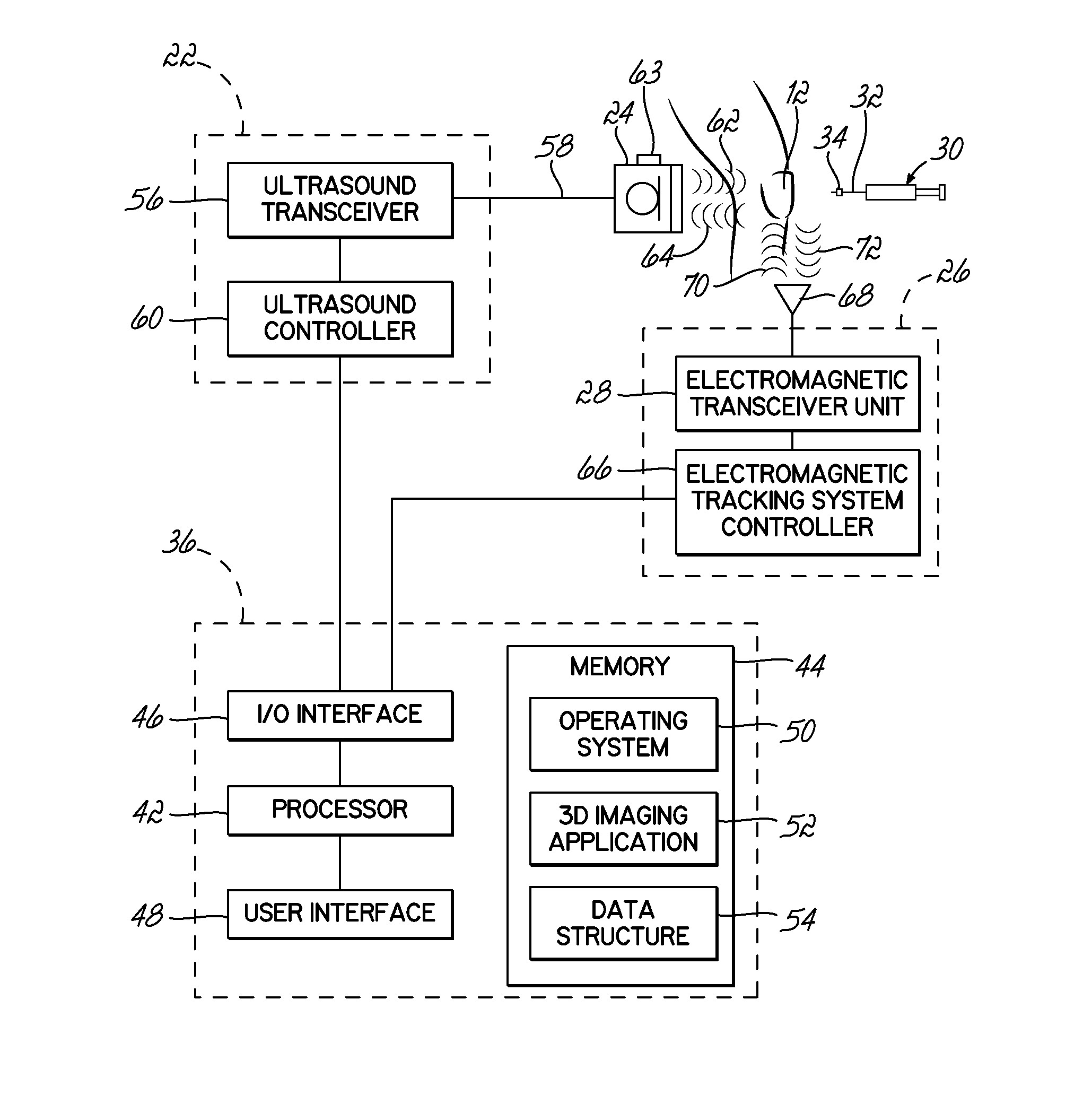
This patent describes an apparatus for treating patients using ultrasound technology. The apparatus can acquire multiple RF signals from a pulse-mode echo ultrasound scan and determine the position of the ultrasound transducer for each signal. It can then generate contour lines from these signals and estimate the 3-D shape and position of anatomical features of the patient based on these lines and transducer positions. This technology can provide valuable information for treatment planning and can improve the accuracy and safety of procedures that require ultrasound guidance.

Problems solved by technology

Joint pain is a major public health problem and is responsible for significant costs and disability in the United States.
Joint pain costs the healthcare system about $37 billion annually.
Many of these treatments involve expensive pre-arthroplasty substances, such as single dose visco-supplements, platelet rich plasma, stem cells, etc.
Failure of these conservative treatments result in an estimated 505,000 knee, 280,000 hip and 42,000 shoulder replacements (arthroplasty) annually in the United States alone.
A major challenge for conservative management of joint pain is the lack of low cost, accurate, non-ionizing joint imaging technology.
Ultrasound is widely accepted as a means to visualize the joint space, but the present technology has significant limitations.
Some surgeons use fluoroscopy to assist with the guidance, which can be harmful to both the patient and the surgeon due to the ionizing X-Ray radiation emitted.
These images are also typically distorted, and require distortion correction.
The resulting corrected 2-D images can be quite blurry.
To limit the amount of radiation exposure, many surgeons do not keep the fluoroscopy machine active to track the needle while the needle is being inserted into the joint.
But, even with these modified techniques, fluoroscopy exposes the patient to X-Ray radiation far in excess of conventional radiographs.
Injections with these modalities are also typically more painful if multiple injection attempts or needle repositioning is needed to correct inaccuracies in the injections due to a lack of real-time imaging to help guide the needle.
MRI scans are conducted under static conditions and are often difficult to interpret.
The inability to allow metal objects near the joint during an MRI, and the confined area in which the patient is placed further limits the ability of MRI to provide real time imaging during the injection.
MRI procedures are also very expensive, and may reveal multiple concerns, which makes it difficult for the physician to make a proper diagnosis.

Method used


Image

  • Real-Time 3-D Ultrasound Reconstruction of Knee and Its Implications For Patient Specific Implants and 3-D Joint Injections
  • Real-Time 3-D Ultrasound Reconstruction of Knee and Its Implications For Patient Specific Implants and 3-D Joint Injections
  • Real-Time 3-D Ultrasound Reconstruction of Knee and Its Implications For Patient Specific Implants and 3-D Joint Injections

Examples


Embodiment Construction

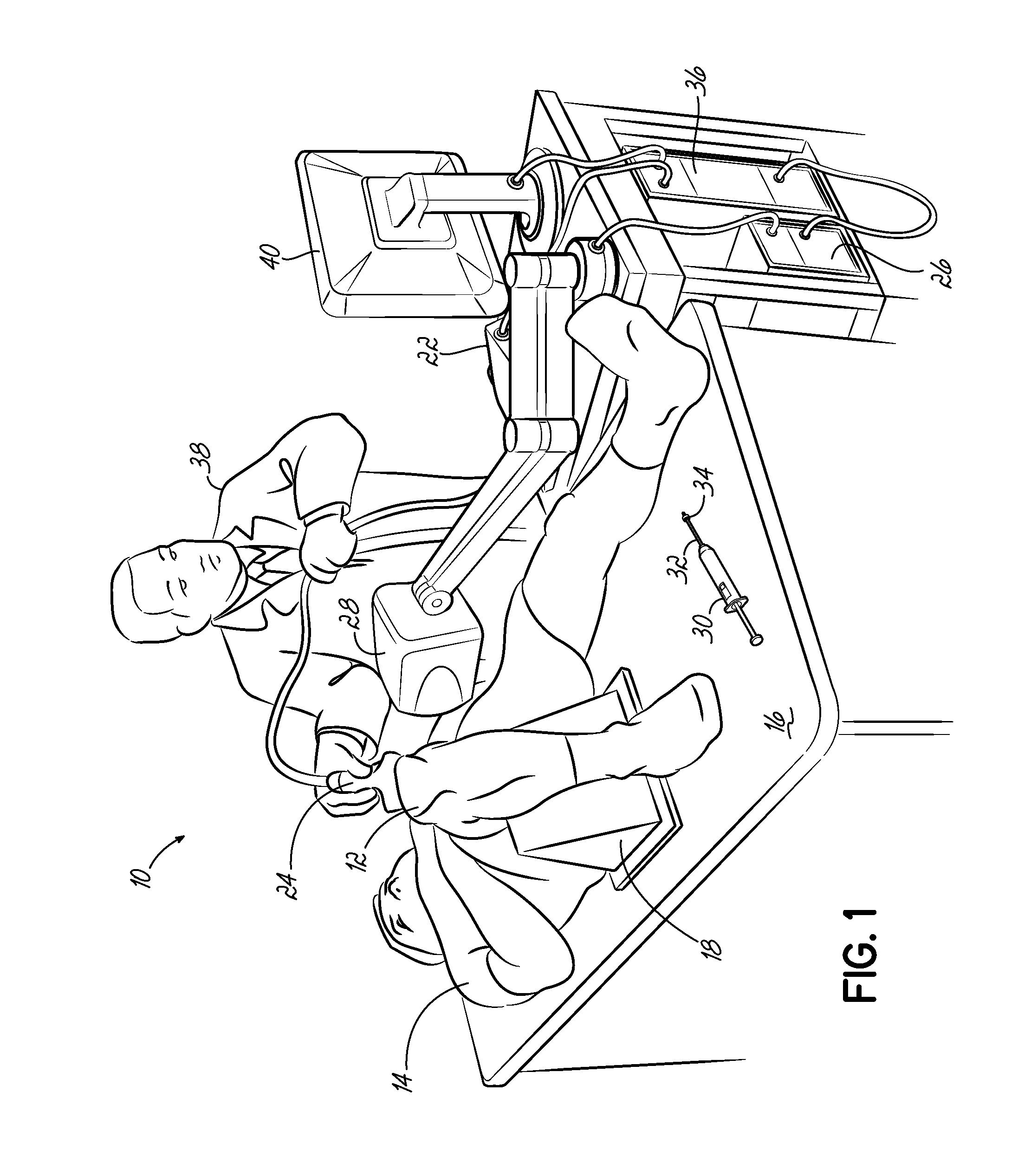
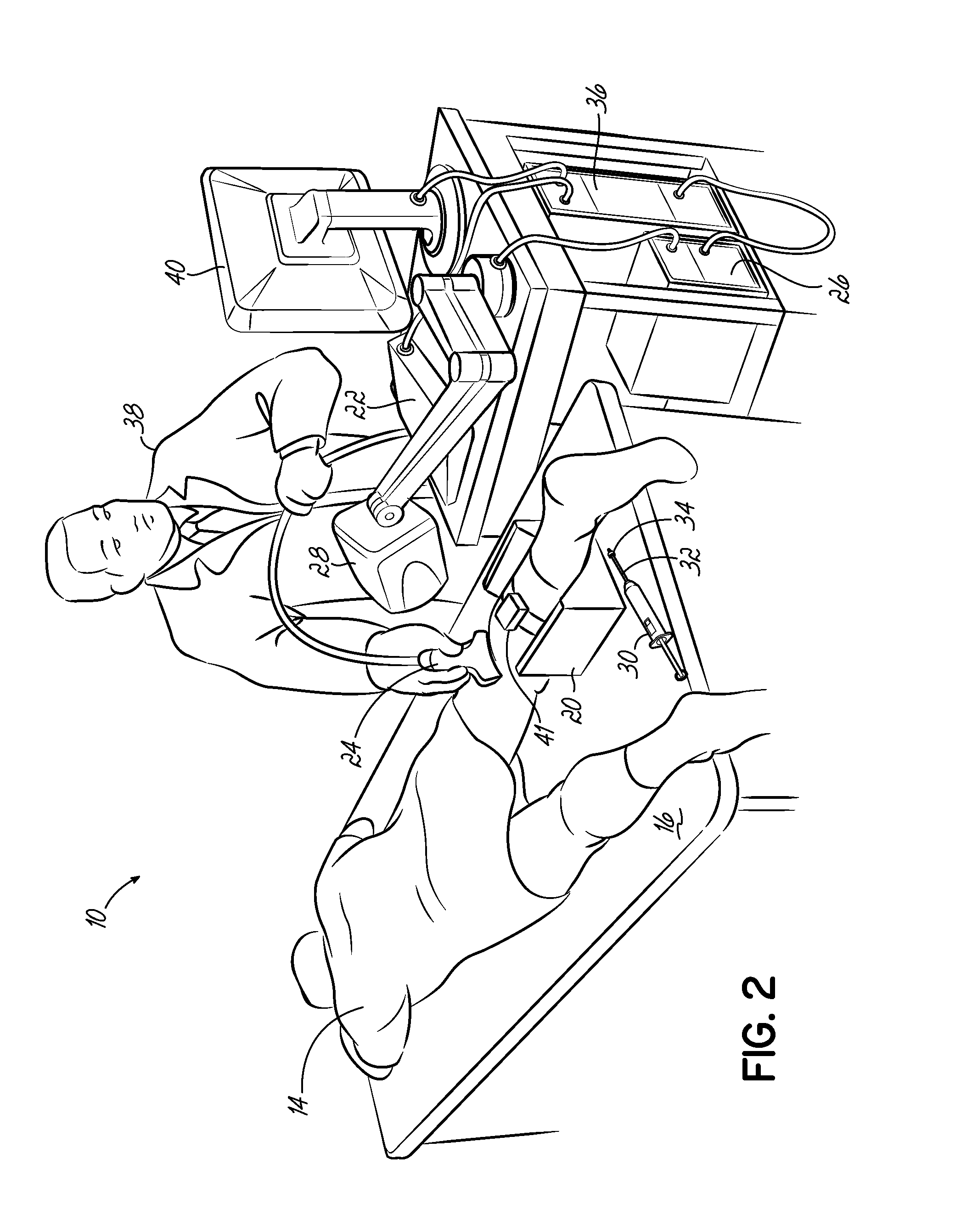
[0031]The present invention overcomes the foregoing problems and other shortcomings, drawbacks, and challenges of conventional joint visualization modalities and injection protocols. Embodiments of the invention provide a patient-specific 3-D view of the joint bones and joint space that reduces the skill level required to perform joint injection procedures. The targeted location for the injection, or desired injection point, can also be designated in the 3-D view. This designation allows for a 3-D vector depicting distance to the target and / or a 3-D distance map to be displayed, allowing for the end of the needle to be precisely placed in an optimal position within the joint. This optimal needle placement will help ensure that the injected material is delivered in a proper position. Needle injection using real-time ultrasound guidance with 3-D joint visualization may improve injection accuracy, reduce time spent on joint injections, reduce the cost and complexity of the process, and...


PUM


Abstract

Methods and apparatus for treating a patient. The method includes acquiring a plurality of radio frequency (RF) signals with an ultrasound transducer, each RF signal representing one or more return echoes from a scan line of a pulse-mode echo ultrasound scan. A position of the ultrasound transducer corresponding to each of the acquired RF signals is determined, and a plurality of contour lines generated from the plurality of RF signals. The method estimates a 3-D shape and position of an anatomical feature, such as a joint of patient based on the generated contour lines and corresponding ultrasound transducer positions. An apparatus, or computer includes a processor and a memory with instructions that, when executed by the processor, perform the aforementioned method.

Description

CROSS-REFERENCE TO RELATED APPLICATIONS[0001]This Application is a continuation of and claims priority to International Application No. PCT / US2012 / 60261, entitled REAL-TIME 3-D ULTRASOUND RECONSTRUCTION OF KNEE AND ITS IMPLICATIONS FOR PATIENT SPECIFIC IMPLANTS AND 3-D JOINT INJECTIONS, filed Oct. 15, 2012 (pending), which claims the benefit of and priority to U.S. Provisional Application Ser. No. 61 / 547,508, filed on 14 Oct. 2011 and entitled REAL-TIME 3-D ULTRASOUND RECONSTRUCTION OF KNEE AND ITS IMPLICATIONS FOR PATIENT SPECIFIC IMPLANTS AND 3-D JOINT INJECTIONS, the disclosures of which are hereby incorporated by reference in their entireties.TECHNICAL FIELD[0002]The invention relates generally to real-time imaging of joints using non-ionizing imaging methods, and more particularly to the use of real-time imaging to plan surgical procedures, including guiding needles during joint injection treatment procedures.BACKGROUND[0003]Joint pain is a major public health problem and is re...

Claims


Application Information

Patent Timeline
IPC IPC(8): A61B8/08A61B8/14
CPCA61B8/14A61B8/0875A61B8/5207A61B8/5223G06T17/00G06T2210/41G06T2207/10028G06T2207/10136G06T2207/30008A61B8/4245G06T7/564G16H50/30
Inventor MAHFOUZ, MOHAMED R.WASIELEWSKI, RAY C.
Owner JOINTVUE
Learn More