
Systems and methods for automated screening and prognosis of cancer from whole-slide biopsy images

a biopsy image and whole-slide technology, applied in the field of biopsy samples computer analysis, can solve the problems of few systems that have addressed the biopsy, misinterpretation of the actual cancer aggressiveness, etc., and achieve the effect of reducing dependency, accurate and unambiguous measurement of prostate cancer aggressiveness

Inactive Publication Date: 2014-08-21
BOARD OF RGT THE UNIV OF TEXAS SYST
View PDF7 Cites 157 Cited by
  • Summary
  • Abstract
  • Description
  • Claims
  • Application Information

AI Technical Summary

Benefits of technology

The patent proposes a way to interpret cancer aggressiveness by using advanced scoring methods that involve measuring patterns in prostate cancer cells. These new scoring methods are designed to provide a more accurate measure of cancer aggressiveness, by simultaneously covering all previous scoring methods and combining all observed patterns into one, continuous-scale score. This method is fast, simple, and can be used by both pathologists and without them. It can also be used for screening large populations and provides a screening test for cancer that doesn't produce false negative or false positive results.

Problems solved by technology

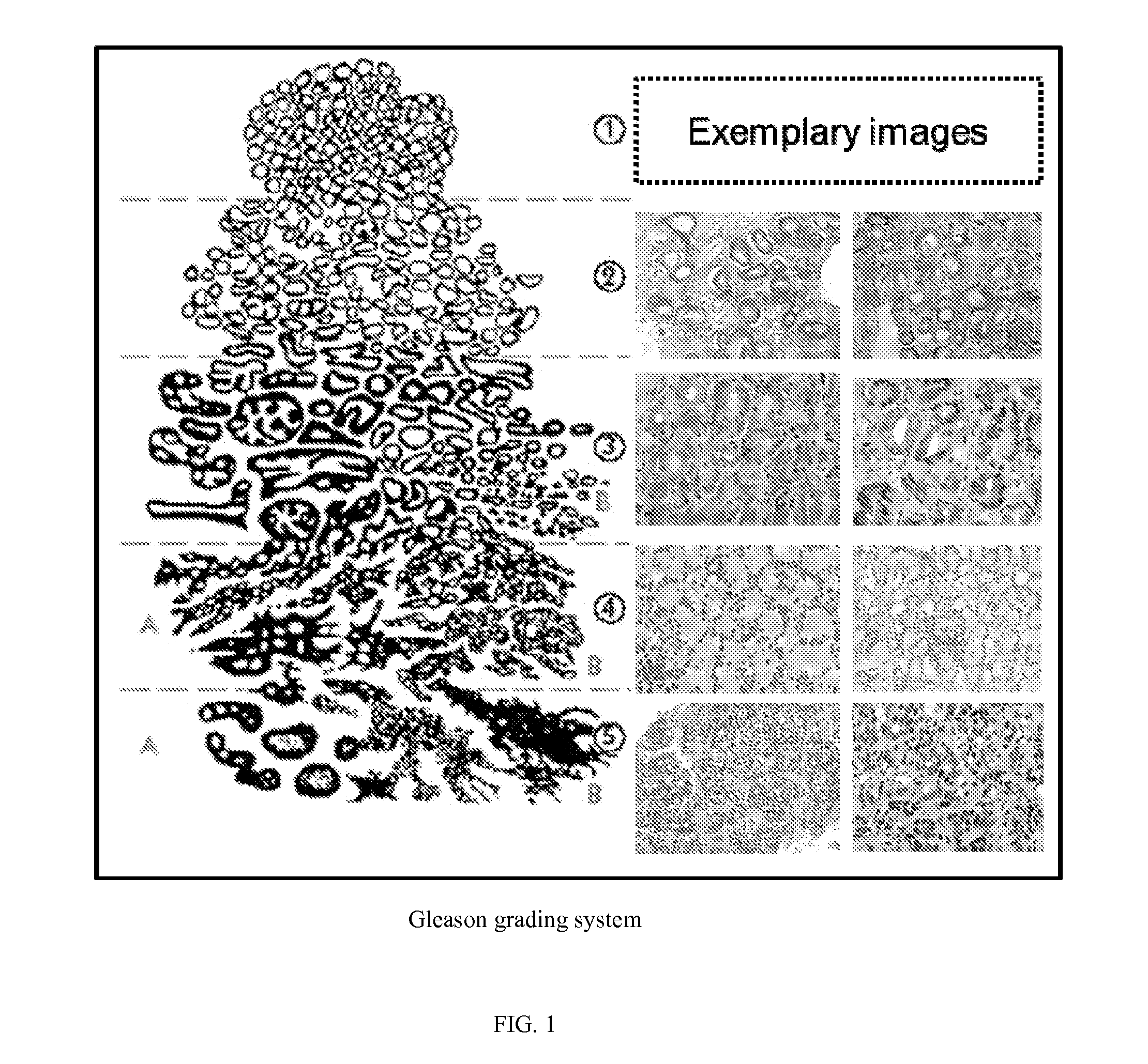
The large number of disparate, differently-scaled Gleason scoring methods can lead to a variety of misinterpretations of reading actual cancer aggressiveness.
Only few systems have addressed the biopsy whole slide processing.
While some of the systems and methods mentioned above are able to classify predefined regions of interest, they do not apply to a biopsy image as a whole.

Method used


Image

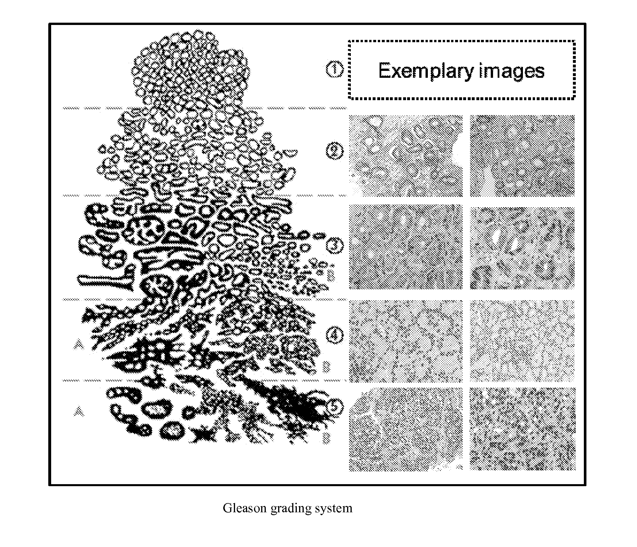
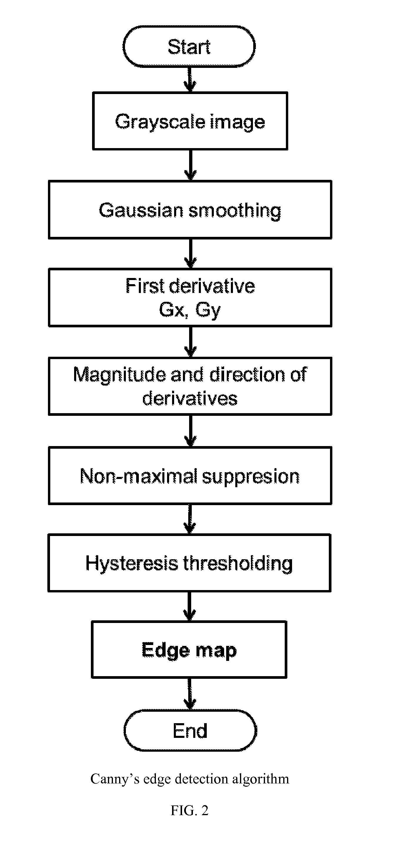
  • Systems and methods for automated screening and prognosis of cancer from whole-slide biopsy images
  • Systems and methods for automated screening and prognosis of cancer from whole-slide biopsy images
  • Systems and methods for automated screening and prognosis of cancer from whole-slide biopsy images

Examples


Embodiment Construction

[0071]It is to be understood the present invention is not limited to particular devices or methods, which may, of course, vary. It is also to be understood that the terminology used herein is for the purpose of describing particular embodiments only, and is not intended to be limiting. As used in this specification and the appended claims, the singular forms “a”, “an”, and “the” include singular and plural referents unless the content clearly dictates otherwise. Furthermore, the word “may” is used throughout this application in a permissive sense (i.e., having the potential to, being able to), not in a mandatory sense (i.e., must). The term “include,” and derivations thereof, mean “including, but not limited to.” The term “coupled” means directly or indirectly connected.

[0072]Embodiments herein relate to automated quantitative analysis and assessment of human or animal tissue images such as liver biopsy, endometrial biopsy, lung biopsy, lymph node biopsy, renal biopsy, bladder biops...


PUM


Abstract

The invention provides systems and methods for detection, grading, scoring and tele-screening of cancerous lesions. A complete scheme for automated quantitative analysis and assessment of human and animal tissue images of several types of cancers is presented. Various aspects of the invention are directed to the detection, grading, prediction and staging of prostate cancer on serial sections / slides of prostate core images, or biopsy images. Accordingly, the invention includes a variety of sub-systems, which could be used separately or in conjunction to automatically grade cancerous regions. Each system utilizes a different approach with a different feature set. For instance, in the quantitative analysis, textural-based and morphology-based features may be extracted at image- and (or) object-levels from regions of interest. Additionally, the invention provides sub-systems and methods for accurate detection and mapping of disease in whole slide digitized images by extracting new features through integration of one or more of the above-mentioned classification systems. The invention also addresses the modeling, qualitative analysis and assessment of 3-D histopathology images which assist pathologists in visualization, evaluation and diagnosis of diseased tissue. Moreover, the invention includes systems and methods for the development of a tele-screening system in which the proposed computer-aided diagnosis (CAD) systems. In some embodiments, novel methods for image analysis (including edge detection, color mapping characterization and others) are provided for use prior to feature extraction in the proposed CAD systems.

Description

BACKGROUND OF THE INVENTION[0001]1. Field of the Invention[0002]The invention generally relates to diagnostic procedures for the detection, analysis, assessment, classification, and grading of cancer from digitized histopathological images. More particularly, the invention relates to the use of computer analysis of biopsy samples to detect and classify / grade and score prostate cancer.[0003]2. Description of the Relevant Art[0004]Prostate cancer is the second killer of American men. Current statistics indicate that 1 in 6 men will suffer from prostate cancer, 1 in 32 men will die from prostate cancer. Although prostate cancer can be diagnosed at early stages, 80% of men diagnosed with prostate carcinoma are above 65 years old.[0005]The risk of developing prostate cancer is commonly attached with the patient's age. In general, doctors recommend males of age 50 or higher to screen for prostate cancer. However, screening could be recommended for males who are at a higher risk of develop...

Claims


Application Information

Patent Timeline
Patent Type & Authority Applications(United States)
IPC IPC(8): G06T7/00G06K9/00
CPCG06T7/0012G06K9/0014G06K9/00147A61B10/0241A61B5/4381A61B5/725A61B5/7267G06T2207/10024G06T2207/10056G06T2207/20036G06T2207/20081G06T2207/30024G06T2207/30096G06T7/13G06T7/181G16H50/30G16H50/70G16H50/20G16H30/20G06V20/69G01N15/1433G06V20/695G06V20/698G06F18/211G06T5/70G06T7/90A61B5/7203A61B5/7257A61B5/726G06T5/20G06T2207/30081
Inventor AGAIAN, SOS S.LOPEZ, CLARA M. MOSQUERAALMUNTASHRI, ALIMETZLER, RICHARD
Owner BOARD OF RGT THE UNIV OF TEXAS SYST
Learn More