
Human disc tissue

a technology of disc tissue and stem cells, which is applied in the field of human disc tissue, can solve the problems of neck and lower back pain from degeneration permanent disruption of the joint, and herniation or protrusion of the disc, so as to prevent, inhibit, or decrease the likelihood of damage or disease of the spinal disc joint, and prevent, inhibit or decrease the likelihood of damage or disease of the cartilage-containing join

Inactive Publication Date: 2014-09-25
DISCGENICS
View PDF0 Cites 2 Cited by
  • Summary
  • Abstract
  • Description
  • Claims
  • Application Information

AI Technical Summary

Benefits of technology

[0020]In another embodiment, the present invention provides a method of preventing, inhibiting, or decreasing the likelihood of damage to or disease of the spinal joint of a subject, comprising the step of administering to said subject an amplified disc stem cell population obtained using a method comprising the steps of: (a) culturing nucleus pulposus cells as an attached monolayer in a first medium comprising serum, basic fibroblast growth factor (bFGF), epidermal growth factor (EGF), and human neonatal foreskin fibroblast (NFF) cell supernatant on gelatin-coated tissue culture plates to confluency; (b) isolating a single cell population of nucleus pulposus cells from said first medium; (c) culturing said nucleus pulposus cells in suspension in a second medium comprising methylcellulose, serum, basic fibroblast growth factor (bFGF), and epidermal growth factor (EGF) on ultra-low binding culture plates until at least one of said cells forms a sphere-like cluster; (d) isolating a population of disc stem cells from said second medium; (e) culturing said disc stem cells in suspension in a third medium comprising serum, basic fibroblast growth factor (bFGF), and epidermal growth factor (EGF) on ultra-low binding culture plates until at least some of said cells form an aggregate cluster; and (f) isolating a population of disc stem cells from said third medium, thereby producing said amplified and enriched disc stem cell population, and thereby preventing, inhibiting, or decreasing the likelihood of damage to or disease of the spinal joint of said subject.
[0021]In another embodiment, the present invention provides a method of preventing, inhibiting, or decreasing the likelihood of damage to or disease of a cartilage-containing joint of a subject, comprising the step of administering to said subject an amplified disc stem cell population obtained using a method comprising the steps of: (a) culturing nucleus pulposus cells as an attached monolayer in a first medium comprising serum, basic fibroblast growth factor (bFGF), epidermal growth factor (EGF), and human neonatal foreskin fibroblast (NFF) cell supernatant on gelatin-coated tissue culture plates to confluency; (b) isolating a single cell population of nucleus pulposus cells from said first medium; (c) culturing said nucleus pulposus cells in suspension in a second medium comprising methylcellulose, serum, basic fibroblast growth factor (bFGF), and epidermal growth factor (EGF) on ultra-low binding culture plates until at least one of said cells forms a sphere-like cluster; (d) isolating a population of disc stem cells from said second medium; (e) culturing said disc stem cells in suspension in a third medium comprising serum, basic fibroblast growth factor (bFGF), and epidermal growth factor (EGF) on ultra-low binding culture plates until at least some of said cells form an aggregate cluster; and (f) isolating a population of disc stem cells from said third medium, thereby producing said amplified and enriched disc stem cell population, and thereby preventing, inhibiting, or decreasing the likelihood of damage to or disease of the cartilage-containing joint of said subject.

Problems solved by technology

Neck and lower back pain from degeneration of the spinal disc joint constitutes a common and significant health and economic burden.
Intervertebral degenerative disc disease contributes to structural weakness of the outer capsule known as the annulus, which leads to herniation or protrusion of the disc into the spinal canal or neuroforamina where it impinges on traversing and exiting nerve roots.
The drawbacks of this approach is the permanent disruption of the joint, and a high risk of chronic low grade arthritis in that joint secondary to the natural biologic response of healthy or normal tissue to tissue damage.
However, non-disc cells do not have the appropriate gene program required to survive in the harsh metabolic environment of the disc space, or produce the appropriate proteoglycan products, or have the intracellular biomechanical cytoskeleton and protein machinery to withstand the constant and significant compressive forces of the disc.
Additionally, nucleus pulposus cells lack the ability to divide more than a few times, and thus are not a sustainable cell source for long term regeneration.
Tissue engineering approaches have not been successful to date, in part due to limitations in knowledge and tissue biology relative to needs of disc tissue engineering and in part, to the harsh biomechanical and biologic environment of the disc, which is inhospitable to most cell types.
Finally, in the absence of a disc tissue stem cell for tissue engineering, the use of other stem cell types such as mesenchymal stem cells and embryonic stem cells has been attempted, but they do not naturally reprogram to functional disc cells successfully.
One of the major obstacles in disc repair using stem cell therapy is the ability to differentiate primary stem cell explants into the appropriate cell type and / or expand them in sufficient amounts for in vivo therapeutics.
Very often, the process of reprogramming or “pushing” a stem cell towards a given fate produces cells that may have some of the properties of the targeted cell type, but are not adequately functional, or a cell population extremely limited in its ability to proliferate further once they are manipulated in vitro.
The stem cells thus obtained are not a viable product, because (a) they are too few; and (b) they lack the proper phenotype and / or function.

Method used


Image

  • Human disc tissue
  • Human disc tissue
  • Human disc tissue

Examples


example 1

Discotek Culture Platform

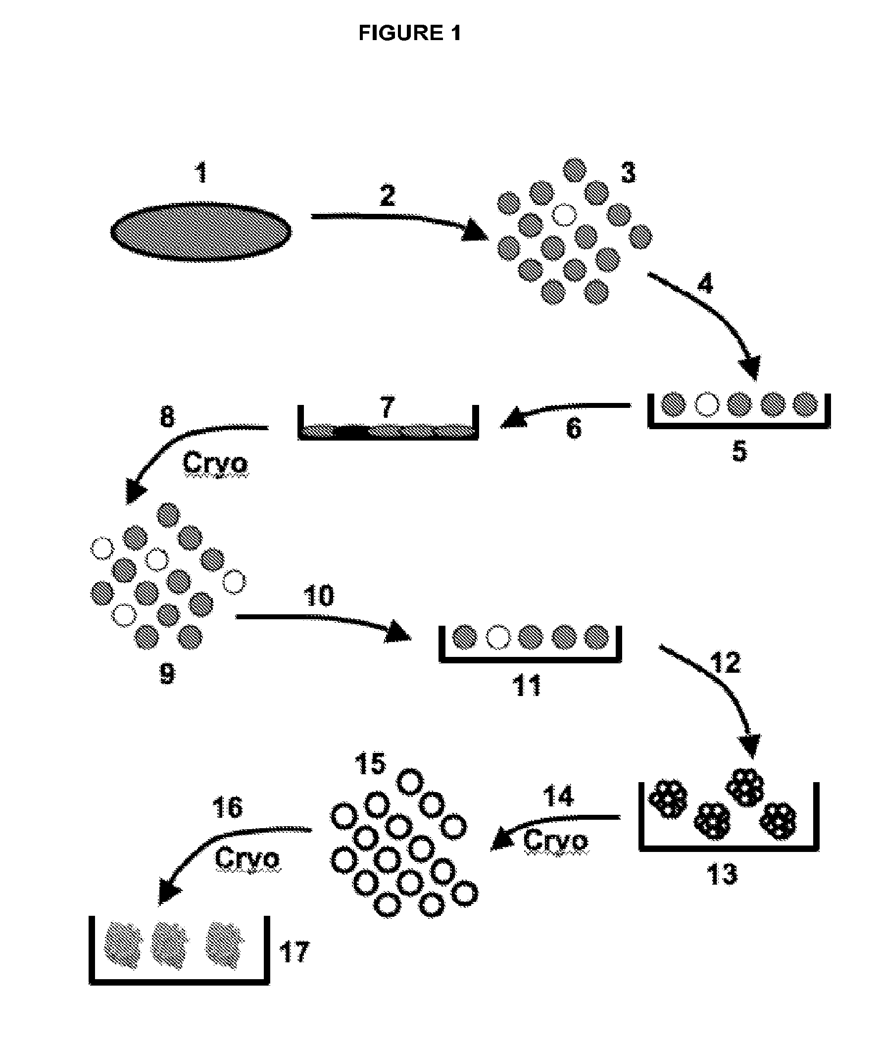
[0315]In one embodiment, the Discotek Culture Platform comprises the steps of (1) preparing a single cell suspension from a disc tissue specimen (FIG. 1, reference numerals 1-3), (2) the Attachment Culture Method (FIG. 1, reference numerals 4-7), (3) the Discosphere Culture Method (reference numerals 8-13), and (4) the Cluster Culture Method (FIG. 1, reference numerals 14-17).

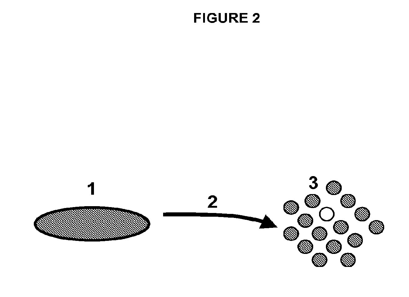
[0316]Disc tissue was procured from a human patient during surgery, or from an experimental animal model (FIGS. 1-2, reference number 1). The tissue was processed by dissection and then enzymatically digested overnight for 24 hours at 37° C. in Collagenase II (FIGS. 1-2, reference number 2). The result was a single cell suspension of all cells that comprise the disc tissue that can be plated into the subsequent culture steps (FIGS. 1-2, reference number 3).

[0317]The single cell suspension was plated into the attachment culture (reference number 4), in which the plates were coated with g...

example 2

Preparation of Single Cell Suspensions

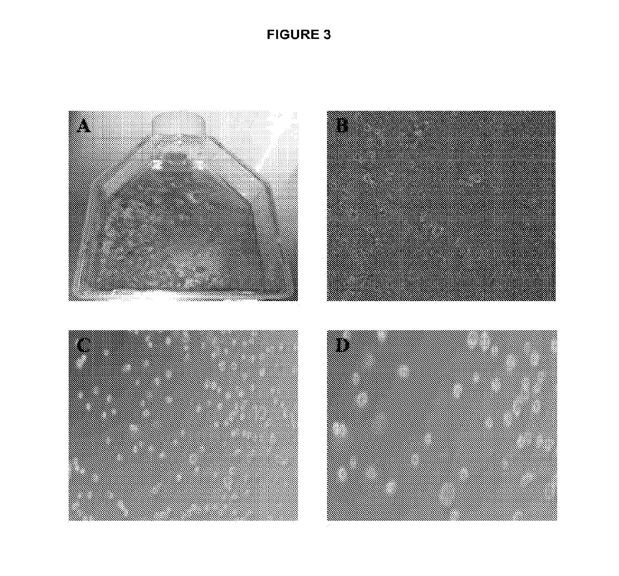
[0320]Human disc tissues were dissected into smaller pieces, and washed thoroughly of blood and other tissue contaminants (FIG. 3A). The dissected fragments were placed in media with Collagenase II at 300 units / ml and incubated for 24 hours at 37° C. (FIG. 3B). A large amount of debris was present that made counting cells difficult and was removed with the washing step. A cell strainer was not used because of the routine presence of cellular clusters that are potentially notochordal clusters that do not break up without further trypsinization and trituration. It was thought that these cell clusters were a critical source of notochordal cells and that they resist digestion due to their extensive extracellular matrix. A single cell suspension was the product of this method (FIGS. 3C-D). This method does not use Trypsin for prolonged digestion, and does not use mechanical disassociation of tissues, to protect the fragile disc stem cell population. ...

example 3

Attachment Culture Method

[0322]The single cell suspension was plated into the Attachment Culture Method (FIG. 5), in which the plates were coated with gelatin, causing the single cell suspension to attach as it settles to the culture flask floor and contacts the surface (FIGS. 1 and 5, reference numerals 4-5). The cultures then grew efficiently as a monolayer reaching confluency in 4-7 days when the starting disc tissue material was derived from young healthy disc tissue specimens. All species and even degenerated tissues grow well with this culture method, but the rate of growth varies and is delayed with tissues from older patients or animals, and degenerated tissues, and certain species (i.e., rat, rabbit, neonatal porcine, young healthy human tissue), as compared to specimens derived from humans that are older, degenerated tissues from any species, or certain other species that are more acellular generally speaking (i.e., adult porcine, cow, etc.) When the cells were confluent, ...


PUM

PropertyMeasurementUnit
concentrationaaaaaaaaaa
timeaaaaaaaaaa
sizeaaaaaaaaaa

Abstract

This invention provides disc stem cells, processes for obtaining and culturing disc stem cells, and methods for repairing damaged or diseased disc tissue comprising the use of the disc stem cells of the invention.

Description

CROSS REFERENCE TO RELATED APPLICATIONS[0001]This application claims priority to U.S. Provisional Patent Application 61 / 442,315, filed Feb. 14, 2011 and U.S. patent application Ser. No. 13 / 113,599, filed May 23, 2011, both of which are incorporated by reference herein in their entirety.FIELD OF THE INVENTION[0002]This invention provides disc stem cells, processes for obtaining and culturing disc stem cells, and methods for repairing damaged or diseased disc tissue comprising the use of the disc stem cells of the invention.BACKGROUND OF THE INVENTION[0003]Neck and lower back pain from degeneration of the spinal disc joint constitutes a common and significant health and economic burden. Indeed, roughly 80% of adults will experience neck and lower back pain at some point in their lives due to intervertebral degeneration. Intervertebral disc degeneration is a product of lifelong, slow, and chronic degeneration that is synchronized with remodeling of the disc and neighboring bony structu...

Claims


Application Information

Patent Timeline
Patent Type & Authority Applications(United States)
IPC IPC(8): C12N5/077
CPCC12N5/0655A61K35/12C12N2500/25C12N2500/46C12N2500/90C12N2501/11C12N2501/115C12N2501/392C12N2533/54C12N2533/78A61K35/32C12N5/066C12N5/0662C12N2500/84
Inventor KUKEKOV, VALERYAKBAR, UMARDUNTSCH, CHRISTOPHER
Owner DISCGENICS
Learn More