Teachable object contour mapping for biology image region partition

a contour mapping and image technology, applied in image enhancement, image analysis, instruments, etc., can solve the problems of limited application, difficult to apply standard image processing software functions to perform biology image recognition, and plug-ins developed in one lab for image recognition rarely work for the application of a second lab, etc., to achieve accurate region partitioning, effective and efficient fitting, and easy tailoring

Inactive Publication Date: 2014-10-16
DRVISION TECH
View PDF12 Cites 11 Cited by
  • Summary
  • Abstract
  • Description
  • Claims
  • Application Information

AI Technical Summary

Benefits of technology

[0010]The current invention provides an object contour mapping method using soft fitting for smooth and stable intensity distribution to enable accurate region partition. The object contour mapping method is teachable so it allows easy tailoring for a broad range of applications. The current invention further uses a multi-resolution approach to encode the spatial correlations and contexts through the spatial integration into low resolution for effective and efficient fitting. The invention also includes a multiple guided partition method for efficient and effective region partition. The teaching image may not be representative of an application. The teaching can be updated by an updated teaching of the current invention. This is important for creating a contour mapping recipe that has stable performance across a broad range of application images
[0011]The primary objective of the invention is to provide teachable object contour mapping method for smooth and stable intensity distribution to enhance all prior art region separation methods. The second objective of this invention is to provide a teachable region partition method for biology image recognition method for broad range of applications. The third objective of the invention is to allow the proper separation of objects even when they overlap and have different sizes. The fou...

Problems solved by technology

It is difficult to apply standard image processing software functions to perform biology image recognition.
As a result the majority of biology recognition is performed either manually or using a simple intensity threshold that has very limited applications.
Yet plug-ins developed in one lab for image recognition rarely work for the application of a second lab.
They are not flexible for broad applications.
The current immature microscopy biology recognition tools impose cost barriers on scientists and the image based scientific discovery process.
The cost in skilled labor for manual recognition and custom script development is high.
A greater cost is that of experiments foregone, or data uncollected, due to problems related to image recognition.
Prior art region partition methods relying on a simple but...

Method used


Image

  • Teachable object contour mapping for biology image region partition
  • Teachable object contour mapping for biology image region partition
  • Teachable object contour mapping for biology image region partition

Examples


Embodiment Construction

[0029]I. Application Scenarios

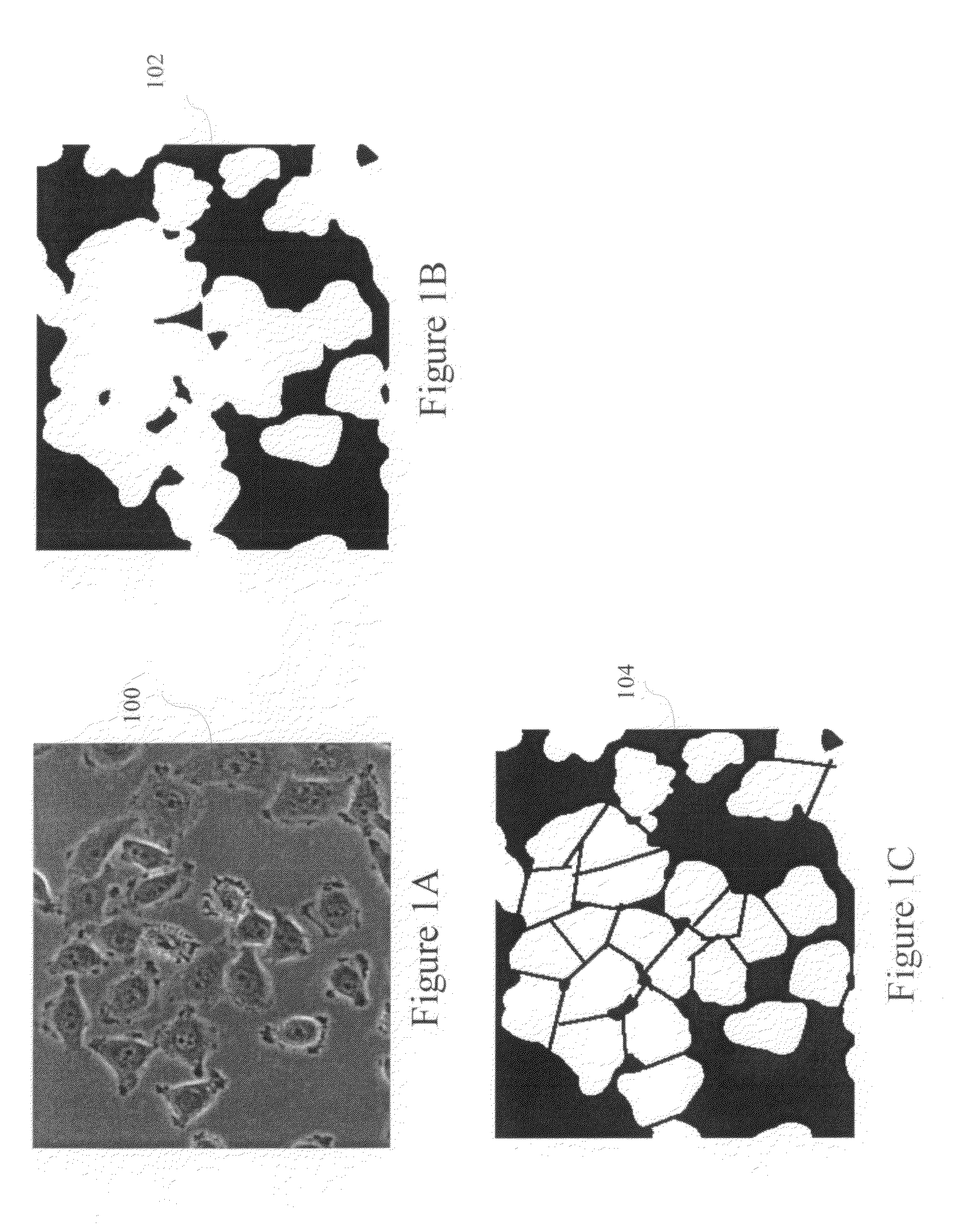
[0030]Biology image region segmentation identifies the regions in computer images where biological objects of interest occupy. Object partitioning in biological image recognition is the process of identifying individual objects in segmented regions. FIG. 1A shows a phase contrast biological image of cells 100 and FIG. 1B shows its biological object segmentation region 102, and 1C shows its region partitioning result 104. The current invention addresses the region partitioning process. Computer image region partition process inputs an image and the segmented objects of interest region and identifies the individual objects among the segmented regions for individual object counting and measurements. The region segmentation step could also create individual object regions from input image directly without the input of the objects of interest region mask.

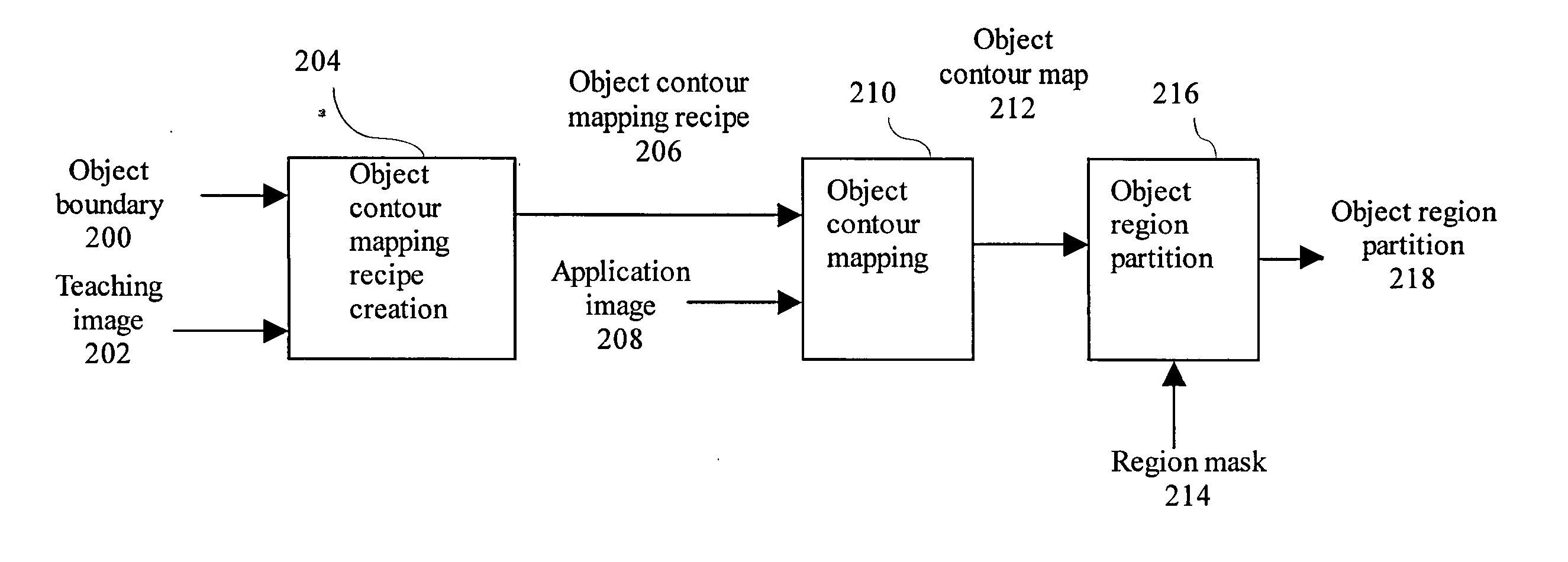
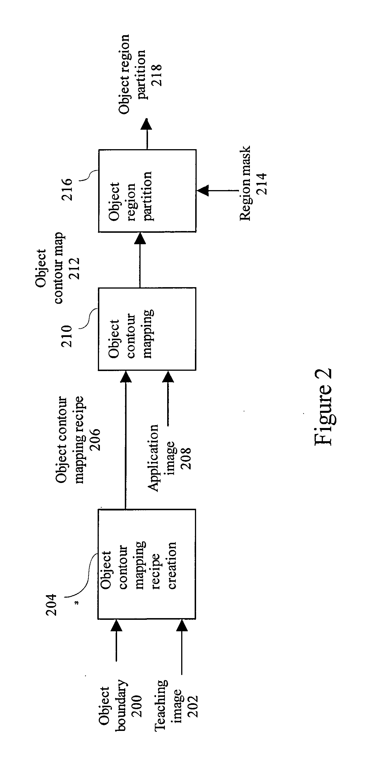
[0031]The application scenario of the teachable region partition method is shown in FIG. 2. It consists o...


PUM


Abstract

A teachable object contour mapping method for region partition receives an object boundary and a teaching image. An object contour mapping recipe creation is performed using the object boundary and the teaching image to generate object contour mapping recipe output. An object contour mapping is applied to an application image using the object contour mapping recipe and the application image to generate object contour map output. An object region partition using the object contour map to generate object region partition output An updateable object contour mapping method receives a contour mapping recipe and a validation image. An object contour mapping is performed using the object contour mapping recipe and the validation image to generate validation contour map output. An object region partition receives a region mask to generate validation object region partition output. A boundary correction is performed using the validation object region partition to generate corrected object boundary output. An update contour mapping is performed using the corrected object boundary, the validation image and the contour mapping recipe to generate updated contour mapping recipe output.

Description

CROSS REFERENCE TO RELATED APPLICATION[0001]This is a divisional of U.S. application Ser. No. 12 / 925,847, filed Nov. 1, 2010.GOVERNMENT INTERESTSStatement as to Rights to Inventions Made Under Federally Sponsored Research and Development[0002]This work was supported by U.S. Government grant numbers 6R44MH075498-03, awarded by the National Institutes of Mental Health. The U.S. Government may have certain rights in the invention.TECHNICAL FIELD[0003]This invention relates to the biology image recognition and the region partitioning step.BACKGROUND OF THE INVENTION[0004]Biology image recognition, the computer extraction of regions containing biological objects such as tissue, cellular and subcellular components, bacteria, viruses of interest in microscopy images, is a fundamental step in quantitative microscopy which has broad applications and markets in basic research, drug discovery, and disease diagnosis. Biology image recognition consists of two major steps (1) a biological region ...

Claims


Application Information

Patent Timeline
IPC IPC(8): G06K9/62
CPCG06K9/6232G06T2207/10024G06T2207/10056G06T2207/20016G06T2207/20096G06T2207/20156G06T2207/30024G06T7/11G06T7/12G06T7/136G06V20/695
Inventor LEE, SHIH-JONG JAMES
Owner DRVISION TECH
Why Eureka
Try Eureka