Due to the dangers associated with the open needle, it is critical that the patient remain still during the
insertion procedure.
If a patient is not still during the procedure, then the suturing process can take considerably longer.
Once the catheter anchor has been sutured to the patient, it is difficult to move or adjust the catheter.
This results in additional wounds in the patient's skin and tissue, consequently multiplying the
risk of infection or the aforementioned other risks to the patient.
There are several safety risks to both the physician and the patient involved with the insertion of a typical sutured catheter anchor.
One risk is a needlestick injury to the physician.
In some cases this may lead to a chronic or life-threatening
disease including HIV infection or
Hepatitis which are passed from one human to another by blood to blood contact.
The risks associated with a needlestick injury can be very serious.
In many hospitals, it is a requirement that a physician undergo expensive testing for possible infection whenever there is a contaminated or suspected contaminated needlestick injury.
The risk of a needlestick injury is significant when using an unprotected needle even when the patient is completely still.
If a patient is agitated or unstable, the risk of a needlestick injury is greatly increased due to the unpredictable motion of the patient.
A patient can also be subject to a needlestick injury if the physician unwittingly punctures his or her skin during the suturing process and then contaminates the patient with the infected needle.
Needlestick injuries have become a very serious
health risk for medical professionals.
Another
significant risk to the patient during and after the insertion of a sutured catheter anchor is infection.
This can lead to significant complications for the patient depending on the type of infection and the patient's health, and in some cases may become life-threatening or lead to death.
Any time a needle enters, exits, and then re-enters the skin, the risk of
contamination increases, and consequently the
risk of infection to the patient increases.
Therefore, the drawing of the exposed needle or suture thread through the patient's skin and out again increases the risk of infection to the patient by both increasing the number of wound sites and by potentially drawing contaminants into the patient's skin which can come in contact with the patient's bloodstream.
The risk of damage done by the insertion of the needle is yet another safety issue.
Even a skilled physician can damage the patient's skin, underlying tissue, nerves, blood vessels, or worse if the patient moves unexpectedly during the time that the needle has penetrated the patient's body.
Given the typical time that is needed to suture a catheter anchor and the numerous types of conditions under which a catheter anchor might be applied, it is not uncommon for the needle to cause an injury to the patient which could be minor or significant.
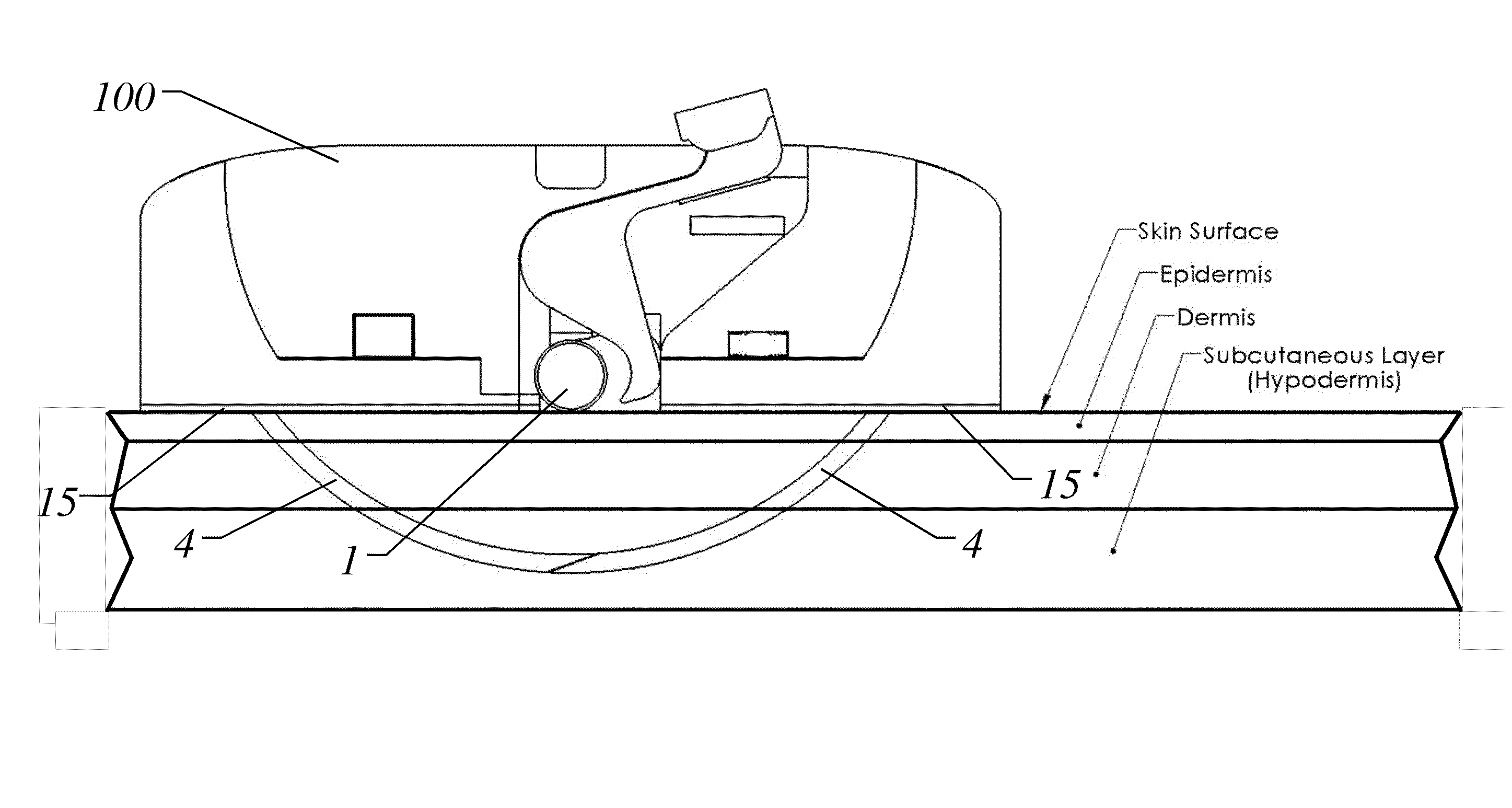
Older patients who have very thin skin (i.e. shallow epidermis and
dermis layers) and minimal fat tissue in the subcutaneous layer of the skin (hypodermis) are particularly at risk for this type of injury especially for catheter insertions in the neck.
Penetrating the skin deep into the subcutaneous layer runs the risk of reaching the underlying
muscle layer or bone depending on the insertion location, and
excessive bleeding may occur due to the presence of larger blood vessels, veins, and arteries.
While no needles or sharps are employed in this methodology, the drawbacks to this type of catheter anchor are significant.
Second, removal of the adhered catheter anchor can cause significant damage to the patient's skin including tearing, and the removal process can be slow and painstaking, sometimes requiring the use of harsh chemicals.
Third,
adhesive-type catheter anchors are difficult to apply to wet, sweaty, or compromised skin.
And the nature of the
adhesive makes it not strong enough to hold most sizes of catheters on the patient's skin, making it suitable only for normally taped applications such as PICC lines.
Some manufacturers of these adhesive-type catheter anchors acknowledge their weaknesses in their own instruction literature and recommend them only for use as a substitute for taped applications, making them unsuitable for catheter applications that normally require sutured catheter anchors.
In these examples and others, the exposed tip of the sharp which exits the skin to engage with the housing during insertion into the patient's skin becomes a
potential risk for infection to the patient.
While the tip of the sharp is exposed to the air (i.e. during the entire time the catheter anchor is attached to the patient) it may be exposed to contaminants.
In order to remove the catheter anchor from the patient, the exposed portion of the sharp is drawn back underneath the skin and through the underlying tissue, potentially exposing the contaminated tip to the patient's bloodstream and increasing the risk of infection over the common suturing methodology.
One significant reason that sharp or needle-based catheter anchors have not been successful in supplanting the sutured catheter anchor is that none have solved the issue of eliminating or significantly reducing needlestick injuries.
These types of catheter anchors can cause needlestick injuries either before, during, or after insertion, and this lack of full protection against needlestick injuries may be responsible for the lack of adoption of these methodologies.
Lerman et al. also claims to have reduced the risk of needlestick injury with its invention, but Lerman lacks any failsafe mechanism to prevent a needlestick injury during insertion or removal.
Furthermore, once the contaminated device has been removed from a patient, there is nothing to prevent the operator or any other person who may come in contact with the device from deploying its sharps and potentially incurring a needlestick injury.
In addition, there is no mechanism that prevents the reuse of the device which could cause grave injury after
contamination.
If the patient is agitated or less than ideal conditions are present, this process can take considerably longer.
When using a sutured catheter anchor the greatest risk from the procedure, particularly one in which the patient is not motionless, is an inadvertent needlestick injury.
Any attempt to speed up the process of inserting the catheter anchor using an unprotected needle (e.g. in a trauma situation where time is of the essence) greatly increases the chances of a needlestick injury.
First, the insertion process of the embodiments nominally takes only about 10-15 seconds in total to fasten the catheter anchor to the patient's skin and to secure the catheter to the catheter anchor.
Second, once the catheter anchor is secured, the catheter can be repositioned as often as needed by releasing the catheter clamping mechanism, repositioning the catheter to its new desired position, and then relocking the catheter clamping mechanism which also takes just a matter of seconds to accomplish.
In any instance where a catheter needs to be repositioned or adjusted after insertion (which happens frequently), the time savings is quite substantial as the sutured catheter anchor would have to be removed and a new one sutured in its place in the new position,
wasting a significant amount of time.
Suturing with a conventional straight or curved needle within the confines of that very shallow and specific depth takes great skill, patience, and dexterity; and this procedure is typically only performed by physicians.
Furthermore, any tools or additional materials (such as the needle or suture) needed to insert the catheter anchor could be lost or misplaced during the procedure, further slowing the process of insertion or extraction.
Proprietary tools or instruments are also likely to increase the cost of the procedure.
Consequently, neither the operator nor anyone else is at risk for a needlestick injury at any time through the use of the device.


