Apparatus and method for ovarian cancer screening

a technology of ovarian cancer and ovarian cancer, applied in the field of ovarian cancer screening, can solve the problems that culdocentesis, which involves pelvic fluid sampling, has not been shown to be accurate in detecting ovarian cancer, and achieves the effects of low complication rate, high volume and quick room turnover

Inactive Publication Date: 2015-02-26
MKT ENTERPRISES
View PDF21 Cites 8 Cited by
  • Summary
  • Abstract
  • Description
  • Claims
  • Application Information

AI Technical Summary

Benefits of technology

[0018]The present apparatus and method advantageously takes advantage of a culdoscopic approach in combination with sampling techniques from within the peritoneal cavity to identify ovarian cancer or precursor cells. The known art only uses imprint cytology, which is a smear of the ovary after ovarian removal (e.g., in the pathology lab), to assist in surgical decision making at the time of ovarian removal.
[0019]The presently described technique can also be provided as a brief screen of women concurrent with a colonoscopy. Women at risk for ovarian cancer are in the same age group as women who are traditional candidates for colonoscopy screening. In addition, the literature suggests a low complication rate for culdoscopy, the most common complication being bowel perforation; and women who present for colon cancer screening have already consented to a procedure with the same risk profile. Colonoscopy suites typically function with high volume and quick room turnover, and the present method could reasonably be performed within 30 minutes.
[0020]Patients identified with cancer would be better able to be counseled preoperatively and indeed may opt to schedule their procedure at a different time or facility so that a gynecologic-oncologist would be readily available. Women who have benign results may also be able to observe pelvic cysts. Additionally, in the case of women with genetic risk for cancer, reassuring results may assist in the decision regarding timing of risk reduction surgery (e.g., many women want to complete their families or avoid menopausal symptoms until later ages).
[0021]Finally, the present apparatus and method provide for direct sampling from the ovary / fallopian tube complex without removal of potentially normal organs.

Problems solved by technology

Unfortunately, it is non-specific.
This is a good tool, but not specific enough to be used as a primary screening tool.
Culdocentesis, which involves sampling of pelvic fluid, has not been shown to be accurate in detecting ovarian malignancy even when the disease is known to be present.

Method used


Image

  • Apparatus and method for ovarian cancer screening
  • Apparatus and method for ovarian cancer screening
  • Apparatus and method for ovarian cancer screening

Examples


example 1

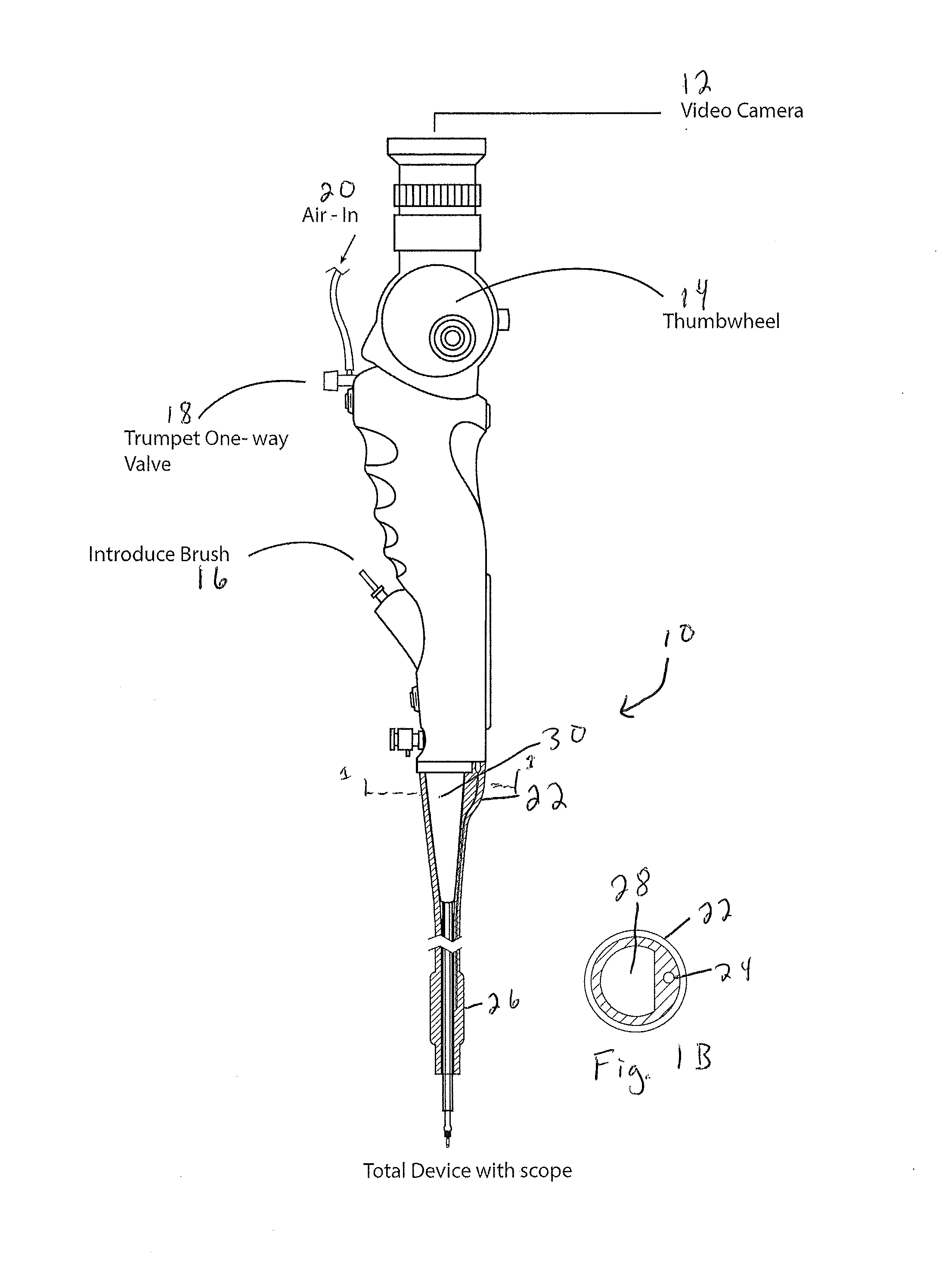
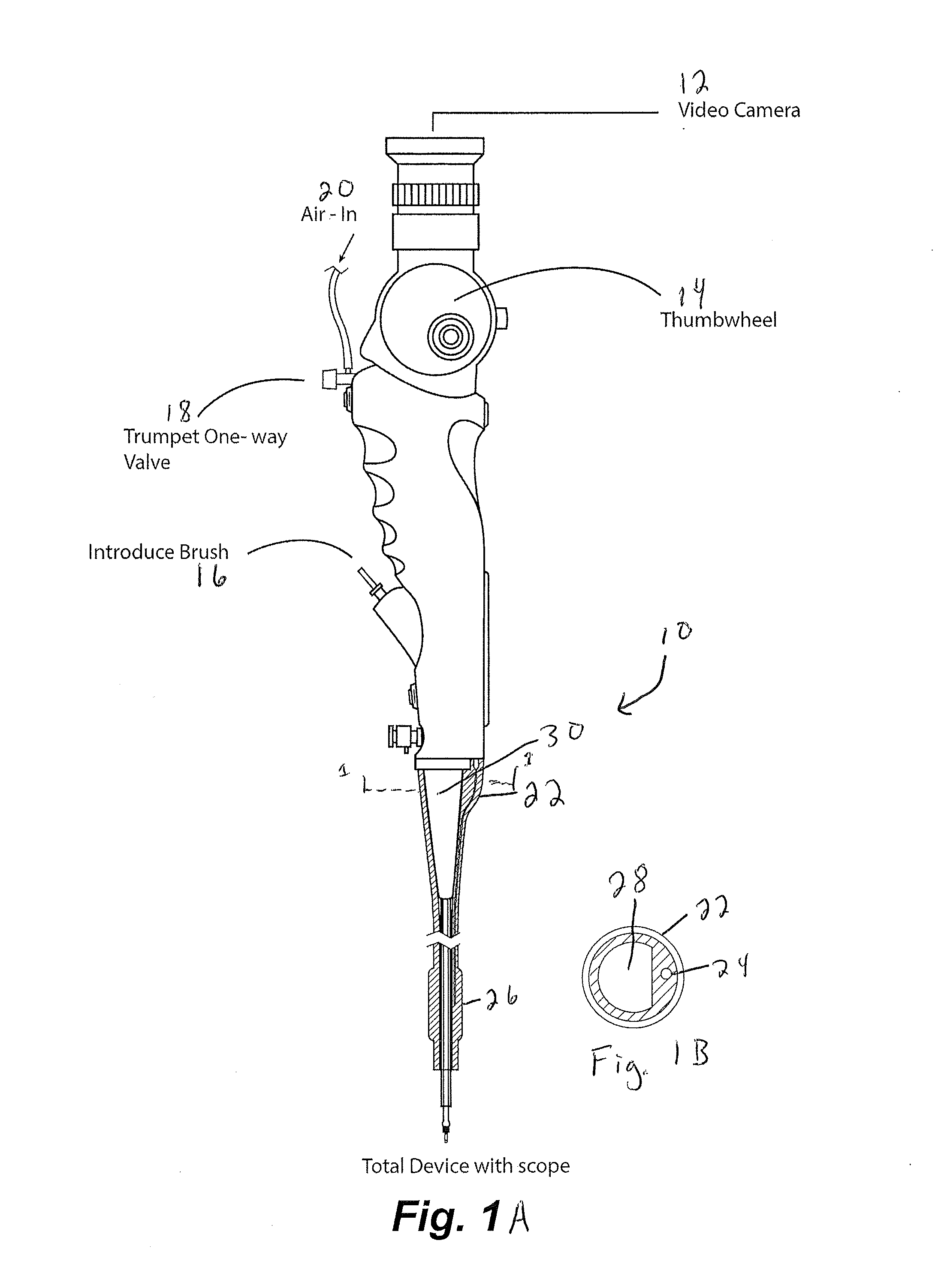
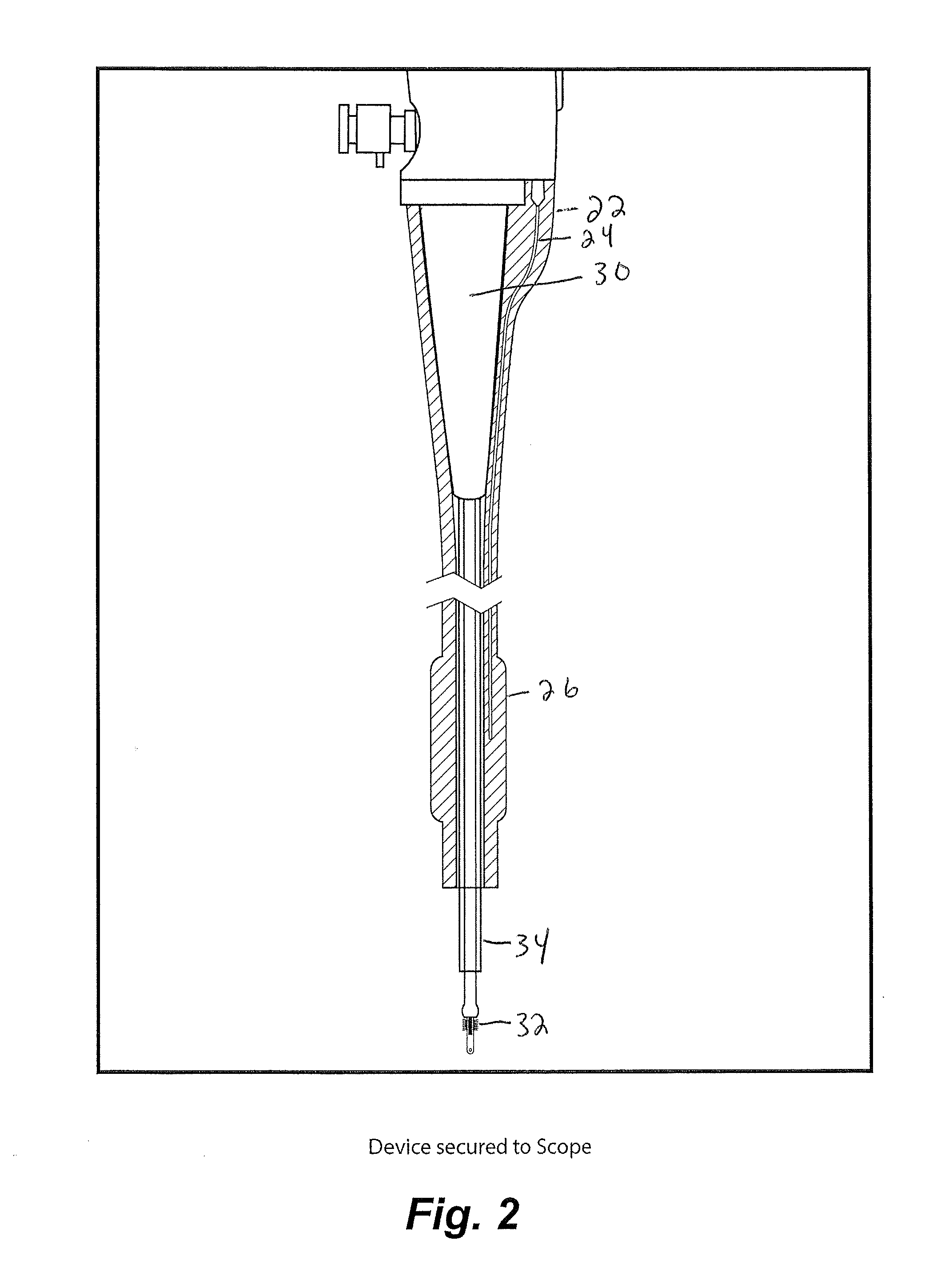
[0078]An exemplary procedure is described immediately below with reference to FIGS. 17-24:

[0079]1. Insert catheter assembly into 5 mm port in the uteroscope;

[0080]2. Insert sampling device into scope working channel (brush end first, the handle is connected on the other end);

[0081]3. Handle should be about ½ inch from touching the working channel;

[0082]4. Insert scope into scope lumen;

[0083]5. Inflate the balloon by attaching a non-depressed standard 30 mL syringe to the stopcock. Once desired inflation is complete close stopcock and disconnect syringe (this is optional but it allows for more room if you remove the syringe.);

[0084]6. Position the end of the uteroscope at the end of one fallopian tube;

[0085]7. Connect depressed 3 mL syringe to luer lock and remove approximately 1 mL of fluid. If required, flush the internal with saline to obtain necessary volumes. Place fluid in standard cup to send to lab;

[0086]8. Move the end of the sampling device to the surface of the ovaries. Cl...


PUM


Abstract

An apparatus and method are provided for sampling the distal tube, fimbria and/or ovary, includes advancing a device into the peritoneal cavity and sampling material on or adjacent to the distal tube, fimbria or ovary.

Description

CROSS REFERENCE TO RELATED APPLICATIONS[0001]This application claims the benefit of U.S. Provisional Application No. 61 / 868,298 filed Aug. 21, 2013, the disclosure of which is incorporated by reference herein in its entirety.BACKGROUND[0002]The present disclosure relates to an apparatus and method for ovarian cancer screening, particularly to an apparatus and method for sampling the distal tube, fimbria and / or ovary via catheter accessing the peritoneal cavity via the cul de sac.[0003]Traditional approaches for ovarian cancer screening have either been though the cervix and into the fallopian tubes or via ovarian removal. For example, U.S. Patent Publication No. 2012 / 0315662 to Linnemeier describes detection of precancerous changes in the fallopian tubes. The described mechanism includes insertion of a catheter through the cervix and into the fallopian tubes, with a cervical brush sampling cells from within the fallopian tubes. The methodology is described as flowing from hysterosco...

Claims


Application Information

Patent Timeline
Patent Type & Authority Applications(United States)
IPC IPC(8): A61B10/02A61B10/04A61M25/10
CPCA61B10/0291A61M25/1002A61B10/04A61M2210/1408A61M2025/105A61M2210/1082A61M2210/1425A61B10/0266A61M2210/1433A61B17/4241A61B2017/4233
Inventor MAZZOLI, JR., THOMAS G.ELLIS, MARIA LYNNEMCDONALD, KIMBERLY ANNFREED, DAVID
Owner MKT ENTERPRISES
Why Eureka
Try Eureka