Method and device for automatic or semi-automatic segmentation of a 3D image data set

a 3d image and data set technology, applied in the field of automatic or semi-automatic segmentation of 3d image data sets, can solve the problems of slow and error-prone segmentation of image data sets from medical imaging apparatuses, and inability to achieve the effect of a large number of user inputs

Active Publication Date: 2015-03-19
SIEMENS HEATHCARE GMBH
View PDF2 Cites 5 Cited by
  • Summary
  • Abstract
  • Description
  • Claims
  • Application Information

AI Technical Summary

Benefits of technology

[0006]An object of the present invention to provide a method for segmentation of a 3D image data set acquired by a medical imaging apparatus, which avoids the disadvantages of the pri

Problems solved by technology

Segmentations of image data sets from medical imaging apparatuses are not reliable for additional image processing nor for a better detection capability and diagnosis.
The model-based segmentation can be implemented quickly, automatically at the press of a button, and is able to function even given image data with reduced image q

Method used


Image

  • Method and device for automatic or semi-automatic segmentation of a 3D image data set
  • Method and device for automatic or semi-automatic segmentation of a 3D image data set

Examples


Embodiment Construction

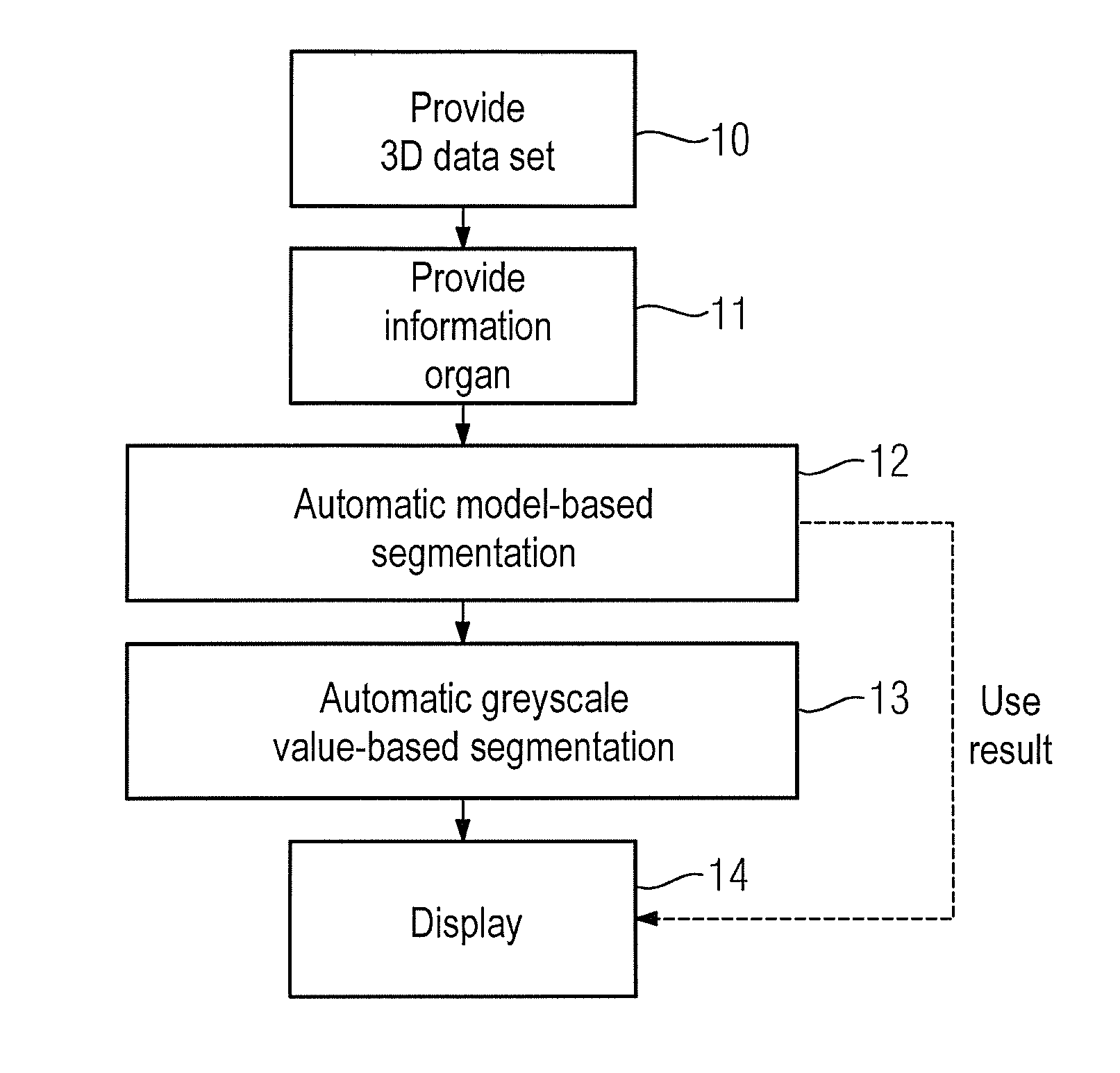
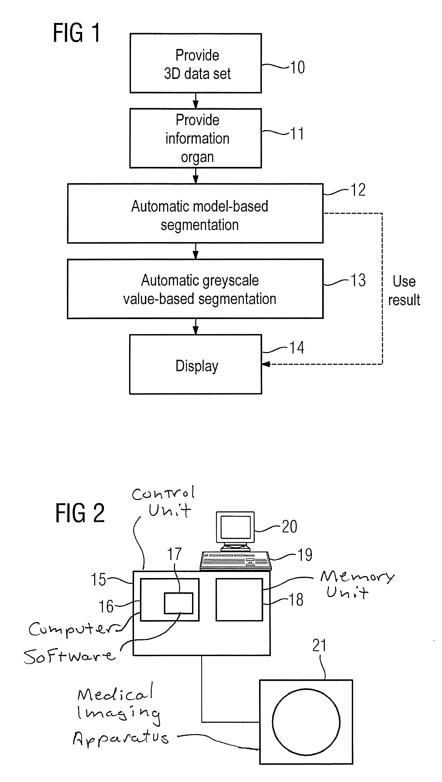
[0023]A sequence of the automatic method according to the invention, with two successively running segmentations on the basis of different segmentation algorithms, is shown in FIG. 1. The method can be implemented by a device shown in FIG. 2, wherein a control unit 15 adopts the control of the method. In a first step 10, a 3D image data set of an examination region is initially provided, the 3D image data set including a depiction of an organ. An organ in this context means a contiguous region of only a portion of the human body, for example hand, foot, brain, heart, left atrium of the heart, aortic root etc. The 3D image data set has been acquired by a medical imaging apparatus, for example a magnetic resonance tomography apparatus, a computed tomography apparatus, or an x-ray apparatus. The acquisition, and possibly a reconstruction of the 3D image data set, can have been implemented immediately before being provided, or the 3D image data set can already be acquired long before an...


PUM


Abstract

In a method and apparatus for automatic or semi-automatic segmentation of a 3D image data set, acquired by a medical imaging apparatus, of an examination region that includes an organ, the 3D image data set is provided to a computer/processor, which is also provided with information with designating the type of organ imaged in the examination region. The 3D image data set is automatically segmented in the computer/processor using a model-based segmentation algorithm, wherein the designated type of organ is used as a basis of the model. The 3D data set is also automatically or semi-automatically segmented using a greyscale value-based segmentation algorithm. At least one of the segmentation results is displayed.

Description

BACKGROUND OF THE INVENTION[0001]1. Field of the Invention[0002]The invention concerns a method for automatic or semi-automatic segmentation of a 3D image data set, acquired by a medical imaging apparatus, and a device to implement such a method.[0003]2. Description of the Prior Art[0004]Segmentations of image data sets from medical imaging apparatuses are not reliable for additional image processing nor for a better detection capability and diagnosis. A segmentation (for example of the left atrium of a human heart) from three-dimensional CT, MR or rotation angiography image data is important for planning and implementation of medical procedures and ablation procedures, since the left atrium of the heart has a high individual variability. Currently, model-based or greyscale value-based segmentation algorithms are used; both types have advantages and disadvantages. The model-based segmentation can be implemented quickly, automatically at the press of a button, and is able to function...

Claims


Application Information

Patent Timeline
IPC IPC(8): G06K9/46G06T7/00
CPCG06K9/46G06T2207/30004G06K2009/4666G06T7/0012G06T7/10G06T2207/10081G06T2207/10088G06T2207/10116G06T7/11G06T7/12G06T7/149
Inventor JOHN, MATTHIASRAHN, NORBERT
Owner SIEMENS HEATHCARE GMBH
Why Eureka
Try Eureka